Genetics Of Periodontitis Notes

Genetics Of Periodontitis

Periodontitis is a dental disorder involving inflammation and infection of the ligaments and bones that support the teeth and results from progressive and uncontrolled gingivitis.

Classification of periodontitis is based on the rate of disease progression. Periodontitis can be divided into two major types, e.g. aggressive (localized aggressive and generalized aggressive) and chronic.

Until recently, periodontitis was thought to be strictly determined by environmental factors like poor oral hygiene, smoking, low socioeconomic condition, bacterial infections of oral cavity, etc. alone. It is a common observation that given the same status of poor oral hygiene and sharing the same environmental factors, some people show severe periodontal disease while some of the same people suffer from mild or no disease.

This indicates the existence of individual differences in susceptibility to the disease and perhaps towards a genetic basis for the susceptibility. Scientists are trying to find out the genes responsible for the disease. It is now well known that periodontal disease is multifactorial (complex) and its susceptibility is influenced both by genetic and environmental factors.

Genes responsible for multifactorial disease are also known as susceptibility genes. However, these genes alone do not produce the disease unless they are exposed to the necessary environmental factors.

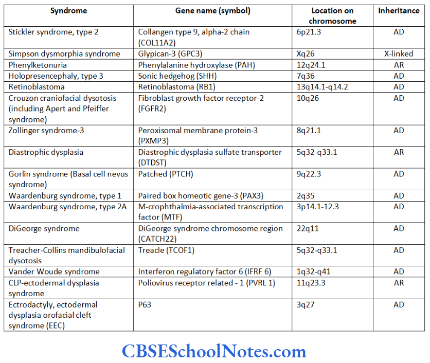
Periodontitis is also associated with many types of syndromes and genetic disorders. Periodontitis presents itself as one of the clinical manifestation of the syndrome. This establishes the genetic basis of periodontitis as these syndromes result due to mutation in a single gene/genes.

Read and Learn More Genetics in Dentistry Notes

Verification of the Genetic Basis of Periodontitis

Following genetic analytical methods have been used to evaluate the genetic basis of periodontitis.

Familial Aggregation Of Periodontal Diseases

Several members of the same family suffering from periodontitis is indicative of a genetic etiology. Many scientific studies have indicated familial aggregation for both aggressive (Boughman et al., 1988; Long et al, 1987; Marazita et al, 1994 and Hart and Korman, 1997) and chronic periodontitis (Hassell and Harris, 1995). Reports of the familial nature of chronic forms of periodontitis are less frequent.

This aggregation within families strongly suggests of a genetic predisposition to the disease. However, the familial aggregation of the disease may also reflect on the common environmental factors prevalent in the family, i.e. levels of oral hygiene, shared transmission of bacteria, same socioeconomic condition, sanitation, etc.

Therefore familial aggregation is only suggestive of a genetic basis but does not prove a definite genetic etiology. To substantiate evidence in favor of a genetic basis of familial aggregation, application of more specific tools such as segregation and twin studies are imperative.

Genetics In Dentistry Genetics Of Periodontitis Patient showing localized aggressive

Genetics In Dentistry Genetics Of Periodontitis Patient showing chronic generalized periodontitis

Segregation Analysis

Segregation analysis is conducted in affected families to determine the mode of inheritance of a disease whether it follows an autosomal-dominant, autosomal- recessive, X-linked dominant, X-linked recessive or multifactorial mode of inheritance.

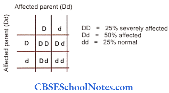
Many studies have been conducted to determine the mode of inheritance of diseases. These studies have indicated many patterns of inheritance in periodontitis. The most definitive segregation analyses in North American families were performed by Marazita and coworkers (1994) who studied more than 100 families segregating aggressive forms of periodontitis and found support for an autosomal-dominant transmission.

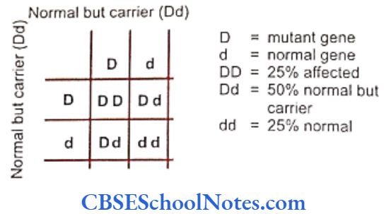
They concluded that autosomal-dominant inheritance with approximately 70% penetrance occurred for both Blacks and nonBlacks. It is believed that various forms of aggressive periodontitis are due to single gene defects that are inherited as autosomal- dominant disorders with incomplete penetrance. Some studies have indicated an autosomal recessive mode of inheritance (Saxen, 1980; Saxen and Nevanlinna, 1984). Few workers have also reported X-linked inheritance (Melnick et al, 1976) in the same context.

Why were these studies unable to point towards a single type of mode of inheritance? It might be because of the multifactorial or polygenic nature of the disease or probably due to the fact that the origin of periodontitis is heterogeneous in nature. There is a common precedent in genetics, with relation to heritable pathologic conditions, to show different modes of inheritance in different families. These findings reflect that different genetic loci are capable of causing the same disease in both dominant and recessive manners.

Even though segregation studies have shown different modes of inheritance, they all support the involvement of genes in the causation of the disease.

Twin Studies

Many human traits are multifactorial (polygenic). The modes of inheritance in which can be manipulated with environmental modifications. Twin studies allow evaluation of the importance of genetic as well as environmental factors in causation of a disease. As early as in 1940, Noack recognized similarities of features in periodontal conditions seen in identical twins. Many twin studies in the past were carried out on monozygotic adult twin groups. These twins were grouped as the reared-together and reared-apart sections. Following conclusions were drawn that:

  • The periodontal conditions in identical twins were similar.
  • A significant genetic influence on various parameters of periodontal diseases was found.
  • Findings in the reared-together and reared-apart groups were similar which strongly suggested a genetic basis of the disease. This finding also indicated that environmental factors had no significant influence on development of the disease.

A study by Corey et al (1993) on monozygotic and dizygotic twins indicated a higher concordance rate for monozygotic twins (23% for monozygotic twins as compared to 8% for dizygotic twins). Concordance implied that both the twins of a pair were suffering from periodontitis. This strongly suggested the involvement of a genetic component in the etiology of periodontitis as we know that monozygotic twins possessed identical genes.

Michalowicz et al (1991) investigated alveolar bone height and probing depth in twins from the Minnesota study and showed significant variations in them, according to the differences in their genotype. The twin groups had similar smoking histories and oral hygiene practices. It was concluded that genetics plays a role in moulding the susceptibility to periodontal disease.

In another study Michalowicz (1994) found that monozygotic twins reared either together or apart have been found to bear a more similar type of periodontal disease experience than found between dizygotic twins. These findings indicate that genetic factors may influence the manifestations of periodontal disease.

In a more recent study (Michalowicz et al, 2000) of another large human twin cohort, hereditary factors accounted for approximately 50% of “adult period- ontitis”. The heritable component for periodontitis was not associated or influenced by behaviors such as smoking, utilization of dental care, and oral hygiene habits. This indicated that development of periodontitis was influenced by genes that mediated biological mechanisms.

Twin studies were also used to know about the effect of host genes on the composition of micro- bacteria in the oral cavity. The long-term colonization of bacteria in the oral cavity may be due to the genetic constitution of the host. Studies have revealed that adolescent twins were more similar in their oral microbiota than pairs of unrelated individuals (Moore et al, 1993).

However, the study of Michalowicz et al (1999) in adult twins indicated that neither the host genes nor the environment played a significant influence on the presence of bacteria in subgingival plaque. These two studies suggested that although the host genes influence initial bacterial colonization but this influences do not persist till adulthood.

Linkage Studies For Periodontitis

Linkage studies are conducted to localize the disease causing gene on a specific chromosome. Till date only very few linkage studies analyzing aggressive periodontitis are reported.

  • Saxen and Koskimies carried out the first linkage study in 1984 on Finnish families to test the claim for association of aggressive periodontitis with HLA antigens. This study concluded that the gene responsible for aggressive periodontitis was not linked with HLA antigen in these families.
  • Boughman et al reported the second linkage study in 1986 for a Maryland population. According to this study the gene responsible for aggressive periodontitis was situated on the long arm of chromosome number 4 (4q 11-13). The inheritance pattern of this gene was determined as autosomal- dominant. The gene was found to cosegregate with the gene responsible for dentinogenesis imperfecta. Both the genes were linked, i.e. present close to each other on the long arm of chromosome number 4.
  • However, another study in 1993 (Hart et al.) on a different population of USA failed to locate the gene for aggressive periodontitis on chromosome number 4. This finding indicated that aggressive periodontitis was heterogeneous in nature, i.e. different genes may be responsible for different forms of aggressive periodontitits.
  • Prepubertal periodontitis (PPP) is a rare and rapidly progressive disease of young children that results in destruction of the periodontal support of the primary dentition. The condition may occur as part of a recognized syndrome or may occur as an isolated finding. Both autosomal dominant and recessive forms of Mendelian transmission have been reported for PPP. Hart et al. (2000) have localized a gene of major effect for PPP on chromosome 11q14. This PPP candidate interval overlaps the region of chromosome 11q14 that contains the cathepsin C gene responsible for Papillon-Lefèvre and Haim-Munk syndromes.
  • Recently in 2004, a linkage study by Li and coworkers mapped the gene responsible for localized aggressive periodontitis on the long arm of chromosome number 1 (1925).

Association Studies For Periodontitis

The susceptibility to a disease depends upon the genetic background of a person. Since periodontitis is an inflammatory disease, it is believed to be affected by genes that are involved in the regulation of inflammatory cell functions. Associations between periodontitis and genes responsible for control of inflammat response are of great value in understanding the genetics of periodontitis.

The immune system controls inflammatory responses against a disease. However, the immune response to a disease varies from person to person and the pattern of immune response in an individual is determined by the genetic make-up unique to the person. The clinical presentation of a disease, therefore, varies in different persons reflecting the diversity of immune response shown by each individual. The same concept is also applicable to developing specific and targeted medical treatment.

Thus if we are able to identify the variations in the genes involved in the control of inflammatory process and ultimately the disease, we can successfully identify the degree of susceptibility, severity and prognosis of the disease and perhaps design medical interventions custom made according to a particular etiology. In recent past many studies have identified the association between periodontitis and variation in the genes (single nucleotide polymorphism) responsible for regulation of immune and inflammatory response.

Association of HLA (Human Leukocyte Antigens) with Periodontitis

The human leukocyte antigen (HLA) complex plays an important role in immune response. Many autoimmune diseases are found to be associated with various HLA antigens. Genes for class I and II antigens are located on chromosome number 6. At present more than 150 HLA antigens are known. The HLA molecules are involved in antigen recognition of periodontal pathogens, interaction between T and B- lymphocytes and in production of IgG. Following positive association between HLA and periodontitis was found:

  • A positive association was reported between aggressive periodontitis and HLA-A9 and B15 antigens (Sofaer, 1990). Persons having these two antigens are at 3.5 times higher risk of developing the disease as compared to those who are negative for these antigens. Thus HLA -A9 and B15 seem to represent susceptibility factors for aggressive periodontitis.
  • Class 2 DR4 antigen is in association with type I diabetes mellitus (Rotter et al, (1992). Since periodontitis is a diabetes related complication, DR4 antigen and periodontitis are said to be in association with each other. The DR4 antigens were found to be more prevalent in patients with aggressive periodontitis than in controls (Katz, Goultschin, Benoliel et al., 1987). However, a few studies have been unable to find any association between the two.
  • HLA-A2 and HLA-B5 antigens appear to be less prevalent in patients of aggressive periodontitis as compared to controls (Kaslick, West, Chasens et al, 1975). This indicates that these HLA antigens are protective in nature.

Though above studies have indicated a positive association, many other studies have also pointed towards a negative association between HLA and periodontitis. The cause of this discrepancy appears to be the involvement of environmental factors and racial differences acting as variables.

Association between Periodontitis and Interleukin-1 (IL-1) Gene Polymorphism

Interleukin-1 plays an important role in the initiation and progression of periodontal disease. Il-1 is found in two different forms; IL-1a and IL-1b. Genes for IL-1 are present on the long arm of chromosome number 2 (2q13). IL-1 is mainly produced by activated monocytes. There is an increased level of IL-1 in periodontal tissue which stimulates bone reabsorption, inhibits collagen synthesis, up regulates matrix metalloproteinase activity and synthesis of prostaglandin.

  • An initial study (Korhman et al, 1997) reported a positive association between polymorphism in the genes encoding for IL-1 and IL-1B (composite genotype) and increased severity of periodontitis. This association was seen in nonsmokers. Genotype positive nonsmokers were 6.8 times more likely to have severe periodontal disease.
  • Another study (Mc Guire and Nunn, 1999) reported that the IL-1 genotype increased the risk of tooth loss by 2.7 times and heavy smoking increased it by 2.9 times. The combined effect of heavy smoking and IL-1 genotype positive increased the risk of tooth loss for 7.7 times.
  • A study carried out in 2001 by Axelsson indicated that genetic polymorphism of IL-1 and smoking seems to have synergistic risk factors. These factors when combined leads not only to tooth loss but also for alveolar bone loss. Nonsmokers are at low risk of tooth loss.

(Thus there are contrasting findings regarding interactions between genetic polymorphism and smoking to affect the risk of periodontitis. Though the IL-1 genotype and smoking habit seem to interact with each other, the nature and direction of this interaction is poorly understood. More studies with large sample size are needed to evaluate the exact correlation between smoking and IL-1 interactions).

  • A variant of the IL-1B coding region with a single nucleotide base pair substitution is associated with a four-fold increase in IL-1B production.
  • Generalized aggressive periodontitis is in linkage disequilibrium with an allele at IL-1B. This suggests that the gene for periodontitis is located close to the gene for IL-1B (Diehl et al, 1999).
  • The IL-1B allele is found to be more prevalent in patients suffering from advance chronic period-ontitis.
  • Though the association between periodontitis and polymorphism of IL-1 gene cluster was observed in about 30% of European population, it was observed so only in 2% of Chinese population (Armitage, 2000).
  • Many studies have indicated the lack of association between interlukin-1 polymorphism and aggressive periodontitis.

Where do we stand now? Studies in both chronic and aggressive periodontitis have yielded mixed results. It is evident that to get a clear picture of the association between IL-1 polymorphism and various types of periodontitis we need more comprehensive studies.

Tumor Necrosis Factor (TNF-α) and Periodontitis

The TNF-a is the proinflammatory cytokinase which is involved in pathogenesis of periodontitis. The TNF-α gene is located on chromosome number 6. Two different polymorphisms have been reported in the promoter region of the gene (Fassmann et al, 2003). This results in increased production of TNF-α. The level of TNF-a molecules is high at the sites of active tissue destruction and low at healthy sites. The TNF-α is the proinflammatory cytokinase

Individuals expressing the TNF-a poly-morphisms manifest greater susceptibility to certain infections. Although till date TNF-a poly-morphism has not directly been linked to susceptibility to periodontitis, there is a strong possibility that such a link exists (Craandijk et al, 2004).

Association between Neutrophil IgG Receptor (FcyR) Polymorphism and Periodontitis

FcyRs are the group of receptors that are expressed on the cell surface of leukocytes binding to IgG antibodies. The interactions between FcyRs and IgG trigger a variety of immune responses like phagocytosis, endocytosis, antibody dependant cellular cytotoxicity and enhancement of antigen presentation.

Polymorphisms in the genes coding FcyRs (especially in FcyRII) receptors (Osborne et al, 1994) are common. The FcyRII receptor is the only receptor that recognizes bacteria opsonized with IgG2. These polymorphisms result in the expression of receptors of high, low and intermediate affinities. The low affinity receptors lead to a decreased immune function and subsequently to increased susceptibility to periodontal pathogen and to severe periodontitis even in presence of high level of antibodies.

Kobayashi et al (2000) found functional poly- morphisms of IgG Fc receptors (FcyR) in Japanese patients with AgP. They found that FcyRIIIb (NA2 allele) was linked with the aggressive form of periodontitis and possibly also linked to an FcyRIIIa allele. Kobayashi et al (2001) reported that 2 FcyRIII alleles may be associated with the degree of severity in chronic periodontitis in a Japanese population.

Meisel et al (2001) analyzed the association between FeyRIIIa (high-affinity receptor) and FcyRIIIb (low- affinity receptor) and chronic periodontitis (CP). Thus a large number of studies have recognized causal association between polymorphisms of FcyR genes and both aggressive and chronic periodontitic conditions.

The alleles of FcyR gene are linked with genes causing periodontitis. The genetic poly-morphism of FeyRII receptor is a promising marker for the susceptibility of periodontitis.

Association between IgG2 Production and Periodontitis

Immunoglobulin G2 (IgG2) is produced in response to periodontal infections. The production of IgG2 is under the control of specific genes. The levels of production of IgG2 vary from person to person. The reduction in the production of IgG2 during the course of periodontal infection may lead to an increased susceptibility to disease. On the other hand an increase in the production of IgG2 provides sufficient protection against the disease. Marazita et al, (1994, 1996) studied families with aggressive periodontitis and found a major locus accounting for approximately 62% of the variance of IgG2 production.

Patients with periodontitis and normal subjects vary greatly in their capacity to produce IgG2. Patients with high titers of IgG2 antibodies have significantly less attachment loss than do patients with low titers. It is observed that patients with localized aggressive periodontitis have high titers of IgG2 compared to patients with generalized periodontitis. This indicates that IgG2 provides sufficient protection against the spread of disease and tries to limit the same.

Associations between Polymorphism in Matrix Metalloproteinase, Cathepsin C and Vit D Receptor with Periodontitis

The matrix metalloproteinase (MMP) are enzymes involved in connective tissue destruction. Single nucleotide polymorphisms are observed in MMP genes and are designated MMP-1, MMP-2, MMP-3 polymorphisms. Very few studies have evaluated the correlation between MMP polymorphisms and periodontitis (Holla et al 2005; Itagaki et al, 2004).

A recent study has indicated a decreased level of Cathepsin C activity in chronic periodontitis. Cathepsin C is a lysosomal enzyme that plays an essential role in immune and inflammatory processes and hence may play an important role in periodontitis (Hewitt et al, 2004). A mutation of the cathepsin C gene leads to Papillon-Lefore syndrome (see later).

Polymorphism in Vit D receptor has been associated with aggressive periodontitis. A very few studies, though, have verified their association.

Syndromic Form Of Periodontitis

It has been observed that there is a clear association between some type of genetically determined syndromes/disorders and periodontitis. These syndromes increase the susceptibility of the individual to periodontitis by interfering with the structural integrity of periodontal tissue or periodontitis may be a concomitant feature of the syndrome. These syndromes are due to mutations in a single gene (monogenic or Mendelian syndromes). Transmission of the disease from one generation to the next follows

Mendelian patterns of inheritance as autosomal- dominant (AD), autosomal-recessive (AR) or X-linked dominant or recessive (XLD or XLR) traits.

As stated above, periodontitis presents itself as a part of a wider spectrum of clinical manifestations of a number of syndromes that occur as a result of mutations in genes. This itself points towards the clearest and most direct clinical evidence that periodontitis has strong genetic etiology. Following is a brief description of some of the genetic disorders where periodontitis is observed as a clinical manifestation.

Neutrophil Functional Dispiders

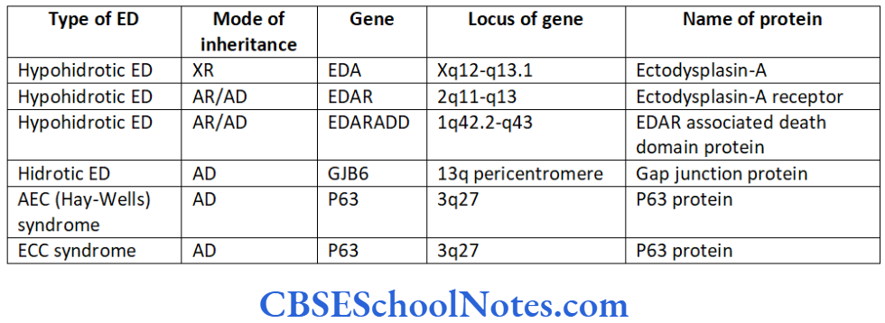
Leukocyte Adhesion Deficiency Syndromes (LAD Syndromes)

Polymorphonuclear leukocytes play an important role in the restriction of the bacterial infection. There are several adhesion receptors on the surface of polymorphonuclear leukocytes. Adhesion receptors are necessary for the proper functioning of leukocytes (phagocytosis and chemotaxis). If the circulating leukocytes have defective or reduced adhesion surface receptors they will not adhere to vascular endothelium.

As a result leukocytes would not be accumulated at the site of inflammation where they are needed to combat the infection. This leads to an increased susceptibility to an infectious disease as exemplified by the leukocyte adhesion deficiency syndrome (LADS). Recurrent bacterial infections, impaired pus formation and impaired wound healing clinically characterize the syndrome. A number of these infections are associated with increased susceptibility to periodontitis. LAD occurs in two forms, LAD syndrome type I and LAD syndrome type 2.

Inheritance: Both types of LAD syndromes are inherited as AR.

Mutation in LAD type 1: The mutation occurs in the B2 integrin chain (ITGB2) gene -21q22.3 (Arnaout et al, 1990). At present more than 20 mutations of the integrin B2 gene are known. The gene mutation causes defects in the integrin receptors of leukocytes. This mutation leads to defects in cell adhesion and chemo- taxis ultimately resulting in increased susceptibility to severe infections including prepubertal aggressive periodontitis.

Mutation in LAD type 2: This is a rare variety of the LAD syndrome and is usually associated with psychomotor retardation (mental retardation, short height and recurrent infection). The syndrome is caused due to a mutation in the guanosine 5′- diphosphate-fucose transporter 1 (SLC 35C1) gene (Lowe et al, 1990; Ishikawa et al, 2005). The infectious episodes and severity of disease is much milder as compared to LAD type I. This syndrome results in chronic periodontitis.

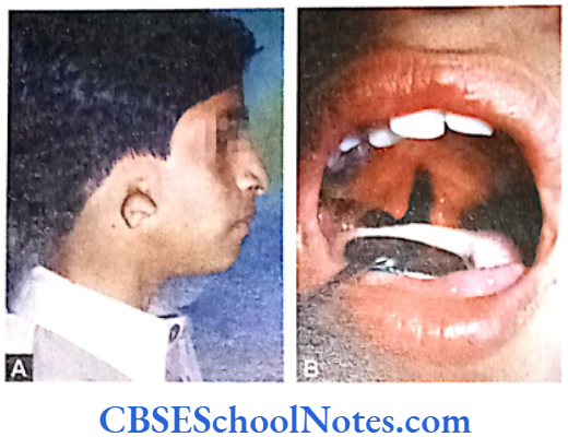
Chédiak-Higashi Syndrome

The syndrome is clinically characterized by a decreased pigmentation of eyes and hair, photophobia, nystagmus and susceptibility to infection.

Inheritance: This syndrome is inherited as an autosomal recessive (AR) trait.

Mutation: The mutation occurs in lysosomal trafficking regulator (LYST) gene-1q42.1-q42.2 (Nagle et al, 1996). This leads to an abnormal transport of vesicles to and from neutrophil lysosomes or to a defect in the ability of cells to produce lysosomes (Charette et al, 2007). Chédiak-Higashi syndrome is associated with severe periodontitis unresponsive to conventional periodontal treatment. However, periodontitis is only seen in its severe form with the syndrome.

Deficiency in Neutrophil Number (Neutropenias)

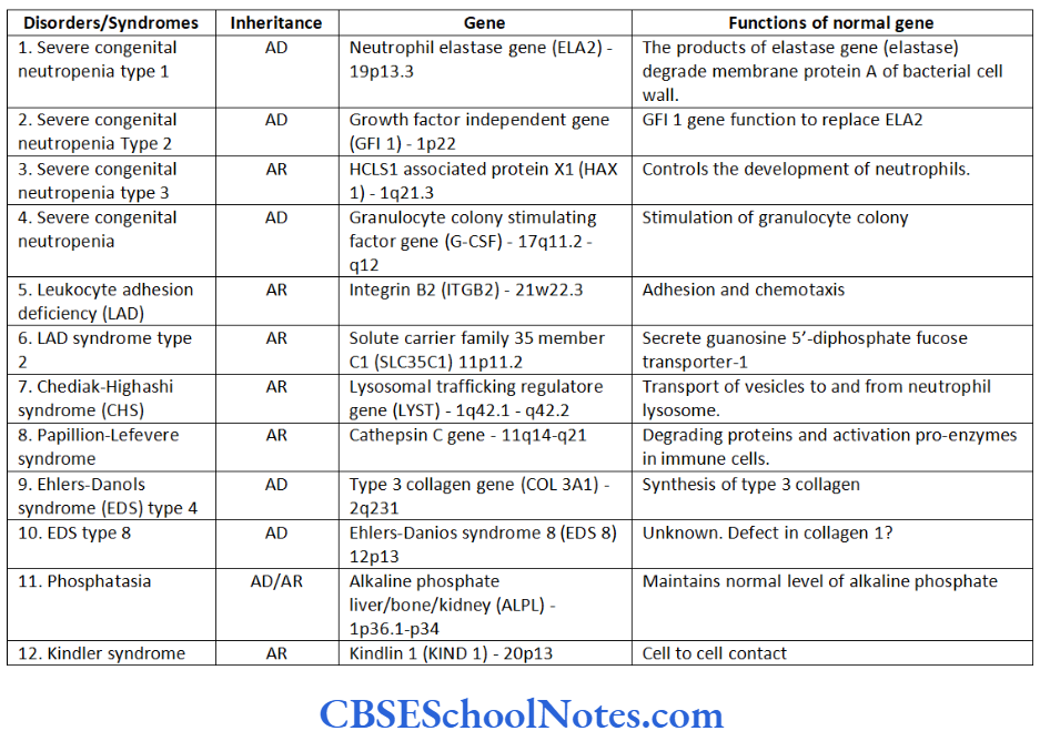
Neutropenia is defined as an abnormally low number of circulating neutrophils. The condition is associated with increased susceptibility to infections such as aggressive periodontitis. Table 14.1- includes various types of neutropenias and associated gene mutations in brief.

Genetic Defects of Structural Components

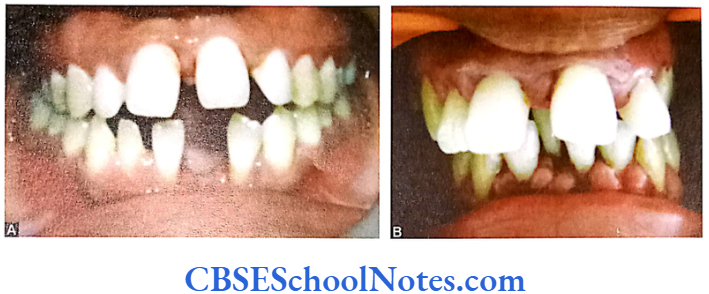
Papillion-Lefèvre Syndrome (PLS)

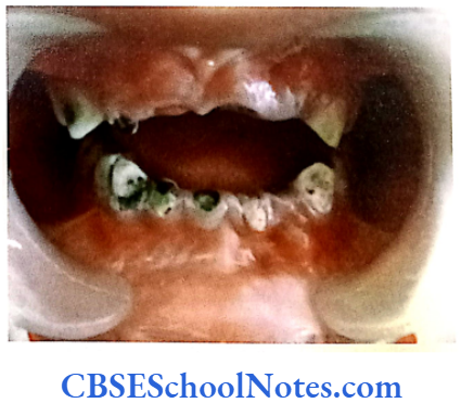
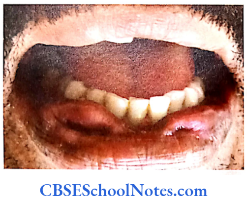
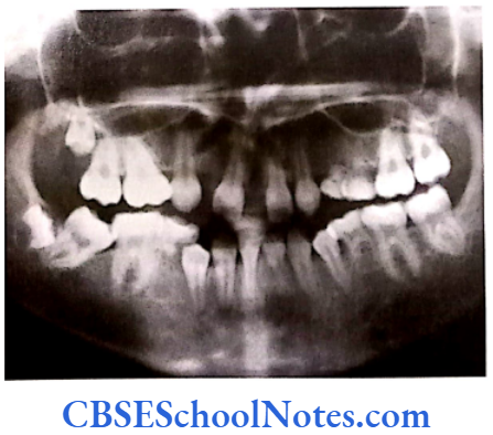
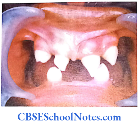
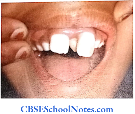
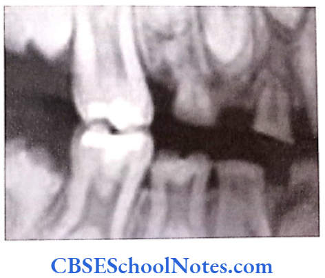
This syndrome is characterized by severe early onset periodontitis, which affects both primary and permanent dentitions and hyperkeratosis of palmar and plantar surfaces.

Inheritance: Autosomal recessive inheritance (AR).

Mutation: It is due to mutations in the cathepsin C gene which is located on chromosome 11 (11q14-q21). At present more than 50 mutations have been identified in cathepsin C gene. Cathepsin C is a cysteine protease that plays a role in degrading proteins and activating proenzymes in immune and inflammatory cells. Inactivation of cathepsin C results in failure to cleave and activate the neutrophil serine proteases cathepsin G, neutrophil elastase and proteinase 3 (Pham et al, 2004).

It was observed that in some of the PLS patients aggressive periodontitis were associated with a kind of virulent microorganism. Elimination of this microorganism prevented the periodontal destruction. This suggested that the periodontitis was not a direct effect of mutation of the gene but the gene mutation increased the susceptibility of a person to infection by the specific microorganism instead.

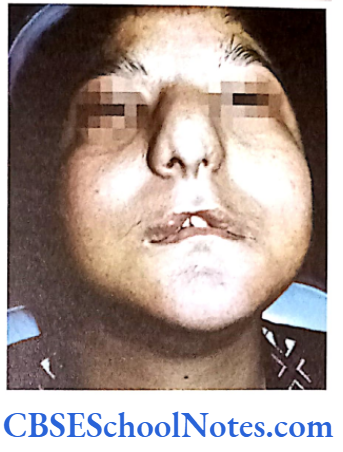
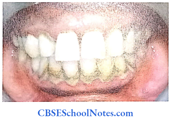
Ehlers-Danlos Syndrome (EDS)

This syndrome is associated with connective tissue disorders. EDS is characterized by defective formation of collagen fibers. The abnormal collagen in the patient leads to fragility and hyperextensibility of the skin, hypermobility of joints, easy bruising and is also associated with early onset of periodontitis. Though at least 17 different types of EDS have been reported but early onset periodontitis has been associated only with two of its subtypes: EDS type 4 and EDS type 8.

Inheritance: Both the types 4 and type 8 EDS are inherited as autosomal-dominant (AD) conditions.

Mutation: The mutation leading to the type 4 EDS was found to be linked to the gene responsible for the synthesis of type 3 (COL 3A1) collagen (Superti-Furga et al, 1988). This leads to the defect in synthesis of collagen 3 resulting in decreased levels of collagen 3.

Genetics In Dentistry Genetics Of Periodontitis Summary of Genetic disorders syndromes associated with periodontitis

Mutations causing type 8 EDS, though, have not yet been identified. This disease has been, nevertheless, linked to chromosome number 12 (12p13). The association of early onset periodontitis with type 8 EDS is more common as compared to that of type 3.
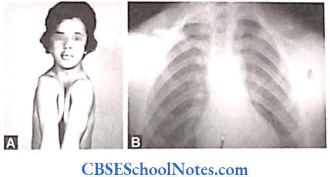
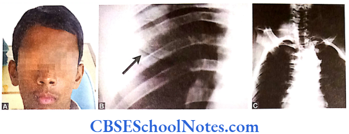
Hypophosphatasia

These patients have decreased levels of alkaline phosphatase in the serum. The condition is associated with abnormal bone mineralization, skeletal abnormalities and cementum hypoplasia. These patients present a severe loss of alveolar bone and premature loss of primary teeth. Pulp chambers may also get enlarged. Lack of connective tissue attachment to bone is responsible for early exfoliation of the primary teeth. Hypophosphatasia may be considered as an etiology of aggressive periodontitis. The condition leads to premature loss of primary teeth and occasionally permanent teeth.

(Mode of) inheritance: Autosomal dominant or recessive.

Mutation: Mutation occurs in the ALPL (alkaline phosphatase, liver/kidney/bone) gene. The gene is localized on the short arm of chromosome 1 (1p36.1- p34). Mutations result in abnormal production of alkaline phosphatase leading to poor mineralization of tissues.
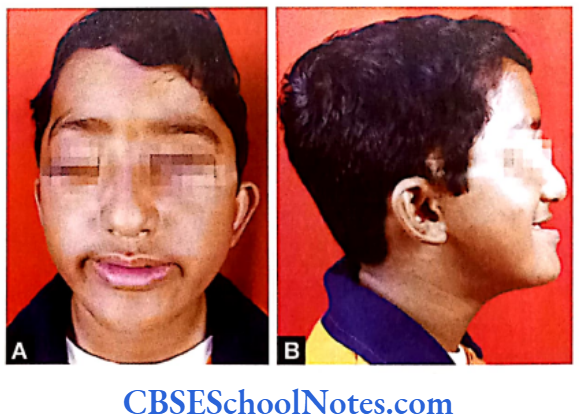
Kindler Syndrome

It is caused due to a mutation in the Kinderlin gene (KIND1) (Jobard et al, 2003). The gene is expressed in many tissues including epidermal keratinocytes. In skin it plays a role in cell adhesion process. Patients of Kindler syndrome show multiple dermatological findings (hyperkeratosis, pigmentation, eczema, skin fragility, etc.), the syndrome is associated with severe aggressive periodontitis both in the primary and secondary dentition.

(Mode of) inheritance: Autosomal recessive (AR) in nature.

Mutation: Mutation occurs in the Kindlin gene (KIND1) situated on the short arm of chromosome number 20 (20p13).

From the above description one can conclude that isolated periodontitits is clearly behaves as a multifactorial disease. Researchers have shown associations between complex form of periodontitis and abnormalities in genes (single nucleotide polymorphism) responsible for regulation of immune and inflammatory responses. Further research is needed to examine the role of both environmental and genetic factors in the causation of periodontitis.

We have also seen that monogenic (single gene) inheritance of periodontitis is possible but these incidences are associated with certain syndromes. We have successfully located many mutant genes (refer table) and understood to some extent the mechanisms of these mutations that disrupt normal homeostasis in the periodontum.

Though a large number of studies are being conducted to identify the genetic basis of periodontitis, we are still far away from determining the risk factors, prevention and treatment of aggressive and chronic periodontitis, genetically.


Genetics Of Dental Caries Notes

Genetics Of Dental Caries

Dental caries is the medical term for tooth decay or cavities. Tooth decay is one of the most common of all disorders of teeth.

Bacteria, that are normally present in the mouth, convert all foods especially sugar and starch into acids. Bacteria, acid, food debris and saliva combine in the mouth to form a sticky substance called plaque that adheres of the teeth. Tooth decay begins if this plaque is not removed throughly and routinely. As stated earlier some of the plaque forming bacteria converts sugar and carbohydrates (starches) in the foods we eat into acids.

These acids dissolve minerals on the surface of the tooth. This erodes the enamel or creates pits on the enamel that are two small to see at first but they get larger over time. Cavities are usually painless until they grow very large and affect nerves or cause a tooth fracture. A tooth abscess can develop. Untreated tooth decay also destroys the internal structure of the tooth (pulp) and ultimately causes loss of the tooth.

The etiology of dental caries has been studied for many years. Multiple factors contribute to a persons risk for caries.

  • Environmental factors: Diet, oral hygiene, fluoride exposure and the cariogenic bacteria.
  • Host factors: Salivary flow, salivary buffering capacity, position of teeth relative to each other, composition of tooth enamel and host immune response.

In spite of all that is known about this disease there are individuals who still appear to be more susceptible to caries and those who are extremely resistant to dental caries regardless of the environmental risk factors to which they are exposed.’

Similar to periodontics, malocclusion and cleft lip or cleft palate, dental caries is also to be a multifactorial (complex) disease. Though dental caries are seemingly caused out of interaction between environmental and genetic factors, yet the disease is to a great extent influenced by environmental factors. Most scientists agree that the genetic component of dental caries has a minor one in comparison to the overall effect of the environment.

Read and Learn More Genetics in Dentistry Notes

Genetics In Dentistry Genetics Of Dental Caries Full mouth case of early childhood caries

Twin Studies

How We Come to Know that Genetics Plays a Role in the Etiology of Caries

Twins studies were carried out to investigate the role of genetics in the etiology of caries. In case of multi- factorial diseases where genetic and environmental factors play important role in the causation of the disease, twin studies can be used as useful tools to evaluate the roles of the genetic component of the disease. Presence or absence of the trait or disease in a large number of the two types of twins (mono and dizygotic varieties) is calculated in percentage.

Diseases in which the percentage of twin pairs, where both the twins of the pair are affected, is greater in the monozygotic group as compared to the dizygotic group, the diseases can be confirmed to have a definite genetic etiology. The genetic basis of a disease is tested in monozygotic twins who are reared together in the same environment or separated after birth and reared in two different environments. All the similarities in these twins would expectedly be due to com- mon genes and all the dissimilarity will be due to environmental factors.

The early twin studies carried out at the beginning of the 20th century provided some indications that inheritance played a role in caries but the evidence were not conclusive. They only pointed that inheritance was only a contributor to the process (Bachrach et al, 1927; Horowitz et al, 1958; Mansbridge et al, 1958 and Goodman et al, 1959). Twin studies conducted later in the century compared the incidence of caries in monozygotic twins and dizygotic twin groups.

Many studies detected a statistically significant genetic component in the susceptibility to caries and demonstrated that caries experience in monozygotic twins had a greater concordance (Bordoni et al, 1973, Hassell et al, 1995; Townsend et al, 1998). These studies concluded that not only environmental factors clearly have a greater influence but that genetic factors also contribute to the causation of dental caries (Niswander et al, 1975).

A major advance in the understanding of the role of inheritance and the incidence of dental caries was achieved by analyzing twins reared apart. These patients had an average age greater than forty and did not share similar environments from shortly after birth until the time of analysis. The analysis of twins raised apart provides the strongest evidence of a genetic contribution to the incidence of dental caries (Borass et al, 1988 and Conry et al, 1993).

The analyses of dental caries incidence in monozygotic and dizygotic twins also indicate that a large number of different genes contribute to the observed outcomes. Although the twin studies provided some strong evidence of genetic contribution to the risk of dental caries, none provided any evidence of linkage to specific genes (Shuler, 2001).

Risk Factors In Dental Dental Caries

The most important components contributing to the risk of dental caries are as under:

  • Microorganisms present in the oral cavity and the host immune response
  • Cariogenic diet
  • Role of saliva in protection against caries • Morphology of tooth and composition of enamel matrix
  • Gene(s) for dental caries.

Many studies have indicated that even if all the environmental factors are identical and monitored to be under controlled and standard conditions, variability in the susceptibility to dental caries still exists and differs within the tested individuals.

This indicates that certain environmental factors are more cariogenic for some individuals as compared to others. This may be explained by the existence of genetic variability among individuals. The genetic influences modify the expression of disease in the individual.

Microorganism Of The Oral Cavity And Host Immune Response

Dental caries will not occur if the oral cavity is free of bacteria. These bacteria are organized into dental plaque. Of the many types of bacteria in the mouth the most caries active appear to be Streptococcus mutans, Lactobacillus spp., Veillonella spp. and Actinomyces spp. These bacteria can be transferred from the mother to child and are present at varying levels in all human mouths. A variety of carbohydrates provide substrates for these organisms to grow on.

Most research on the bacteriology of dental caries has focused on the ubiquitous S. mutans and its ability to ferment sucrose (Loesche, 1986). This organism preferentially ferments sucrose to produce significant amounts of acid and extracellular polysaccharide (plaque). However, most researchers now agree that other organisms present in the mouth are capable of plaque formation and acid production from a variety of fermentable carbohydrate substrates besides sucrose which is present in the normal mixed diet.

The individual’s genotype may influence the likelihood of intraoral colonization of cariogenic bacteria. Scientists have shown that persons may be caries-resistant or caries-susceptible for a particular strain of bacteria. Even if cariogenic bacteria are present in the oral cavity the caries-resistant person usually doesn’t develop caries. This confirms the presence of important genetic elements influencing susceptibility to dental caries.

Streptococcus mutans was first isolated from human carious lesions in 1924 by Clark. This indicated that dental caries had a bacterial etiology and were transmissible infections. S. mutans isolates have been divided into eight serotypes. Human isolates represent serotype c, e and f. The S. mutans serotype c being the most prevalent streptococci isolated from human dental plaque.

Salivary immunoglobulin A (sIgA) is the major antibody present in the saliva. It is the host’s first line of immune defense against Streptococcus mutans. Salivary IgA is synthesized and secreted by plasma cells located in salivary glands. The host’s immune system protects individual from caries by producing various types of antibodies. In humans majority of these antibodies are of the IgA type but types IgG and IgM are also present in the saliva. Salivary IgA acts in the following ways to restrict infection:

  • It neutralizes bacterial exotoxins.
  • It neutralizes enzymes contributing to the disease processes such as glucosyltransferases. (Caries only form when this enzyme acts in presence of sucrose. Glucosyltransferases breakdown sucrose into its components like simple sugars known as fructose and glucose. Mutans streptococci use these sugars to form plaque. The enzyme glucosyltranferase could be used in making effective vaccines for the reduction in dental caries. Certain components of these enzymes when injected into an individual produce antibodies against the enzymes and inactivate bacterial activity to reduce plaque formation. In the past this enzyme was used as an antigen and was injected in the salivary glands of rats. The immunized rats produced the antibody IgA in their saliva and had fewer dental caries. Thus in future this enzyme may be used as vaccine in humans against caries).
  • Inhibits the attachment of bacteria on epithelial or tooth surface.

Because of the above function of the salivary IgA most of the caries vaccines induce salivary IgA antibody response to S. mutans antigens (Han, 2007).

Human studies in the past have measured the levels of antibodies present in blood serum and saliva of caries patients. Some of these studies found a negative association between IgA and caries activities while other reports have shown a positive association. Few reports found no correlation between the two. Thus the association between the levels of IgA and the development of dental caries has been studied with conflicting results.

However, the number of papers is growing that report increases in immunoglobulin IgA contents in the saliva in cases with high caries experience (Weyna et al, 1979 and Dens et al, 1995). One group of research workers studied pair-matched patients between a group of IgA deficient patients and a section of immunocompetent normal subjects by age and plaque index. During a two years period they observed less caries experience in immunodeficient patients than found in normal controls (Robertson et al, 1980). Another study observed that IgA-deficient children showed caries scores lower than those of healthy children (Fernandes et al, 1995).

Association between HLA Antigens and Susceptibility to Dental Caries

Current evidences support the relationship between immune complex genes (HLA) and caries and the association of different levels of cariogenic bacteria and enamel defects. Many studies are now available which show the association between increased risk for caries and immune complex (HLA) genes.

One study reported the strong association between HLA DRw6 loci and DMFS index (Lehner et al, 1981). The same immune complex locus showed a low dose response to Streptococcus mutans antigens. However, few studies could not detect a relationship between HLA DR type and dental caries (De Vries et al, 1985 and Acton et al, 1999).

Celiac disease is an autoimmune disorder of the small intestine that occurs in genetically predisposed people of all ages. Symptoms include chronic diarrhea, weight loss and fatigue. The vast majority of celiac patients have one of the two types of HLA DQ. This gene is a part of the MHC class II antigen-presenting receptor.

The gene is located on the short arm of the sixth chromosome and as a result of this linkage the locus has been labeled CELIAC1. Celiac disease patients exhibit an increased incidence of dental caries. This might be due to the fact that these patients have defective enamel that predisposes the tooth to dental caries (Aine et al, 1990; Aine, 1996 and Aguirre et al, 1997).

However, the cause of this defective enamel in celiac disease patients has not been well-understood. These patients show significant positive correlation between their HLA type and presence of the enamel defect. The HLA-DQ2 and DQ 8 is strongly associated with enamel defects and dental caries in celiac patients.

The association between HLA complex and caries has indicated that a few genes in the HLA complex are responsible for dental caries resulting from altered enamel development and also due to low dose response to cariogenic bacteria (Lehner et al, 1981), i.e. less aggressive immune response to bacterial invasion.

We still do not know whether specific genes dedicated to the development of enamel or responsible immune response to cariogenic bacteria are located close enough (linked) to certain HLA complex. It is expected that the existence of any such association would be determined in the near future.

It is well known that individuals with immune deficiency diseases are susceptible to dental caries and have a greater frequency of harboring S. mutans than do normal persons, e.g. as seen in HIV infection.

Cariogenic Diet

The dietary components that contribute most to the process of caries formation are fermentable carbohydrates. These need to be retained in the mouth long enough to be metabolized by oral bacteria (principally Streptococcus mutans) to produce acid. The acid attacks the tooth enamel and gradually dissolves it (demineralization). A repair process known as remineralization offsets this demineralization process. The balance between remineralization and demineralization deter- mines the occurrence of caries.

The presence of fat in experimental diets has been shown to affect cariogenicity of sugars. The effects have been ascribed to enhance clearance of sugars from the mouth. It is also conceivable that several fatty acids express a potent antibacterial effect. The presence of calcium and phosphorus has been shown to influence the cariogenicity of foods; the effect, however, is restricted to the food containing the minerals. Evidence suggests that pyridoxine (vitamin B6) may exert a cariostatic (stopping caries) effect by enhancing decarboxylation activity in dental plaque.

Studies have indicated that the persons suffering from hereditary fructose intolerance are free from caries (Newburn et al, 1980 and Saxen et al, 1989). They are free from caries because there is absence of sugar in their diet and not because a hereditary fructose intolerance provides some kind of resistance to the production of caries.

Very few studies are available that has investigated the heritability of caries in relation to sucrose. A twin study in 2003 was aimed to determine heritability estimates for dental caries traits and sucrose sweet-ness preference. Results indicated that variations in dental caries traits and sucrose sweetness preferences have a significant genetic contribution mediated independently (Bertz et al, 2003).

Many studies have revealed that higher and more frequent sugar intake may increase the risk of caries formation in children. The high sugar intake reflects a preference for sweet substances. Inherited behavior and taste thresholds may play an important role in the frequency of carbohydrate intake. Genetic sensitivity to taste may be associated with a preference for or rejection of some food by children.

Many studies indicate that children belonging to the group “non- tasters” (with high threshold for taste) were sweet likers and prefer strong tasting food (Verma et al, 2006) while children belonging to group “tasters” are sweet dislikers and preferred weak tastes. The incidence of dental caries was significantly higher in nontasters as compared to tasters.

Role Of Saliva In Protection Against Caries

Saliva is body’s natural protective mechanism against decay. It contains salivary proteins that adsorb strongly onto the teeth, protecting enamel against acid dissolution. This adsorbed protective layer is referred to as the pellicle. Salivary proteins also act as antibacterial agents. Saliva is the primary resource of calcium, phosphate and fluoride; materials used to remineralize the enamel.

Saliva also acts quickly to clear away food debris from the mouth and to buffer the organic acids that are produced by the bacteria. Saliva is therefore a very vital and complex material in the prevention of dental caries. Salivary dysfunction can lead to rapid deterioration of dental enamel. Salivary dysfunction may occur as a result of certain medications or as side effects of medical treatments such as radiotherapy.

There is a strong correlation between the composition of saliva and the production of caries. The formation of dental plaque is the result of interactions between environmental and genetic factors. The caries- susceptible plaque is formed due to presence of certain chemicals in saliva. A group of saliva proteins known as proline-rich proteins (PRPs) are responsible for early plaque and pellicle formation (Mayhall, 1970 and Bennick et al, 1983).

At present eight different kinds of PRPs are known that are thought to be produced by a cluster of genes located on the short arm of chromosome number 12 (Goodman et al, 1985 and Mamula et al, 1985). People show variations in the type of PRPS produced in their saliva due to variations in their genotype at these regions. Some people with certain protein genotypes (especially Pa+ and Pr22) are more susceptible to dental caries (Yu et al, 1986), On other hand individuals with genotypes Pa- and Pr11 are resistant to the dental caries. Similarly low levels of salivary calcium and phosphate have been shown to be associated with the increased risk of caries.

Different individuals respond in a different way to specific biochemical differences in oral environment depending on their genetic constitutions. For example there is significant difference between monozygotic and dizygotic twins in terms of salivary flow, pH, and salivary amylase activity when compared between the two groups.

On the contrary, both the monozygotic twins of a pair will respond similarly (because of same genetic constitution) to these factors whereas such similarity may be lacking between the two individuals of a dizygotic pair (because of difference in their genetic constitutions).

Literature is nearly equally divided both in favor of and against the anticaries role of salivary immuno- globulins, especially sIgA. Many studies have indicated inverse relations between sIgA and caries (Camling et al, 1987 and Rose et al, 1994). Few studies have also reported increased levels of sIgA (Prakash et al, 1994). However, some studies have indicated no correlation at all (Kristila et al, 1994).

Xerostomia or decreased secretion of saliva (due to pathological dysfunction of salivary glands) has been demonstrated to be responsible for increased rate of caries. Studies indicate that a low salivary flow rate (less than 1 ml/min after salivary stimulation) is associated with an increased risk of caries. Sjögren’s syndrome is an autoimmune disorder in which abnormally activated immune cells attack and destroy exocrine glands that produce tears and saliva.

The primary and secondary variants of Sjögren’s syndrome are found to be associated with increased caries risk (Ravald et al, 1998). This is due to the fact that Sjögren’s patients have a decreased flow rate of saliva. Similarly, the condition of scleroderma related xerostomia is also associated with caries tooth (Wood and Lee, 1988).

Several medical conditions including therapeutic radiations administered to the head and neck regions (Nasman et al, 1994) and pharmacological agents with xerostomic side effects (Ryberg et al, 1990) lower salivary flow rate dramatically to pathological levels and elevate the patient’s risk of caries. The evidence therefore indicates that normal salivary flow rate is strongly protective against caries and clinicians should identify individuals with reduced salivary output to modify their treatment.

Dental caries has a higher prevalence rate in females as compared to males. A recent study indicated that biochemical composition of saliva and salivary flow rates are modified in women due to hormonal fluctuations during events such as puberty, menstruation and pregnancy. This makes the oral environment significantly more cariogenic for women when compared to men.

Saliva is a major carrier of topical fluoride. The concentration of fluoride in the ductal saliva, as it is secreted from salivary glands, is low. This concentration of fluoride is not likely to resist cariogenic activity. Drinking fluoridated water, brushing with fluoride toothpaste or using other fluoride dental products, on the other hand, can raise the concentration of fluoride in the saliva present in the mouth to about 100- to 1,000-folds.

Saliva and the extraneous sources thus serve as important sources of fluoride that gets concentration in the plaque and aid tooth remineralization. Fluoride concentrated in plaque and the saliva inhibits demineralization of the sound enamel and enhances the remineralization or recovery of demineralized enamel (Featherstone, 1999). Fluoride also inhibits dental caries by impairing cellular mechanisms of cariogenic bacteria.

High caries prevalence has been reported for individuals suffering from the deletion at 22q11; the High caries prevalence has been reported for 22q11 Deletion Syndrome (22q11 DS). It was observed that patients with 22q11 DS had impaired salivary secretion rates, higher numbers of cariogenic bacteria, increased salivary protein concentrations and reduced output of electrolytes in the saliva compared to the controls. This indicated that the salivary function is affected in 22q11 DS explaining increased caries risk seen in these subjects (Klineberg et al, 2007).

Morphology Of Tooth And Composition Of Enamel Matrix

The morphology of teeth related to their shapes, sizes, pit and fissure morphology, enamel structure and composition, arch forms, dental spacing and order of the teeth are some of the important factors that regulate the “washing” effects of saliva and thereby may profoundly influence the production of caries.

These factors are in fact largely determined by hereditary factors. Both kinds of twin studies (“twins reared together” and “reared apart”) have indicated that the morphologies of teeth, arch forms, dental spacing, malocclusion, etc. have strong genetic contributions.

Teeth are composed of a thin layer (1-2 mm) of dental enamel, which forms the hard protective coating over the tooth. This layer mainly consists of calcium, phosphate and other ions in a structure known as “hydroxyapatite”. Dental enamel is porous and is susceptible to acid dissolution during the process of demineralization. Many genes are known which are active in the formation of enamel (AMELX, ENAM, KLK-4 and MMP20 which encode various proteins like ameloblastin, amelogenin, enamelin, tuftelin- 1, and tuftelin interacting protein 11).

Certain variations in some of these enamel matrix genes may be associated with enhanced caries susceptibility. Some of these genes are linked to specific syndromes where the process of tooth development itself is altered. This is due to an alteration in the proteins responsible for biomineralization of the enamel matrix. Patients with alterations in the morphology of teeth and formation of the enamel are susceptible to caries. Syndromes, which are associated with caries and well-defined altered craniofacial phenotypes, are usually determined by mutation in a single gene.

These syndromes are Turner’s syndrome (Takala et al, 1985), the fragile X syndrome (Shellhart et al, 1986), ectodermal dysplasia, cleft lip and/or cleft palate (Dahllot et al, 1989), diastrophic dysplasia, etc. We do not know much about specific genes responsible for these disorders yet.

Nonsyndromic forms of Amelogenesis Imperfecta (AI) present with abnormal formation of the enamel. The enamel is composed mostly of minerals that are modified and regulated by the activities of the proteins in it. AI is caused due to malfunction of proteins in the enamel: Ameloblastin, enamelin, tuftelin and amelogenin (Refer Chapter 10).

People afflicted with amelogenesis imperfecta have teeth with an abnormal color. The teeth have a higher risk for dental cavities. The AMELX, ENAM, KLK-4 and MMP20 genes provide instructions for making proteins that are essential for normal development of tooth. Mutations in the AMELX, ENAM, MMP20, and KLK-4 genes have been found causative for amelogenesis imperfecta (nonsyndromic form).

Mutations in any of these genes alter the final structure of these proteins or completely prevent synthesis of any protein at all (Refer Chapter 10). As a result tooth enamel is abnormally thin or soft and has high a risk of developing dental caries.

The Search Of Candidate Gene(s) For Dental Caries

Very recently (postgenomic) quite a few studies have tried to locate the candidate gene for caries but with little success. A candidate gene is a gene known to be located in a region of interest in the genome. Product/s of the candidate gene has/have biochemical or other properties the presence or absence of which can be directly related to a disease. Findings form the following significant studies are as under:

  • A study on mice indicated that major gene(s) responsible for the regulation of susceptibility to dental caries or resistance are located on chromosomes number 1, 2, 7 and 8 (Nariyama et al, 2004).
  • Genome wide genotype data and DMFT scores in a large number of families were evaluated (Vieira et al, 2008). Low caries susceptibility loci were found on chromosomes number 5 (5q13.3), 14 (14q11.2) and X (Xq27.1). The high caries susceptibility genes were identified on chromosome number 13 (13q31.1) and 14 (14q24.3). The presence of genes for caries on the X chromosome may account for the sex differences observed in the incidence of caries. This study was the first of genome wide scans introduced for dental caries.
  • In a recently concluded study single nucleotide polymorphism (SNP) assays were performed for 6 candidate genes. The candidate genes selected for this study were the amelogenin (AMELX), ameloblastin (AMBN), tuftelin (TUFT1), enamelin (ENAM), tuftelin-interacting protein (TFIP11) and kallikrein 4 (KLK4) genes. There were no significant associations concluded between single candidate genes and caries susceptibility. A significant interaction between tuftelin and S. mutans was however observed (Slayton et al, 2005).
  • The Osteopontin (OPN) gene plays an important role in mineralization. In a recently conducted study OPN was chosen as candidate gene with respect to caries susceptibility as OPN gene was found to be associated with incidences of enamel hypoplasia in primary dentition. Results indicated an association between the OPN gene and caries in the primary dentition (Willing et al, 2006).
  • A new study applied scanning of single- nucleotide polymorphism (SNP) markers with relation to selected candidate genes (ameloblastin, amelogenin, enamelin, tuftelin-1 and tuftelin interacting protein 11) that influence enamel formation. One copy of a rare amelogenin allele was found to be associated with caries experience. This result suggested that variations in amelogenin may contribute to caries susceptibility (Deeley et al, 2008).

In conclusion one may say that the search for genetic variables in the etiology of dental caries has just begun.


Genetics Of Cleft Lip And Cleft Palate Notes

Genetics Of Cleft Lip And Cleft Palate

Molecular Regulation Of The Development Of The Palate

The palate is formed by the fusion of one median palatine process (primary palate) and two lateral palatine processes. The median palatine process is formed by the fusion of right and left medial nasal processes. The fused medial nasal processes form median part of upper lip, the part of upper jaw (which carries four incisors) and the primary palate.

Cleft Lip And Palate Risk Factors

The two lateral palatine processes are formed by shelf-like outgrowths from the maxillary processes in the 6th week of development. The growth of shelf-like process depends upon the interaction between ectoderm and mesenchyme. The following important genes play important role in the development of the palate. The sonic hedgehog (SHH), bone morphogenetic proteins (BMP), fibroblast growth factors (FGF) and members of the transforming growth factor B (TGF B) gene superfamily determine the formation of the palate (Carlson, 2004).

Cleft Lip And Palate Inheritance Pattern

  • The mesenchyme of the palatal shelf expresses MSX-1 that stimulates BMP-4 signaling in the mesenchyme.
  • This leads to expression of SHH signaling in the apical ectoderm.
  • SHH further induces BMP-2 signaling in the underlying mesenchyme.
  • Both BMP-2 and BMP-4 stimulate mesenchyme proliferation leading to the growth of the shelf like palate.
  • Epidermal growth factor (EGF) stimulates glycosaminoglycan production within the palatal

Genetics In Dentistry Genetics Of Cleft Lip And Cleft Palate Coronal section passing through developing nasal and oral cavities

Genetics In Dentistry Genetics Of Cleft Lip And Cleft Palate At the site of fusion between two lateral palatine

Genetics In Dentistry Genetics Of Cleft Lip And Cleft Palate Patient showing cleft palate

As the right and the left palatal shelves start fusing with each other in the midline, they are covered by epithelium. Some of these fused midline epithelial cells soon disappear by the process of apoptosis, while some other cells transform themselves from epithelial to mesenchymal cells.

Cleft Lip And Palate Risk Factors

Read and Learn More Genetics in Dentistry Notes

This transformation of cells is mediated due to the release of transforming growth factor-83 (TGF-B3). It is well known that TGF-B3 is expressed in the epithelium just before fusion. Mutation of TGF-B3 gene leads to formation of isolated cleft palate.

(The TGF-B3 knock out mouse exhibits an isolated cleft palate due to failure of palatal shelf fusion. Although the palatal shelves otherwise develop normally, they show a marked reduction in the filopodia present on the midline epithelial cell surface and show a down-regulation of chondroitin sulphate proteoglycan on the apical surface of the epithelia. Both of these are required for efficient midline epithelial adhesion).

Cleft Lip And Palate Risk Factors

Thus, it is clear that a tight control of a cascade of genes is required to complete normal palatogenesis. Anteriorly each palatal shelf joins with the primary palate. The fusion between two palatal shelves occurs anteroposteriorly and the process is completed by the 11th or 12th week.

Etiology Of Cleft Lip And Cleft Palate

Cleft lip with or without cleft palate (CL/P) and cleft palate (CP) are common birth defects that represent a major public health burden both in the social and medical contexts. The prevalence of CL/P varies from 0.3 to 2.5 per 1000 livebirths Worldwide. Cleft lip and palate (CLP) and cleft lip (CL) comprise 45 and 25% of all children respectively born with an oral cleft.

Environmental Causes Of Cleft Lip And Palate

The population-based studies have shown that 50 to 70% of CL/P births had no other major malformation while upto 30% are associated with other anomalies. 10 to 15% of all CL/P cases report a positive family history. The median cleft lip and lateral facial cleft are rare conditions.

The etiology of CL/P is complex and thought to involve genetic influences with variable interactions from environmental factors. The etiological factors of cleft lip and cleft palate can be grouped as under:

Genetics In Dentistry Genetics Of Cleft Lip And Cleft Palate Patient showing bilateral cleft-lip

Genetics In Dentistry Genetics Of Cleft Lip And Cleft Palate The median cleft lip

Genetics In Dentistry Genetics Of Cleft Lip And Cleft Palate Unilateral lateral facial cleft

Nongenetic-This includes various environ- mental (teratogenic) risk factors which may cause CL/P.

Genetic-Genetic causes include:

  • Syndromic: Here the cleft is associated with other malformations. Usually, it is due to a single gene (monogenic or Mendelian) disorder. The clefting may also occur due to other chromosomal abnormalities due to multiple gene involvement.
  • Nonsyndromic: Here the cleft is mostly an isolated feature and occurs in the vast majority of individuals having a cleft-lip or palate (up to 70% cases). In this form of cleft neither a recognized pattern of malformation nor a known cause for the disorder can be identified.

Overall, environmental factors are considered much less important agents than genetic factors in the etiology of oral clefts.

Environmental Causes Of Cleft Lip And Palate

Nongenetic (Environmental) Risk Factors

Scientists have investigated many nongenetic environmental factors (teratogens) that increase the possibility of clefting. The lack of total concordance found in monozygotic twins (Spritz, 2001) indicated environmental etiology. Following is a list of these teratogens:

  • Drugs
    • Vasoactive drugs – Pseudoephedrine, aspirin, ibuprofen, amphetamine, cocaine.
    • Anticonvulsant drugs – Phenobarbitol, trimethadione, valproate, and dilantin.
    • Anticancer drug – Aminopterin.
    • Antinausea or vomiting drug – Diphenhydrinate.
    • Others – Corticosteroids, isotretinoin (Accutane), sulfasalazine, naproxen.
  • Maternal smoking-Leads to embryonic hypoxia (Wyazynski et al, 1997).
  • Alcohol intake-May also lead to fetal alcohol syndrome (Jones et al, 1973; Goslin et al, 2001).
  • Maternal fever
  • Exposure to chemicals-Xylene, toluene, acetone, benzene and pesticides.
  • Nutrition-Folic acid and vitamin A deficiency during pregnancy (Hartridge et al, 1999). 7. Altitude Hypoxia (Castilla et al, 1999).
  • Traumatic stress by releasing hydrocortisone.

Syndromic Form Of CL/P And CP

It is monogenic (Mendelian) form of clefting. This form of cleft is associated with syndrome (linked to varieties of other congenital anomalies). Since these syndromes are determined by the mutation in single gene they are of a high-risk type. Over 300 syndromes are known to be associated with cleft lip or palate as an associated feature. The syndromic CL/P can be subdivided into following categories (Table 12.1):

Environmental Causes Of Cleft Lip And Palate

  • CL/P associated with syndromes caused by mutation in single gene.
  • CL/P associated with syndromes caused by chromosomal abnormalities.
  • CL/P associated with syndromes caused due to known teratogens.
  • CL/P associated with syndromes whose cause is not known.

About 300 syndromes are known to have clefting of the lip or palate as an associated feature. The following table describes in brief a few well-known syndromic forms of CL/P.

Genetics In Dentistry Genetics Of Cleft Lip And Cleft Palate Syndromic Forms of CL P or CP

Besides above-mentioned syndromes, X-linked Optiz syndrome (due to mutation in MIDI gene on chromosome Xp22) and mutation in the MSX1 gene and mutation in TBX22 genes are also known to be responsible for syndromic form of clefting.

Genetic Mechanism for Syndromic Form of Clefting

We have started, though recently, to understand genetic mechanisms implicated in some of the syndromic forms of CL/P.

DiGeorge syndrome (arising due to a deletion in chromosome 22q11) results from the failure of migration of neural crest cells in third and fourth branchial arches. These individuals also have associated CP (Seambler, 2000). We now know that the mutation in TBX22 gene encompasses almost all the common features of DiGeorge syndrome (Lindsay et al, 2001).

  • The geneTCOF1 in Treacher-Collins syndrome encodes the protein called treacle. The exact function of treacle is unknown at present. It is speculated that treacle is involved in shuttling proteins between nucleolus and cytoplasm of the cell (Marsh et al, 1998). At present we do not know how this function can cause various malformations observed in Treacher-Collins syndrome.
  • The mutation of SHH (Sonic hedgehog) gene leads to holoprosencephaly (Belloni et al, 1996). Holoprosencephaly is associated with midline defects like incomplete or deformed forebrain division, cyclopia, etc. The SHH signaling peptides play the role in midline patterning of human embryo.
  • The genetic mechanism of CP in Stickler syndrome can be explained on the basis of defective collagen biosynthesis. Stickler syndrome (type I) occurs due to mutation in COL2A1 gene which encodes for type II collagen (Snead et al., 1999). Stickler syndrome (type II) has also been noted to occur due to mutation in the COL11A1 gene encoding type XI procollagen.

Nonsyndromic Clefting

Nonsyndromic cleft lip and/or palate (NSCLP) is a malformation characterized by an incomplete septation between the nasal and oral cavities without any associated anomalies. The distinction between syndromic and nonsyndromic clefting is very easy. In case of syndromic clefting there is always the presence of other associated anomalies in addition to CL/P and CP.

Role Of Genes In Cleft Lip And Palate

In cases of NSCLP the clefting is not associated with any other anomaly. Nonsyndromic orofacial clefts (OFC) are one of the most common malform- ations among livebirths and are composed of two separate entities: cleft lip with or without cleft palate (CL/P) and isolated cleft palate (CP).

The nonsyndromic clefting is said to be polygenic in nature. It is produced out of interaction between a number of genes; each producing a small effect that add up together to create the clefting. In other words, a cleft occurs when the total genetic liability of an individual reaches a certain minimal level termed the threshold.

It should be noted that every individual carries some genes that predisposes cleft formation but if the liability due to these genes is less than the threshold or the critical levels, no cleft results.

Role Of Genes In Cleft Lip And Palate

NSCLP is also said to be a complex multifactorial trait with interactions between genetic and environmental factors playing an important role in its causation. As the etiology of NSCLP is complex, many reports in the literature are contradictory (Murray, 1995).

Cleft Lip And Palate Inheritance Pattern

The genetics of nonsyndromic clefting has been investigated by following methods:

  • Familial and Segregational Analysis: Many studies have shown an increased risk of clefting in siblings. The risk was found to be 30% higher in siblings as compared to normal population prevalence. This clearly shows that NSCLP is genetically determined. The mode of inheritance of nonsyndromic cleft lip with or without cleft palate (NSCLP) is still a matter of dispute. Many studies have indicated towards a multifactorial mode of inheritance while others are of opinion that the inheritance has a mixture of monogenic and multifactorial patterns.
  • Twin Studies: NSCLP has shown higher concordance rates in monozygotic twins. The concordance rate in monozygotic twins is approximately 25 to 45% as opposed to 3-6% in dizygotic twins. On the other hand, lack of complete concordance was also found similar to any other multifactorial trait. This suggested involvement not only of genetic but also of environmental factors in the causation of non- syndromic clefting.
  • Linkage Analysis: Linkage analysis is used to map a disease (mutant) gene to specific region on a chromosome. Today we know that mutations in many candidate genes or loci and their chromosomal locations are associated with non- syndromic clefting. (A candidate gene is a gene known to be located in a region of interest in the genome. The product(s) of a candidate gene has/ have biochemical or other properties suggesting that it may be the gene being related to a condition).
  • Association studies: Both kinds of the approaches, i.e. candidate gene approach and genome wide association studies are reported for nonsyndromic clefting. Association studies have identified many genes for the clefting.

The following candidate genes have been identified as etiological factors of nonsyndromic CL/P.

It has been observed that some of the genes that are responsible for causing syndromic forms of clefting are also responsible for causing nonsyndromic form of clefting. These overlapping genes are MSX-1, IRF 6, PVRL1, CATCH22, TGFß3 and TBX22. It is now becoming apparent that the same genes contribute to the population of nonsyndromic clefts perhaps through variable penetrance or through the action of different modifiers.

Role Of Genes In Cleft Lip And Palate

In conclusion, one may say that genetic analysis of CL/P is quite confusing as mutation screening of specific candidate genes, association studies and even genome-wide scans have largely failed to reveal the exact molecular basis of human clefting. There is also overlapping of genes determining syndromic and non- syndromic forms of CL/P.

Greater efforts are necessary in order to have a complete picture of the main factors involved in lip and palate malformation. These elements will permit us to better understand and better treat patients affected by clefting.

Genetics In Dentistry Genetics Of Cleft Lip And Cleft Palate Possible genes whose mutation may result in the nonsyndromic clefting


Genetics Of Craniofacial Disorders And Syndromes Notes

Genetics Of Craniofacial Disorders And Syndromes

Molecular Regulation Of The Development Of Face

During the beginning of the 4th week of embryonic development the face is represented by stomatodeum situated beneath the developing brain. The stomato- deum is separated from primitive gut by bucco- pharyngeal membrane. This membrane breaks down at the end of 4th week. Surrounding structures of stomatodeum ultimately form the face.

Genetics Of Craniofacial Disorders

These structures are the frontonasal process, the medial and lateral nasal processes, the maxillary and mandibular processes. All these facial processes are covered by the ectoderm beneath which lies the mesoderm (mesenchyme). The mesenchyme of upper face comes from neural crest cells from the forebrain and midbrain areas while that of the mandible come from neural crest cells of midbrain and hindbrain region.

Genetics Of Craniofacial Disorders

The genetic control of the early development of the face is guided as per sequential events given below.

  • The frontonasal process is formed by the synthesis of retinoid acid in the ectodermal cells covering the forebrain. Retinoic acid is responsible for the maintenance of the fibroblast growth factor -8 (FGF-8) signals and sonic hedgehog (Shh) signals (in the forebrain and in frontonasal ectoderm) (Carlson, 2004).
  • Shh and FGF-8 molecules stimulate neural crest cells to proliferate in the frontonasal process.
  • After the 5th week of development the proli- feration of frontonasal process slows down and maxillary, mandibular, medial and lateral nasal processes start growing rapidly. Growth of all these processes results from interactions between the overlying ectoderm and underlying mesoderm. Here again the active signaling molecules in the ectoderm are FGF-8 and Shh. These signals stimulate growth of the mesenchyme.

Genetics In Dentristry Genetics Of Craniofacial Disorders And Syndrome Formation of face, early stage

  • The growth of the maxillary process is due to establishment of a signaling center in the mandibular arch. FGF- 8 is the molecular signal for maxillary process formation.
  • Next the homeobox containing Msx-1 gene is expressed in the mesenchyme of all the facial processes.
  • The transcription factor Otx-2 is expressed in the first arch (maxillary and mandibular processes). This gene characterizes the precursors of the first arch. (It should be noted that the Hox genes are not expressed in the first arch. However, they are expressed in all the other pharyngeal arches).
  • Further development of the mandibular process is strictly under genetic control. The medial region of the mandibular process responds to FGF-2 and FGF-4 local epithelial signals and stimulates growth of the underlying mesenchyme. These signals are mediated through Msx-1 factors. Growth of lateral region of mandibular process is due to FGF-8 signals. These signals are mediated by bone morphogenetic proteins- Bmp-4 and Bip-7 which are produced in the lateral regions of the mandibular process.
  • The development of the mandibular arch in the proximal to distal direction depends upon the expression on Dlx group of transcription factors.
  • Dlx-1 and Dlx-2 are expressed most proximally in the mandibular process; Dlx-5 and Dlx-6 are expressed more proximally and Dlx-3 and Dlx-7 are expressed most distally.
  • Dlx-1 and Dlx-2 are also expressed in the maxillary process.

Read and Learn More Genetics in Dentistry Notes

During the 7th week the maxillary processes increase in size and move medially. The maxillary process now fuses with the medial nasal process on each side. Hence, the upper lip is formed by the fusion of two maxillary and two medial nasal processes. Stomatodeum is now bound above by the upper lip.

The two mandibular processes grow medially and fuse in the midline to form the lower lip and lower jaw. The fused mandibular processes now form the lower margin of stomatodeum.

To begin with, the lateral nasal process and maxillary process are separated by a deep furrow, the nasolacrimal groove. This groove extends upto the developing eye. The ectoderm of the floor of nasolacrimal groove forms a solid epithelial cord. The cord gets detached from the surface epithelium and gets canalized to form lacrimal sac and nasolacrimal duct at a later stage.

Genetics In Dentristry Genetics Of Craniofacial Disorders And Syndrome Formation of medial and lateral nasal processes

Genetics In Dentristry Genetics Of Craniofacial Disorders And Syndrome Signals expresses in medial and lateral regions of the mandibular process

Genetics In Dentristry Genetics Of Craniofacial Disorders And Syndrome Fusion of medial nasal process and maxillary process

After formation of the basic facial structures by the end of 7th embryonic week, these structures are invaded by mesenchymal cells of the 1st and 2nd pharyngeal arches. The first arch mesenchymal cells form muscles of mastication and are innervated by the trigeminal (5) nerve. The mesenchymal cells of second arch form the muscles of facial expression and are thus innervated by the facial (7) nerve.

Ectodermal Dysplasia

Ectodermal dysplasias (EDs) are heritable conditions characterized by abnormal development of two or more ectodermal structures such as the hair, teeth, nails and sweat glands. Besides these defects related to embryonic ectoderm, an affected person may also show defects of cranial and facial structures, digits and some other parts of the body.

The disease is caused due to defects in the cutaneous and oral embryonic ectoderm and as such it may simultaneously affect many structures that are derived from the ectoderm. Therefore, each person with ectodermal dysplasia may have a different combination of ectodermal defects.

One may have hair and nails affected while another may involve sweat glands and teeth. Each combination is considered a distinct type of ectodermal dysplasia. More than 192 distinct disorders have been defined till date.

Incidence: 1 case per 10,000 births to 1 in 17,000 births.

Pathophysiology

Sweat gland defects-People with ectodermal dysplasia may not sweat or may have decreased sweating due to the lack of sweat gland development. Children with the disease may have defective body mechanisms that control fevers because the skin cannot sweat and control temperature properly. Affected adults are unable to tolerate a warm environment.

Defects of other glands derived from ectoderm– Hypoplasia of the salivary and lacrimal glands may occur. In some patients mucous glands may be absent in the upper respiratory tract and in the bronchi, esophagus, and duodenum. These defects may result in oral dryness, absence of tears and difficulty in swallowing.

Hair defects: Affected individuals tend to have sparse scalp and body hair (hypotrichosis). The hair is often light-colored, brittle and slow-growing.

Abnormal nails-Abnormal nail plate formation may result in brittle, thin, ridged or grossly deformed nails.

Abnormal teeth-Abnormal morphogenesis or absence of multiple teeth may occur.

Additional features-This includes a prominent forehead, thick lips and a flattened bridge of the nose. Sometimes affected person may also show thin, wrinkled and dark-colored skin around the eyes; chronic skin problems such as eczema and a bad- smelling discharge from the nose (ozena).

Genetics In Dentristry Genetics Of Craniofacial Disorders And Syndrome Patient of anhidrotic ectodermal dysplasia

Following are few very common EDs:

Hypohidrotic ED

This is also known as EDA (anhidrotic ED) or Christ- Siemens-Touraine syndrome. It is the most common form of ED. The condition is mostly (in 95% of cases) inherited as an X-linked recessive trait. As a consequence only males are affected and females are carriers. However, due to lyonization and mosaic expression of the abnormal X-chromosome, females may show mild features of EDA.

Less commonly (in 5% cases) the condition is inherited as an autosomal dominant and autosomal recessive disoder.

Hypohidrotic ED Clinical Features

The affected person shows typical facial features represented by a prominent forehead, thick lips, broad nose, sunken cheeks, low set ears and wrinkled and dark eyelids. Teeth are either less in number or completely absent. If present, teeth are conical or pegged-shaped. Patients may have very less, dry and light hair. The skin is dry due to lack of sweat gland formation and these patients may acquire eczematous conditions of the skin. Tears are often absent.

Genetics of Hypohidrotic ED

The EDA, EDAR, and EDARADD genes provide instructions for manufacturing proteins that work together during embryonic development. These proteins form the parts of a signaling pathway that is critical for interaction between the two cell layers, the ectoderm and the mesoderm.

In the early embryo these cell layers form the basis for the genesis of many of the body’s organs and tissues. Ectoderm- mesoderm interactions are essential for the formation of several structures that arise from the ectoderm including the skin, hair, nails, teeth, and sweat glands.

The EDA gene provides instructions for making a protein called ectodysplasin A (Bayes, et al 1998 and Monreal, et al 1998). This protein is part of a signaling pathway that plays an important role in the development of ectodermal appendages (hair, teeth and sweat glands) before birth. The ectodysplasin-A has an important role to play in ectodermal- mesodermal interactions during embryonic development.

Defects in the molecular structure of this protein inhibit the action of enzymes necessary for normal development of ectoderm (Chen, et al 2001). More than 60 mutations have been identified in the EDA gene. The gene is located on the long (q) arm of the X-chromosome between positions 12 and 13.1 (Xq12-q13.1). The hypohydrotic ED is inherited as an X-linked recessive trait.

The EDAR gene provides instructions for making a receptor protein called the ectodysplasin A receptor. This protein is a part of the signaling pathway that plays an important role in development before birth. The ectodysplasin A receptor interacts with a protein called ectodysplasin A (produced from the EDA gene).

Ectodysplasin A attaches to this receptor on the cell surface like a key in its lock. When these two proteins are connected they trigger a series of chemical signals that affect cell activities such as division, growth, and maturation. Gene EDR is located on long arm of chromosome number 2 between 11 and 13 positions. Mutations of this gene cause hypohidrotic ectodermal dysplasia which is inherited as autosomal recessive trait (Shimomura, et al 2004).

Genetics In Dentristry Genetics Of Craniofacial Disorders And Syndrome Genetics of common ectodermal dysplasia

The EDARADD gene provides instructions for making a protein called the EDAR-associated death domain protein (Headon, et al 2001). This protein is part of a signaling pathway active in developmental events before birth. The EDARADD protein interacts with another protein, called the ectodysplasin A receptor which is produced from the EDAR gene. This interaction occurs at a region called the death domain.

This domain configuration is present in both the proteins. The EDARADD protein acts as an adapter, which means it assists the ectodysplasin A receptor in triggering chemical signals within cells. These signals affect cell activities such as division, growth and maturation. Before birth this signaling pathway controls the formation of ectodermal structures such as hair follicles, sweat glands, and teeth.

The EDARADD gene is located on the long arm of chromosome number 1 at 43th position (1q43). Mutation of this gene causes hypohidrotic ED, which is inherited as autosomal recessive inheritance.

Mutations in the EDA, EDAR, or EDARADD gene prevent normal interactions between the ectoderm and the mesoderm and thus impair the normal develop ment of hair, sweat glands and teeth. The improper formation of these ectodermal structures leads to the characteristic features of hypohidrotic ectodermal dysplasia.

Some mutations in the EDA gene represent alterations or substitutions in single DNA building blocks (base pair parameters) whereas other mutations insert or delete genetic material in the gene. These changes lead to the production of a nonfunctional version of the ectodysplasin A protein.

This abnormal protein cannot trigger chemical signals needed for normal interactions between the ectoderm and the mesoderm. Without these signals hair follicles, teeth, sweat glands and other ectodermal structures do not form properly leading to the characteristic features of hypohidrotic ectodermal dysplasia.

Hidrotic ED

This condition is also known as the Clouston syndrome. It is inherited as an autosomal dominant disease. The gap junction proteins help in the communication and interaction between cells. The hidrotic ED mutation is present in the GJB6 gene located in the long arm of chromosome number 13 in its pericentriolar region (13q). This gene encodes the gap junction protein connexin 30 (Cx30) (Jerome, et al 2000 and Guilherme, et al 2004).

Hidrotic ED Clinical features

Scalp hair is very sparse, fine and brittle. Alopecia is common. Eyebrows are thinned or absent. Nail dystrophy is commonly seen. Persistent paronychial infections are frequent. Fingers and toes are abnormal, i.e. they are either more in number or fused with each other. Bulbous fingertips may be present. Patients may have normal face, normal teeth and normal sweating.

AEC (Hay-Wells) Syndrome

Its full name is ankyloblepharon-ectodermal dysplasia- clefting. It is inherited as an autosomal dominant trait of variable expressivity. It is due to a mutation in the p63 gene, which is located on the long arm of chromosome number 3 at position 27. The syndrome is caused by heterogenous missense mutation in p63 (McGrath, et al 2001).

AEC (Hay-Wells) Syndrome Clinical Features

Patients exhibit characteristic facial features like ankyloblepharon (congenital adhesion of the upper and lower eyelids by fibrous bands), sunken maxilla, broad nasal bridge and cleft palate. Absence or sparse hair in the scalp, absence or malformation of nails and pegged teeth are presented in the disease. Mild hypohydrosis is also common.

EEC Syndrome

EEC is an acronym of ectrodactyly-ectodermal dysplasia-cleft lip/cleft palate syndrome. It is inherited as an autosomal dominant trait of low penetrance and variable expressivity. The disease is due to a mutation in the gene p63 located on long arm of the chromosome number 3 at position 27 (Bokhoven, et al 2001).

Genetics In Dentristry Genetics Of Craniofacial Disorders And Syndrome EEC syndrome

EEC Syndrome Clinical Features

Affected persons may show severely malformed hands and feet (lobster claw deformity). Some may show hypoplastic metacarpals and metatarsals. Cleft lip and palate are very common.

Other features include dry and coarse hair; abnormal nails, mild hypohydrosis, enamel hypoplasia and microdontia.

Associated defects include blepharophimosis, deafness and abnormalities of the genitourinary tract.

EEC Syndrome Treatment

  • Wigs can be used or special hair care taken for hair loss.
  • Patient should always remain in a cool environment to prevent dryness of skin. Skin care products should be used to prevent dryness of the skin and for prevention from exposure to heat.
  • Dental evaluation should be conducted every 6 to 12 months. Dental treatment like simple restoration of dentures or dental implants may become necessary in many patients.

Holoprosencephaly

The face and the forebrain (prosencephalon) of an embryo normally begin to develop in the 5th and 6th weeks of pregnancy. The development of face is related to the development of the forebrain. If the forebrain fails to segment into normal right and left hemispheres, it may result in deformities of the face. The abnormal development of brain and face is known as holoprosencephaly (HPE).

Following three types of the HPES are described in the literature:

Alobar-This type of HPE is features complete failure of separation of right and left cerebral hemispheres. There is single lobe and a single ventricle. This is a very severe form of HPE and the affected usually die during intrauterine life or in their early infancy. This variety is associated with severe craniofacial abnormalities.

Semilobar-This type of HPE is characterized by incomplete separation of cerebral hemispheres. This is associated with milder facial abnormalities.

Lobar-It is marked by substantial but still incomplete separation of the hemispheres. Separation is seen posteriorly but part of frontal lobe may remain fused together. Sometimes patients may have a nearly normal brain.

The diagnosis of the various types of HPE can be easily made with the help of a CAT scan or an MRI.

Holoprosencephaly Signs and Symptoms

The most severe forms of holoprosencephaly produce seizures and mental retardation. Midline structures of brain like the corpus callosum and septum pellucidum are not developed. The two thalamic lobes may be fused into one. Olfactory tracts and bulbs may remain absent in some cases. The circle of Willis is not well developed usually.

Typical facial defects involve the eyes, nose and upper lip. In some cases the nose may be entirely missing. However, there is no strict association between the types of HPE and the degree of severity of the facial defects in each of the types. Yet some associations are commonly seen:

  • Facial defects associated with alobar type of HEP-The most severe of the facial defects is cyclopia; an abnormality characterized by the development of a single eye located in the area normally occupied by the root of the nose and a missing nose or a nose in the form of a proboscis (a tubular appendage) located above the eye. Premaxillary agenesis with median cleft lip, ocular hypotelorism, flat nose and sometimes bilateral cleft lip may be present with cyclopia.
  • Facial defects associated with semilobar type of HPE-Bilateral cleft lip with the median process representing the philtrum-premaxilla anlage. There may be midline cleft lip and/or palate. Nose may show a flat nasal tip, absence of nasal septum and/or a flat nasal bridge. Ocular hypotelorism is very common. Microcephaly is also seen in many affected persons. Sometimes a person affected with semilobar type of HPE may have relatively normal facial appearance.
  • Facial defects associated with lobar type of HPE-The least severe presentation in the spectrum of facial anomalies is the median cleft lip which is also called premaxillary agenesis. Bilateral cleft lip with a median process, ocular hypotelorism and a flat nose may be the only facial abnormalities. Person may also present with relatively normal facial appearance.

Microforms of HPE that can be observed in relatives of probands with HPE include the following:

  • Microcephaly/Single central maxillary incisor/ Ocular hypotelorism/Anosmia/hyposmia (resulting from absence of olfactory tracts and bulbs)/Iris coloboma/Absent superior labial frenulum/Midface hypoplasia.

Causes of HPE

Etiology of HPE is heterogeneous. It is due to the fact that both environmental and genetic factors have been identified as causative agents.

HPE Environmental Causes

  • Maternal diabetic mellitus during pregnancy • Alcohol
  • Retinoic acid
  • Hypocholesterol.

HPE Genetic Causes

  • Chromosomal Abnormalities
    • Trisomy 13 (Patau’s syndrome) and Trisomy 18 (Edward’s syndrome)
    • Deletion and duplication of various chromosomes.
  • Single Gene Abnormalities
    The mutation in a single gene causes two types of HPE, viz. Syndromic HPE (associated with various syndromes) and nonsyndromic HPE.

Nonsyndromic HPE

The nonsyndromic gene mutations responsible for HPE are usually inherited as autosomal dominant traits. The following table indicates genetics of autosomal dominant nonsyndromic HPES

SHH-The human sonic hedgehog gene (SHH) encodes a secreted protein (sonic hedgehog protein). This protein is involved in establishing cell fates at several points during development. It is expressed in the Hensen’s node and floor plate of the neural tube. Thus it is one of the primary inducers of the ventral neural tube.

Various types of mutations of the SHH gene are known (frame shift, heterozygous deletion, miss-sense mutations, etc). Mutation of sonic hedgehog gene leads to faulty production of sonic hedgehog protein that ultimately results in abnormal development of the forebrain and face (Roessler, et al 1996).

ZIC2-This gene encodes zinc finger protein 2. It plays a major role in mediating the response to sonic hedgehog protein signaling. Mutation of ZIC2 causes holoprosencephaly (Brown, et al 1998).

SIX3-This gene encodes the homeobox protein SIX3. It participates in midline forebrain structuring and in formation of eyes. It is present in the anterior region of the neural plate and midline ventral forebrain. Its mutation interferes with the development of brain and eye (Wallis, et al 1999).

TGIF-It modulates the TGIF beta pathways. Thus TGIF links the NODAL signaling pathway leading to the bifurcation of the human forebrain and the establishment of ventral midline structures (Gripp, et al 2000).

Genetics In Dentristry Genetics Of Craniofacial Disorders And Syndrome Genetics of HPE

PTCH 1 – This gene encodes for the patched 1 receptor protein. This protein is the receptor for SHH.

Treatment of HPE

The treatment of persons suffering from HPE is basically the management of its symptoms.

  • Hormonal replacement in case of pituitary dysfunction.
  • Antiepileptic drugs for seizures.
  • Surgical repair of abnormalities associated with face such as cleft lip/palate.

Mandibulofacial Dysostosis (Treacher Collins-Franceschetti Syndrome)

The syndrome is characterized by disorders of craniofacial developmental events. The disease is hereditary in nature.

Mandibulofacial Dysostosis Incidence: From 1 in 40,000 to 1 in 70,000 live births.

Mandibulofacial Dysostosis Clinical Features

Some of the important clinical features are as under:

  • Down slanting palpebral fissure, coloboma of lower eyelids.
  • Hypoplasia of malar bones and occasional absence of palatine bones.
  • Hypoplasia or sometimes agenesis of mandible. Microstomia.
  • Cleft palate and malocclusion of teeth.
  • Malformation of the external ear. Sometimes middle and internal ears (auditory ossicles, cochlear and vestibular apparatus) are also affected. This leads to deafness.
  • Occasional heart defects.
  • Patients are of normal intelligence and have normal reproductive life.

Mandibulofacial Dysostosis Genetics

Inheritance: The inheritance shows an autosomal dominant (AD) pattern with complete penetrance and variable expressivities.

The syndrome is due to the mutation in the gene called the Treacher Collins-Franceschetti syndrome 1 (TCOF1) gene. It is located on the long arm of chromosome number 5 (5q31.3-q33.3). Approximately about 150 mutations have been identified in the TCOF 1 gene.

Genetics In Dentristry Genetics Of Craniofacial Disorders And Syndrome Mandibulofacial dysostosis

The TCOF1 gene codes for a protein called treacle which is required for normal craniofacial development. Mutation of gene leads to the absence or reduced production of treacle in the cells (Edwards, et al 1997). Researchers believe that a loss of this protein triggers certain apoptotic signals in cells important for the development of facial bones that self-destruct themselves (undergo apoptosis).

This abnormal cell death leads to Treacher Collins syndrome. As per other views towards the syndrome, it occurs due to the failure of migration of neural crest cells in the 1st and 2nd branchial arches. This leads to dysplasia of musculoskeletal derivatives of these arches.

Cleidocranial Dysplasia

Cleidocranial dysplasia (CCD) is a disorder exhibiting defective endochondral and intramembranous bone formation.

Cleidocranial Dysplasia Incidence: 1 per million individuals worldwide.

Cleidocranial Dysplasia Clinical Features

Following abnormalities of bone formation are Inheritance: The CCD is an autosomal dominant observed.

Genetics In Dentristry Genetics Of Craniofacial Disorders And Syndrome A 14 years old patient of cleidocranial dysostosis

Skull

Closure of the fontanels is delayed with the presence of many sutural bones are in the cranial sutures. Though head is brachycephalic in shape, bossing of frontal, parietal and occipital regions gives the skull a large globular shape.

Shoulder Girdle

Clavicles are either completely or partially absent. The patient can approximate the two shoulders in front of the chest (Fig. 11.8). Sometimes anomalies in other bones of the body may also be seen, e.g. in the pelvis, vertebral column, etc. with shortening of metatarsals, metacarpals and long bones.

Face

Maxilla, lacrimal and zygomatic bones are under- developed. Cleft palate is occasionally present. Paranasal air sinuses are underdeveloped. Sometimes the patients may have enlarged mandible as compared to the normal.

CCD patients may show a delayed eruption of permanent teeth. They may have supernumerary or impacted teeth. Patients are of normal intelligence but slightly short in height as compared to other normal members in the family.

Genetics

Inheritance: The CCD is an autosomal dominant (AD) disorder.

Narahara et al (1995) observed CCD in association with a translocation defect involving chromosomes 6 and 18. It is now believed that CCD is due to a mutation of the gene called core binding factor alpha-1 (CBFA1). This gene belongs to the RUNT transcription factor family and hence the other name of this gene is also runt related transcription factor 2 (RUNX2).

The gene CBFA1 is located on the short arm of chromosome number 6 (6p21) (Mundlos, et al 1995 and Mundlos, et al 1997). CBFA1 gene controls differentiation of precursor cells into osteoblasts and is thus essential for membranous as well as endochondral bone formation.

Researchers believe that the CBFA1 protein acts as a “master switch” regulating a number of other genes involved in the development of cells that build bones (osteoblasts). Thus CBFA1 gene plays an important role in osteoblastic differentiation.

Some mutations result in change of just a single building block (amino acid) of the CBFA1 protein (nucleotide alteration). Other mutations introduce a premature stop signal that result in an abnormally short protein. Occasionally the entire gene may be missing due to an anomaly.

This leads to the shortage of the functional protein (haploinsufficiency of CBFA1) that interferes with normal bone and cartilage development resulting in the signs and symptoms of cleidocranial dysplasia. Thus it may be concluded that CCD is caused due to mutations in the CBFA1 gene and that a heterozygous loss of function (autosomal dominance) is sufficient to cause the disease.

Apert Syndrome (ACROCEPHALOSYNDACTYLY)

It is a congenital genetic defect, which affects the first branchial arch (the precursor of maxilla and mandible). This syndrome is characterized by:

  • Craniosynostosis (premature fused cranial suture).
  • Craniofacial anomalies like midface hypoplasia
  • Syndactyly (fused fingers and toes).

Apert Syndrome Incidence: 1 per 2, 00,000 live births.

Apert Syndrome Clinical Features

  • Craniosynostosis involves the coronal suture resulting in a prominent forehead, flat occiput and brachycephaly.

Genetics In Dentristry Genetics Of Craniofacial Disorders And Syndrome Apert's syndrome

Genetics In Dentristry Genetics Of Craniofacial Disorders And Syndrome Supernumerary teeth in Apert syndrome

  • Down slanting palpebral fissure.
  • Low set ears with occasional loss of hearing. • Hypertelorism and exophthalmos.
  • Depressed nasal bridge and a short wide nose.
  • Maxilla is not well-developed. High arched palate.
  • V-shaped maxillary dental arch, malocclusion, supernumerary teeth (Fig.11.10), cleft palate or bifid uvula.
  • The digits of hands and feet are either webbed or fused (syndactyly). The severity of the fusion varies with a minimum 3 of digits on each hand the premature fusion of the coronal, sagittal and and foot fused.
  • Cardiovascular and GIT anomalies are common.
  • Patients may be of normal intelligence but usually retarded.

Apert Syndrome Genetics

The inheritance of the syndrome is autosomal dominant (AD) in nature or the disorder arises fresh due to new mutations in an individual.

Apert syndrome results from mutations in the fibroblast growth factor receptor 2 (FGFR2) genes. This gene is located on the long arm of chromosome number 10 (10q 26). 98% of mutations of FGFR2 genes are substitution mutations such as Ser 252 Trp (between Serine and Tryptophan), Pro 253 Arg (Proline and Arginine) (Wilkie, et al 1995).

Such fibroblast growth factor receptor 2 mutations cause an increase in the number of precursor cells of osteogenic pathways. This again leads to increased subperiosteal bone formation and premature ossification of the calvaria ending in early closure of the cranial sutures.

Syndactyly of Apert syndrome may be due to keratinocyte growth factor receptor (KGFR) mediated effect. The optic disk pallor is more severe in Pro 253 Arg mutation while cleft palate and visual impairment is seen more in the Ser 252 Trp substitution.

Sometimes Apert syndrome may also be caused by deletion/translocation of the short arm of chromosome 2 to long arm of chromosome 11 or 12.

Some other syndromes like Pfeiffer syndrome and Crouzon syndrome (craniofacial dysostosis) also show features of craniosynostosis. These are allelic disorders with overlapping features between the related disorders (Wilkie, et al 1995).

Crouzon Syndrome (Craniofacial Dysostosis)

Crouzon syndrome or craniofacial dysostosis is similar to the Apert syndrome except that it is not associated with syndactyly. Crouzon syndrome is a genetic disorder also known as branchial arch syndrome. Specifically this syndrome affects the first branchial (or pharyngeal) arch; structures that are precursors of the maxilla and mandible.

The main feature of this syndrome is its craniosynostosis or occasionally the lambdoid sutures. The pathological fusion begins in the first year of life and is completed by the second or third year. The order and rate of fusion in the sutures determine the degree of deformity and disability.

Crouzon Syndrome Incidence: 1 in 25000 persons.

Crouzon Syndrome Clinical Features

  • Signs of the disease originate from the early closure of cranial sutures.
  • Coronal and sagittal sutures are obliterated.
  • Short and broad head (brachycephaly).
  • Bulging eyes (exophthalmos) due to shallow eye sockets after early fusion of surrounding bones of the orbit.
  • Hypertelorism (greater than normal distance between the eyes), divergent squint.
  • Progressive optic nerve atrophy results from raised intracranial tension which leads to subsequent visual impairment.
  • Under-development of maxilla (insufficient growth of the midface results in protrution of the chin despite normal growth of the mandible). Due to maxillary hypoplasia Crouzon patients generally have a considerable and permanent underbite and subsequently cannot chew using their incisors (they do not use their incisors to take a bite from a sandwitch). For this reason the Crouzon patients eat in an unusual way.

Genetics In Dentristry Genetics Of Craniofacial Disorders And Syndrome Crouzon syndrome

  • They may show a narrow/high-arched palate, posterior bilateral crossbite, hypodontia (missing teeth), malocclusion and an increased spacing between the teeth.
  • Most Crouzon patients also have noticeably shorter humerus and femur bones in proportion to the rest of their bodies when compared to members of the general population.
  • Deafness.
  • Mental retardation is a frequent feature.

Crouzon Syndrome Genetics

Crouzon Syndrome Inheritance – Autosomal dominant (AD). Sometimes the disorder arises due to new mutations.

The syndrome is due to a mutation in the fibroblast growth factor receptor genes (FGFR-2) which is mapped to the chromosome locus 10q25-10q26. This gene provides instructions for furnishing a protein called the fibroblast growth factor receptor 2.

Among its multiple functions this protein signals transfor- mation of immature cells into bone cells during embryonic development. Mutations in the FGFR2 gene probably over-stimulate signaling by the FGFR2 protein and result in the bones of the skull fusing prematurely (Rutland, et al 1995).

Crouzon syndrome with acanthosis nigricans is always due to an Ala 391Glu mutation within the transmembrane region of the FGFR3 gene. Acanthosis nigricans is a brown to black, poorly defined, velvety hyperpigmentation of the skin.

Crouzon syndrome exhibits locus heterogeneity with causal mutations in FGFR2 and FGFR3 in different affected individuals.

Crouzon Syndrome Treatment

Surgery is typically used to stop the closure of sutures and prevent damage to the growing brain by compression exerted by the non-expansible skull. Blindness and mental retardation are typical outcomes in neglected cases. Once treated for the cranial vault symptoms, Crouzon patients generally live a normal lifespan.

Pfeiffer Syndrome

Pfeiffer syndrome is a genetic disorder characterized by premature fusion of certain skull bones (cranio- synostosis). This early fusion prevents the skull from growing normally and affects the shape of the head and face. Pfeiffer syndrome also affects bones in the hands and feet.

Pfeiffer Syndrome Incidence: 1 in 100,000 individuals.

Pfeiffer Syndrome Clinical Features

  • Pfeiffer syndrome result from the premature fusion of the skull bones.
  • High forehead.
  • Wide set eyes.
  • Underdeveloped upper jaw (maxillae).
  • Dental problems due to crowded teeth and often a high palate.
  • A beaked nose.
  • Poor vision.
  • Hearing loss in about 50% of children.
  • The thumbs and great toes are wide (broad) and bend away from the other digits.
  • Unusually short fingers and toes (brachydactyly) are also common and there may be some webbing or fusion between the digits (syndactyly).

Pfeiffer syndrome is divided into three subtypes as type 1, type 2 and type 3. Type 1 or “classic” Pfeiffer syndrome includes individuals with mild manifes- tations including brachycephaly, midface hypoplasia and finger and toe abnormalities. Most individuals with type 1 have normal intelligence and a normal lifespan.

Types 2 and 3 are more severe forms of Pfeiffer syndrome often involving problems with the nervous system. Type 2 consists of a cloverleaf-shaped skull, extreme proptosis, finger and toe abnormalities, elbow ankylosis or synostosis, developmental delay and neurological complications.

Type 3 is similar to type 2 but without a cloverleaf skull. Clinical overlap between the three types may occur.

Pfeiffer Syndrome Genetics

Pfeiffer Syndrome Inheritance: It is inherited as an Autosomal Dominant (AD) entity.

Mutations in the FGFR1 and FGFR2 genes cause Pfeiffer syndrome.

FGFR1 gene is located on the short arm of chromosome number 8 (8p11.2-p11.1) and FGFR2 is located on the long arm of chromosome number 10 (10q26) (Lajeunie, et al 1995).

The FGFR1 and FGFR2 genes play an important role in signaling the cell to respond to its environment perhaps by division or cell maturation. A mutation in either of the genes causes a prolonged signaling promoting an early maturation of bone cells in a developing embryo and premature fusion of bones in the skull, hands and feet.

Type 1 Pfeiffer syndrome is caused by mutations in either the FGFR1 or FGFR2 genes. Types 2 and 3 are affected by mutations in the FGFR2 gene. Mutations in the FGFR1 usually present a milder phenotype of the disease.

Pfeiffer Syndrome Treatment

Treatment includes multiple-staged surgery of craniosynostosis. Midfacial surgery is performed to reduce the exophthalmos and the midfacial hypoplasia.

Differential Diagnosis

The main differential diagnoses include syndromes that are characterized by craniosynostosis. Mutations in the same FGFR (FGFR1, FGFR2 or FGFR3) can result in different variants of craniosynostosis syndromes thereby implicating a common pathological mecha- nism with a common FGFR gain of function mecha- nism resulting in Pfeiffer, Apert, Muenke, and Beare- Stevenson syndromes.

  • Pfeiffer and Apert syndromes are noteworthy for some similarities between them but the two disorders essentially are genetically distinct entities.
  • Crouzon syndrome has similar phenotype as the Pfeiffer syndrome but lack the hand and foot anomalies.
  • Phenotypic overlap occurs with Muenke syndrome which is caused by a specific FGFR3 mutation.

Cherubism

Cherubism is a rare genetic disorder characterized by abnormal bone tissue in the lower part of the face.

Cherubism Incidence: The condition is very rare.

Cherubism Clinical Features

The disease begins in early childhood. Swelling of the lower face starts around the third or fourth year of life and progresses until the late teens. The lower jaw (mandible) and upper jaw (maxilla) become enlarged as the bone is replaced by painless cyst like growths. These growths give the cheeks a swollen and rounded appearance. X-ray reveals multilocular cystic changes in the mandible and maxilla.

The abnormal growths are gradually replaced with normal bone in early adulthood. As a result many affected adults have a normal facial appearance. The deciduous dentition may be shed spontaneously and prematurely. The permanent teeth are also defective in their number that may be more or less than the normal. Most people with cherubism have almost no signs and symptoms affecting other parts of the body.

Cherubism Genetics

Cherubism Inheritance: The mode of inheritance in cherubism is autosomal dominant (AD). Usually the affect person has only a single affected parent or some time the disease is due to new (de novo) mutation.

Cherubism is due to mutation in the SH3BP2 (SH3-domain binding protein 2) gene located on the short arm of chromosome number 4 at the 16.3 position (4p16.3). About 11 different mutations are known in the SH3BP2 gene which give rise to cherubism (Ueki, et al 2001).

Mutations in the SH3BP2 gene lead to the production of an overactive protein. The overactive protein likely causes inflammation in the jaw bones and triggers the production of osteoclasts, which are bone eating cells that breakdown bone tissue during bone remodeling.

An excess of these osteoclasts contributes to the destruction of bone in the upper and lower jaws. A combination of bone loss and inflammation likely underlies the cyst-like growths so characteristic of cherubism.

Cherubism Treatment

Treatment of cherubism is not well-established. It is usually a self limiting disease. Surgical treatment should be designed on individual basis depending on the functional and esthetic need of the patient.

Van Der Woude Syndrome

Van Der Woude Syndrome syndrome is sometimes also known as lip-pit IRF6 protein is active in cells that give rise to tissues syndrome.

Van Der Woude Syndrome Clinical Features

  • People with this disorder are born with a cleft lip, a cleft palate or both.
  • Affected individuals usually have depressions (pits) near the center of the lower lip which may appear moist due to the presence of salivary and mucous glands in the pits.
  • Some individuals may show mucous cysts of lower lip.
  • It is a common form of syndromic cleft lip and palate and accounts for 2% of all cleft lip and palate cases.
  • Affected individuals may have hypodontia and usually possess normal intelligence. Incidence: The incidence of this syndrome is 1 in 35,000 to 1,00,000 individuals worldwide.

Van Der Woude Syndrome Genetics

Inheritance: Van Der Woude syndrome (VDWS) is inherited as an autosomal dominant (AD) trait or sometimes of sporadic inheritance. The syndrome has 80% penetrance and variable expression.

It is caused by mutations of the IRF6 (interferon regulatory factor 6) gene located on the long arm of chromosome 1 (1q32-q41). About 60 different mutations of IRF6 gene are known currently. The IRF6 protein is active in cells that give rise to tissues in the head and face. It is also involved in the development of other parts of the body including the skin and genitals.

Mutations in the IRF6 gene that cause van der Woude syndrome prevent one copy of the gene in each cell from producing the functional protein. A shortage of the IRF6 protein affects the development and maturation of tissues in the skull and face. These abnormalities underlie the signs and symptoms of van der Woude syndrome that include mouth) and pits or mounds in the lower lip.

The cleft lip, cleft palate (an opening in the roof of the action of certain modifier genes on IRF6 function. marked phenotypic variation may be caused by the Intriguing linkage studies have suggested that a 17p11.2-p11.1 may influence the degree of phenotypic second modifying gene mapped to chromosome expression of a gene defect at this locus.

Genetics In Dentristry Genetics Of Craniofacial Disorders And Syndrome Van Der Woude Syndrome

Most reported cases of VWS have been linked to chromosome 1q32-q41 (VWS1) but a second VWS locus (VWS2) has been mapped to 1p34 (Koillinen, et al 2001). Direct sequence analyses of genes in this region gene encoding interferon regulatory factor-6 (IRF6).

(1932-q41) have identified specific mutations in the Mutation analysis demonstrated that popliteal pterygium syndrome can be caused by mutations in syndrome. Many scientists have also reported that the same gene and is therefore allelic to van der Woude VDWS can also be caused due to a microdeletion involving 1q32-q41 in families with this syndrome.

Van Der Woude Syndrome Treatment

  • Examination and genetic counseling by a pediatric geneticist (dysmorphologist) should be suggested. Surgical repair of cleft lip and cleft palate or other anomalies may be required.
  • Surgical excision of lip pits is often performed for cosmetic reasons even in less severely affected individuals.

Gorlin-Goltz Syndrome

Jarisch and White first reported the Gorlin syndrome in 1894 that was later described in detail by Gorlin in 1960.

Genetics In Dentristry Genetics Of Craniofacial Disorders And Syndrome Gorlin syndrome patient

Gorlin-Goltz Syndrome Clinical Features

The clinical features that are considered for diagnosing Gorlin syndrome are classified into two groups of criteria; major criteria and minor criteria.

Gorlin-Goltz Syndrome Major Criteria

  • Two or more basal cell carcinomas in persons cell growth and division. As a result cells proliferate younger than 20 years.
  • Odontogenic keratocysts of the jaw.
  • Bifid, fused ribs.
  • Calcification of the falx cerebri.
  • Palmar or plantar kits.

Gorlin-Goltz Syndrome Minor Criteria

  • Syndactyly of digits.
  • Macrocephaly.
  • Hypertelorism, frontal bossing, cleft lip or palate. is affected with Gorlin syndrome.
  • Scoliosis, vertebral anomalies.

Gorlin-Goltz Syndrome Incidence: The prevalence of Gorlin syndrome is estimated to be 1 in 57,000 people.

Gorlin-Goltz Syndrome Genetics

Mutations in the PTCH1 gene cause Gorlin syndrome. This gene encodes for a protein called Patched-1 which functions as a receptor protein. The Patched-1 receptor proteins have specific sites into which certain other proteins called ligands (e.g. Sonic Hedgehog) fit like keys into locks. Ligands and their receptors together trigger signals that affect cell development and function.

Patched-1 prevents cell growth and division PTCH1 gene acts as a tumor suppressor gene which (proliferation) until Sonic Hedgehog is attached. The means it keeps cells from proliferating too rapidly or in an uncontrolled way. Mutations in this gene prevent the production of Patched-1 or lead to the production of an abnormal version of the receptor.

An altered or missing Patched-1 receptor cannot effectively suppress cell growth and division. As a result cells proliferate uncontrollably to form tumors that are characteristic of Gorlin syndrome.

The genetic locus for Gorlin syndrome is located on chromosome bands and subbands 9q22.3-q31. More than 50 germline mutations in PTCH are described. About 40% of cases of Gorlin syndrome represent new mutations in affected individuals.

Inheritance: Gorlin syndrome is inherited in an autosomal dominant trait. Usually one of the parents is affected with Gorlin syndrome.

Waardenburg Syndrome (WS)

It is an inherited disorder often characterized by varying degrees of hearing loss and changes in skin and hair pigmentation.

Waardenburg Syndrome (WS) Clinical Features

  • Eyes are of two different colors. One eye is usually brown and the other blue. Sometimes one eye has two different colors.
  • Difference in eye color is associated with hearing impairment.
  • People with WS may also have distinctive hair coloring such as a patch of white hair or premature gray hair as early as age 12.
  • Some individuals may show white patches on the skin.
  • Cleft lip and cleft palate may also be associated with WS.
  • Some individuals may show intestinal or spinal disorders.

Waardenburg Syndrome (WS) Incidence: About 1 in 10,000 to 1 in 20,000 individuals. mutations in the PAX3 gene. Mutations in the MITF

Types of WS or Types of Waardenburg Syndrome (WS)

Though there are about four different types of Waardenburg syndrome but most common types of WS identified by scientists are Type I and Type II. The type of WS is determined by the distinctive features present in a person.

Type 1 WS-It is represented by persons having wide- set eyes (hypertelorism) due to a prominent, broad nasal root (dystopia canthorum). Hearing impairments occur in about 20% of individuals with this type of Waardenburg syndrome.

Type 2 WS-Persons who do not have a wide set eyes but who have many other WS characteristics are described as having Type II WS. About 50% of persons with WS Type II have a hearing impairment or are deaf. Type II WS can be further classified into 4 different sub types.

Waardenburg Syndrome (WS) Genetics

Inheritance: This condition is usually inherited in an autosomal dominant (AD) pattern. A small percentage of cases result from new mutations in the gene. Some cases of type II Waardenburg syndrome appear to have an autosomal recessive pattern of inheritance. Most often the parents of a child with an autosomal recessive disorder are not affected but are carriers of the disease.

The genes that cause Waardenburg syndrome are involved in the formation and development of several types of cell including pigment-producing cells called melanocytes. Melanocytes make a pigment called melanin that contributes to the coloration of skin, hair, and eye and also plays an essential role in the normal function of the inner ear.

Mutations in any of these genes disrupt the normal development of melanocytes leading to abnormal pigmentation of the skin, hair, and eyes and problems with hearing. In addition to melanocyte development these genes are important for the development of nerve cells in the large intestine.

Mutations in any of these genes result in hearing loss, changes in pigmentation, and intestinal problems Some individuals may show intestinal or spinal related to Hirschsprung disease. disorders.

Type 1 Waardenburg syndrome is caused by and SNAI2 genes are responsible for type 2 Waardenburg syndrome.

Paired box 3 (PAX3) gene is active in cells called neural crest cells. These cells migrate from the Paired box 3 (PAX3) gene is active in cells called developing spinal cord to specific regions in the embryo. The protein made by the PAX3 gene directs the activity of other genes (such as MITF) that signal neural crest cells to form specialized tissues or cell types such as limb muscles, bones in the face and skull (craniofacial bones), some nerve tissue and pigment-producing cells called melanocytes.

Melanocytes produce the pigment melanin, which contributes to hair, eye, and skin color. Melanocytes are also found in certain regions of the brain and inner ear.

Microphthalmia-associated transcription factor (MITF) gene helps to control the development and function of pigment-producing cells called melano- cytes. The official name of this gene is “micro- phthalmia-associated transcription factor” and “MITF’ is the gene’s official symbol. The MITF gene is also known by a few other names listed below. The Snail 2 (SNAI2) gene probably plays a role in the formation and survival of melanocytes.

The mapping analysis by Farrer, et al (1994) indicated that WS type I is linked to PAX gene situated on chromosome number 2; WS IIA is linked to mutation in gene MITF located on short arm of chromosome 3 (3p): WS IIB is due to mutation in gene located on short arm of chromosome 1 (1p) and WS IIC is due to mutation in gene situated on short arm of chromosome number 8 (8p).

Waardenburg Syndrome (WS) Treatment

No effective treatment is available for persons with Waardenburg syndrome.

Osteogenesis Imperfecta

The osteogenesis imperfecta (OI) is a serious inherited disorder. The bones formed are defective, i.e. they are brittle and easily fractured. This condition results due to an abnormality in the formation of type 1 collagen. It is formed either in less quantity or poor quality. 90% of body collagen is type 1. This type of collagen fibers is found in bones, capsule of organs, cornea, dentin, sclera, tendon, dermis, fascia, the dura and ligaments.

Incidence: the disease is observed in 7 per 100,000 persons. Type 2 and 4 are more common than the rest.

The disorder is classified in various types, i.e. OI type 1 to OI type V8. The genetic cause of OI type 5 and 6 are not known. The types 1 to 4 are inherited as autosomal dominant and type 7 and 8 as autosomal recessive traits. Clinical features of only first four types of OI are described in brief:

OI, type 1-It is the mildest form and may remain undetected till the occurrence of the first fracture in a child. The collagen formed in this type of OI is of normal quality but produced in insufficient quantity resulting in the bones getting fractured easily and the spine showing slight curvature defects. Joints are loose or sub-laxed.

The sclera looks blue (sometimes purple or gray) instead of white as the sclera is very thin and the choroidal veins present beneath the sclera gives a blue look to the sclera. Sometimes Type 1 OI is associated with DGI characterized by opalescent teeth.

OI, type 2-This is also known as congenital OI. It is the most severe form of OI and it is lethal at birth or shortly thereafter. As most cases die within first few Mutation of COL1A1 Gene years, this type of OI may not be encountered in dental practice. The collagen produced in neither of good quality nor of sufficient quantity. This leads to severe bone deformity and small stature in persons who survive the disease.

OI, type 3 – This type shows progressive deformities with increasing age, i.e. neonates present with mild symptoms that bloom into severe symptoms with age. The collagen produced is of sufficient quantity but of poor quality. Bone deformities are severe and bone gets fractured easily. Joints are loose. Sclera shows discoloration.

OI, type 4-Collagen produced is of sufficient quantity but poor quality. Affected person is of short stature with deformed and fractured bones and spinal curve disorders. There may be early loss of hearing.

Genetics of Ol or Osteogenesis Imperfecta

The formation of collagen begins as procollagen molecules. Each rope-like procollagen molecule is made up of three chains, the two proalpha 1 chains and one proalpha 2 chain.

The proalpha 1 chain is produced by gene COL1A1 (Collagen type 1 alpha 1) and proalpha 2 chain is produced by gene COL1A2 (collagen type 1 alpha 2) (Byers, et al 1991 and Byers, 1993). Gene COL1A1 is located on the long arm of chromosome number17 between 21.3 and 22.1. Gene COL1A2 is located at 7q22.1.

Two other genes are also involved in the synthesis of collagen type I, i.e. CRTAP (Cartilage associated Two other genes are also involved in the synthesis protein) and LEPRE 1 (Leucine proline-enriched proteoglycan 1). The CRTAP gene codes instruction for making a protein called cartilage associated protein. This protein is critical for normal folding and assembly of collagen. It plays an important role in bone development.

This gene is located on short arm of chromosome number 3 at position 22.3. The LEPRE1 gene is also known as P3H1. It provides the code for making an enzyme called prolyl-3 hydroxylase 1. This enzyme modifies an amino acid called proline in collagen molecules. Proline is necessary for proper folding and assembly of collagen. This gene is located on the short arm of chromosome number 1 at position 4.1.

Mutation of COL1A1 Gene

The mild mutation of this gene leads to OI type 1. There is a reduction in the production of proalpha 1 chain thus reducing the amount of collagen 1 produced. A severe mutation of this gene leads to production of type 2 to type 4 varieties of OI. Deletion of segments of DNA from COL1A1 gene results in nonfunctional proalpha1 chain.

Some mutations may replace the amino acid glycine with some other amino acid. Sometimes mutation may also interfere with the assembly of collagen molecules. Thus the mutation on gene COL1A1 leads to the formation of abnormal types 1 collagen.

Mutation of COL1A2 Gene

This gene codes for proalpha 2 chain of collagen molecule. The mutation of COL1A2 gene produces abnormal collage type 1. The mutation of this gene leads to severe types of OI (type 2, type 3 and type 4).

Mutation of CRTAP Gene

Mutation of this gene leads to type 7 variant of OI. Cartilage associate protein is not formed due to the underlying mutation. This results in the production of abnormal cartilage.

Mutation of LEPRE 1 Gene

The mutation leads to abnormal production of enzyme prolyl-3 hydroxylase 1 which ultimately leads to incorrect folding and assembly of collagen molecules. The mutation of this gene is associated with OI type 8.

Osteogenesis Imperfecta Modes of inheritance-Type 1 to type 4 OI are inherited as autosomal dominant entities while type 7 and 8 are inherited as autosomal recessive ones.

Osteogenesis Imperfecta Treatment

No genetic cure is available at present. Putting metal rods in these bones can prevent fractures of long bones.

Genetics Of Developmental Disorders Of Teeth Notes

Genetics Of Developmental Disorders Of Teeth

Molecular (Genetic) Control Of Development Of Tooth

Each tooth has a distinct morphology and a specific location and determination of both the factors is under strict genetic control. Following description discusses the sequential molecular events that control the development of tooth and bring forth definite morphological stages during such development.

Our knowledge of molecular control of tooth development is mainly based on the studies conducted in mouse embryos. Most of the investigations and experiments on these embryos were carried out between embryonic age 9.0 and 17.0 days (E9.0- E17.0). These studies dealt with the expression of a number of transcription factors, signaling molecules, growth factor receptors and extracellular matrix molecules.

Teeth develop on the mandibular and maxillary arches through a series of interactions between the oral epithelium and underlying mesenchyme (i.e., the ectomesenchyme of first arch which is derived from the neural crest). Following are the main steps in the formation of a tooth.

  • A thickening of the oral epithelium appears at E11.5 in mice. This is the first morphological sign of tooth development. The thickened oral epithelium is now called as dental lamina.
  • The dental lamina now grows into the underlying mesenchyme of the first branchial arch and forms the epithelial bud at E13.5. During the formation of epithelial bud the mesenchymal cells get condensed around the developing bud and form dental papilla. The dental papilla later forms tooth pulp and odontoblasts. The dentine is formed by these odontoblasts.
  • On the embryonic day 14.5 (E14.5) the epithelial bud changes to a cap shaped structure. This stage is called as the cap stage. The cap undergoes further folding and forms a bell shaped structure (bell stage, E15.5). The epithelium of the bell eventually gives rise to ameloblasts, which form enamel.
  • The central portion of the epithelium of cap forms a special signaling center called as enamel knot. The enamel knot is involved in the formation of tooth cusps.

Read and Learn More Genetics in Dentistry Notes

As stated earlier the formation of tooth begins with the interaction between the oral epithelium and the mesenchyme of the first branchial arch. It is believed that the oral epithelium of the first branchial arch sends signals to underlying mesenchyme. Under the influence of these signals the mesenchyme starts responding by expressing various regulatory genes.

How is the Oral-Aboral Axis Formed?

The oral epithelium of the first branchial arch from the day E9 starts expressing the gene Fgf-8 that secretes the fibroblast growth factor (FGF). This growth factor leads to the expression of Lhx-6 and Lhx-7 genes on the ectomesenchyme just beneath the oral epithelium.

Lhx genes are the Lim-homeobox domain genes which are expressed as transcription factors and control the pattern of tooth formation. The expression of Fgf-8 is restricted to the oral epithelium and matches very closely to the expression domain of Lhx-6/7 in the ectomesenchyme of the first branchial arch. The expression of these genes (Fgf-8, Lhx-6/7) thus establishes the oral-aboral axis.

How are the Sites of Formation of Tooth Germ Decided?

Many genes are involved in the formation of tooth germs. These are mentioned as under:

  • The fibroblast growth factor encoded by the Fgf- 8 gene that acts on the underlying mesenchyme of the first branchial arch and induces the expression of Pax-9 gene (Carlson, 2004). The Pax-9 gene is the member of Pax gene family and encodes a paired domain containing transcription factor. This gene defines the localization of tooth germs. Thus the exact site of appearance of the tooth germ is decided by the expression of Pax-9 in the mesenchyme region at the prospective sites of all teeth.

Genetics In Dentistry Genetics Genetics Of Developmental Disorders Of Teeth Some Important genes involved in tooth development

Genetics In Dentistry Genetics Genetics Of Developmental Disorders Of Teeth Initiation of development of tooth

  • Pax-9 gene is expressed first in the prospective molar region (at E10) and then in the prospective incisor sites.
  • The expression of Fgf-8 is widespread (not limited to only those areas where future tooth will form). However, the expression of Pax-9 is limited only to those sites, in the mesenchyme, where the future teeth germs would form. It means that there should be some mechanism by which the expression of Pax-9 is inhibited in mesenchyme where tooth is not destined to be formed.
  • Both BMP-4 and BMP-2 molecules (Bone morphogenetic proteins) are expressed by Bmp- 4 and Bmp-2 genes. BMP-4 and BMP-2 proteins are able to inhibit the ‘Pax-9 inducing activity’ of Fgf-8 in the tooth mesenchyme. At quite early stages, Bmp-4 is expressed only in the epithelium but at later periods is expressed in mesenchyme also.
  • The expression of Pax-9 is inhibited in those areas where teeth are not designed to develop.

Genetics In Dentistry Genetics Genetics Of Developmental Disorders Of Teeth Fgf 9 activates

Genetics In Dentistry Genetics Genetics Of Developmental Disorders Of Teeth Future teeth are formed at sites

  • Therefore the tooth germs develop only in those areas where Pax-9 expression is induced in the mesenchyme by the Fgf-8 expression in the overlying epithelium. Tooth germ does not develop in those areas where Bmp-4 signaling inhibits the ‘Pax-9 inducing activity’ of Fgf-8.
  • Bmp-4 signaling also stimulates mesenchymal expression of Msx-1 and Bmp-4 itself. The Msx-1 (muscle specific homeobox like gene) expression is also induced by Fgf-8. Consequently, both Msx-1 and Pax-9 have similar functions i.e., both are essential in the mesenchyme for tooth morphogenesis. Thus, whereas Fgfs and Bmps have opposite effects on the expression of Pax-9, they both stimulate the expression of Msx-1.
  • At E 11.5 the potential to direct the tooth development shifts from epithelium to mesenchyme. The expression of Bmp-4 also shifts now from the epithelium to mesenchyme. The Pax-9 and Msx-1 genes are co-expressed in the mesenchyme where the function of both the genes is required for the expression of Bmp-4.
  • After E11.5 Pax-9 expression is not under the control of epithelium signals. Similarly, Bmps are no longer able to inhibit Pax-9 expression in mesenchyme.
  • The Pax-9 and Msx-1 are essential for the establishment of the odontogenic potential of the mesenchyme.
  • Lef-1 is first expressed in the dental epithelium (during dental lamina stage). Expression of this gene then shifts to mesenchyme during the bud stage. The mutation of Lef-1 gene (or Lef-1 knockout mice) shows the arrest of all dental development at its bud stage.
  • The expression of Shh is localized in the dental epithelium at E11 and thus Shh is considered to be a good signaling candidate for tooth initiation. Shh stimulates epithelial cell proliferation at the sites of tooth development. Gli-2 and Gli-3 genes are two downstream mediators of Shh action. In case of Gli-2 and Gli-3 double mutation, embryos fail to produce any recognizable tooth bud.

How do Genes Control the Formation and Function of the Enamel Knot?

The enamel knot is the transient signaling center of the epithelium and directs the next phase of tooth development. The enamel knot is responsible for the formation of the tooth cusps that later give each individual teeth its characteristics surface.

  • The development of the enamel knot is regulated by signals originating in the mesenchyme. One of the important functions of Pax-9 and Msx-1 is the maintenance of mesenchymal Bmp-4 expression. The Bmp-4 signaling is involved in the formation of the enamel knot.
  • Bmp-4 induces the expression of p21, Bmp-2 and Msx-2 in the epithelium. These genes are associated with programmed cell death. The enamel knot is formed at the cap stage of dental development and secretes factors responsible for the apoptosis in the knot itself.
  • At the same time the enamel knot secretes Fgf4 and Fgf9 which stimulate proliferation of certain neighboring cell compartments (enamel epithelium and dental papilla) (Fig. 10.4). As Fgf receptors are not present on the enamel knot cells, they do not respond to the Fgfs and thus fail to show proliferation.
  • It is believed that Lef-1 gene (Hmg box containing transcription factor) mediates Fgf/BMP signaling in the epithelium at the late bud and early cap stage of tooth development. In the absence of Lef-1 tooth formation is arrested at the bud stage. The expression of Lef-1 has been shown to be inducible by Bmp-4. Thus Lef-1 is the mediator of Bmp-4 signaling in the epithelial tooth bud and is necessary for the establishment of enamel knot.

Genetics In Dentistry Genetics Genetics Of Developmental Disorders Of Teeth Mesenchymal Bmp 4 activates

  • Besides the above-mentioned genes, many other genes (i.e., Bmp-2, Bmp-7, Fgf-9, Slit-1 and Shh) are also expressed by cells of enamel knot. Shh expression is needed for the determination of bud morphology at E 13.

How are the Tooth Type and their Correct Position Determined?

Most of the genes for dental patterning are expressed in the dental mesenchyme before E11. It has been suggested that there exists an odontogenic homeobox gene code that might determine the identity of each individual teeth. These homeobox genes include Msx-1, Msx-2, Dlx-1, 2, 3, 5, 6, and 7, Barx-1, Otlx-2, Lhx-6 and 7. These genes are expressed in specific spatial pattern in the mesenchyme of the first branchial arch which is derived from the neural crest cells of the mid-brain region.

  • The instructions for tooth type formation are due to temporal regulation of homeobox genes expression in the ectomesenchyme, which is induced by ectodermal signals before E11.
  • The release of Fgf-8 from the epithelium induces the expression of the homeobox gene Barx-1 in the mesenchyme of the proximal part (postrior part) of the first branchial arch. Presumptive molars develop in the proximal part of the first arch. Dlx-1 and Dlx-2 genes are also expressed in this region of the branchial arch and along with Barx-1 they are also involved in the formation of molar teeth. Though the Dlx-1 and Dlx-2 are expressed in both mandibular and maxillary arch but Dlx-5 and Dlx-6 are only expressed in mandibular arch. The mutation of both Dlx-1 and Dlx-2 leads to the absence of maxillary molars only. This is because the expression of Dlx-5/6 is responsible for the formation of mandibular molars. Thus Dlx-5/6 compensate for the loss of Dlx-1/2.
  • The release of BMP-4 from the epithelium induces the expression of Msx genes in the distal part (anterior part) of the mesenchyme of first arch where presumptive incisor forms. Thus the expression of Msx-1 and Msx-2 in the distal mesenchyme is induced by BMP-4 and leads to the formation of the incisor teeth.
  • It is believed that the formation of canine and premolars in human results due to overlapping domains of gene expression, i.e. overlapping expression of Msx and Dlx genes.
  • The Msx genes are considered as incisor genes and Barx-1 as molar genes. The formation of canine and premolars in humans may be under the control of overlapping expression of Msx and Dlx genes.

Tooth Agenesis

Tooth agenesis is defined as deficiency in tooth number and it is one of the most common developmental anomalies in humans. The non- syndromic tooth agenesis is observed in various forms, i.e., partial or total (generalized). In mild form only one or few teeth are absent. While in its severe forms, many teeth are absent.

It should be noted that agenesis of third molars is much more common than the absence of other teeth (one or more third molars fail to develop in up to 35% of people in various population groups of the world). On the other hand the agenesis of deciduous teeth is quite rare; (below 1%) in various populations.

If we exclude the agenesis of the third molars, the absence of more than two teeth is observed in only about 1% of population. The absence of more than six teeth is very rare (0.1-0.3 % of population).

Genetics In Dentistry Genetics Genetics Of Developmental Disorders Of Teeth Msx 1 gene is responsible for formation of incisors

Tooth agenesis is usually an isolated anomaly (non- syndromic) in which tooth agenesis itself is the primary condition. However, it is also observed in association with oral clefts (Boogaard et al, 2000) and various malformation syndromes. When tooth agenesis is associated with syndromes it is usually severe in form.

Following is the list of syndromes and other congenital malformations in which tooth agenesis is found in conjunction with other developmental anomalies:

  • Rieger’s syndrome
  • Wolf-Hirschhorn syndrome • Williams syndrome
  • Kabuki make up syndrome
  • Cleft lip / cleft palate
  • Ectodermal dysplasia
  • Chondroectodermal dyspalsia
  • Achondroplasia
  • Holoprosencephaly.

The following description of tooth agenesis is confined to isolated or nonsyndromic agenesis.

Classification of Tooth Agenesis

Tooth agenesis is observed in following three forms:

Hypodontia refers to the developmental lack of a few teeth. The population frequency is over 5% (missing of wisdom teeth excluded).

Oligodontia refers to the developmental lack of more than six teeth (wisdom teeth not included). The population frequency is low especially for cases when the absence of teeth is the only malformation (“isolated” cases). Most often oligodontia appears as part of a congenital syndrome that affects several organ systems (ectodermal dysplasia, achondroplasia, chondroectodermal dysplasia, Rieger syndrome, etc.).

Anodontia refers to complete lack of teeth, which is very rare in occurrence.

Shapes and positions of existing teeth may also be abnormal in association with missing teeth. The features often seen include “peg-shaped” maxillary lateral incisors, “taurodontism” of molars and “mal- positions”.

Genetics In Dentistry Genetics Genetics Of Developmental Disorders Of Teeth A patient of hypodontia

Genetics In Dentistry Genetics Genetics Of Developmental Disorders Of Teeth Congenital partial oligodontia

Genetics In Dentistry Genetics Genetics Of Developmental Disorders Of Teeth Patient of ectodermal dysplasia showing developmental lack of teeth

Etiology of Tooth Agenesis

Though many environmental etiological factors for tooth agenesis have been identified, there is definitive proof that genetic factors play a major role in their etiology

Environmental factors that are implicated are: maternal systemic diseases (maternal diabetes, hypothyroidism, rubella infection during pregnancy), anticancer treatment during childhood (radiotherapy and chemotherapy).

Genetics of Tooth Agenesis

Pax-9 and Msx-1 are two key genes involved in the embryological development of teeth and their mutation leads to tooth agenesis (Mostowska et al., 2003: Matalova et al., 2008 and Kapadia, et al. 2007). Pax-9 is situated on chromosome 14 (14q21) and belongs to the Pax gene family that encodes a group of transcription factors playing a major role in early development.

Pax proteins are defined by the presence of a DNA-binding domain, the ‘paired domain’, which makes sequence-specific contact with the target DNA region. Msx-1 is a homeobox gene involved in numerous epithelial-mesenchymal interactions during vertebrate embryogenesis and appears to be incredibly significant during early tooth development. It is situated on the short arm of chromosome 4.

Pax-9 and Msx-1 encode transcription factors that are known to be essential for the switch in the odontogenic potential of developing tissues in the epithelium and the mesenchyme. These molecules play an important role in the maintenance of mesenchymal Bmp4 expression responsible for the formation of the dental organ.

Pax-9 is able to regulate Msx1 expression directly and interact with Msx-1 at the protein level to enhance its ability to transactivate Msx1 and Bmp4 expression during tooth development. Pax-9 and Msx1 act as partners in a signaling pathway that involves Bmp4.

Furthermore, the regulation of Bmp4 expression by the interaction of Pax-9 with Msx-1 at the level of transcription and through formation of a protein complex determines the fate of the transition from the bud to cap stage during tooth development.

Till date seven Msx-1 mutations as well as some whole gene deletions have been discovered in tooth agenesis patients. Msx-1 frameshift mutation is responsible for autosomal-dominant oligodontia without clefting or nail dysplasia. The mutation involves duplication of the guanine nucleotide at position 62 in exon 1 of the Msx-1 gene. This mutation in Msx1 is usually associated with the absence of multiple permanent teeth including all second bicuspids and mandibular central incisors.

A number of mutations (upto 15) have been identified in the Pax genes that include nonsense, missense, frameshift and deletion types of defects. Mutation in the initiation codon of Pax-9 causes severe or complete inhibition of Pax9 translation at one allele resulting in a reduced amount of Pax-9 transcription factor, representing a haploinsufficiency for Pax-9. This functional insufficiency or absence of Pax-9 protein produced from Pax-9 gene ultimately results in tooth agenesis.

The Msx1 and Pax9 kindred’s have a high but equal probability of missing the third molars and hence the absence of third molars is not a useful indicator of the particular gene (Msx1 or Pax9) that is likely to be affected in a given kindred. Mutations in Msx-1 and Pax-9 genes may cause different types of oligodontia (different sets of teeth are missing in different gene mutations).

For example all individuals with a mutation in Msx-1 lack all second premolars and third molars (and a variable number of other permanent teeth). Typically mutations in the Pax-9 cause agenesis of most permanent molars (and again, a variable number of other permanent teeth). These differences presumably reflect different functions of these genes during development.

Very recently it has been shown that oligodontia and predisposition to cancer are caused by a nonsense mutation in the Axin-2 gene. The Axin-2 gene is located on the chromosome 17. The Axin-2 is a Wnt-signaling regulator. Wnt signaling regulates embryonic pattern formation and morphogenesis of most of the organs. Wnt-signal activity is necessary for normal tooth development.

During tooth development Axin-2 is expressed in the dental mesenchyme, the odontoblasts and the enamel knot. Aberrations of regulation of Wnt signaling may lead to cancer. The nonsense mutation of Axin-2 is not only associated with tooth agenesis but also with colorectal cancer.

Dlx1 and Dlx2 genes play an important role in odontogenic patterning. These genes are important in the development of maxillary molars in mice. But these genes are not required for development of incisors and mandibular molars. The mutation of Dlx1 and Dlx2 in mice leads to failure of development of maxillary molars.

Mode of Inheritance of Tooth Agenesis

The heritability of developmental missing teeth has been shown in many studies. The responsible genetic factors may be of dominant, recessive or multifactorial (genetic and environment) patterns in terms of inheritance.

Both hypodontia and oligodontia due to mutation in Pax9 and Msx1 genes have autosomal dominant mode of inheritance. However in both the cases the degree and identity of missing teeth may vary between relatives. The variability is probably caused by other genetic and environmental factors and in some cases the etiology is similar to multifactorial traits. Many studies have suggested that most cases of hypodontia have a polygenic inheritance pattern.

In some cases of hypodontia autosomal recessive and X-linked inheritance have also been reported.

Supernumerary Teeth Or Hyperdontia

Hyperdontia is the condition of having supernumerary teeth or teeth which appear in addition to the regular number of teeth.

The most common supernumerary tooth is a mesiodens which is a malformed, peg-like tooth that occurs between the maxillary central incisors. Fourth and fifth molars that form behind the third molars are another kind of supernumerary teeth. Another rare type of supernumerary teeth is a “third set of teeth” that forms underneath and pushes out the second set of teeth, much like the second set that is formed underneath and pushes out the first set of teeth.

Hyperdontia can be syndromic (i.e. associated with Gardner’s syndrome, cleft lip and cleft palate and Hyperdontia can be syndromic (i.e. associated with cleidocranial dysostosis) or nonsyndromic (isolated). Supernumerary teeth in deciduous dentition are less common than seen in permanent dentition.

The etiology of supernumerary teeth is not completely understood. It is thought that the super- numerary tooth is created as a result of a branching of the tooth bud or from fragmentation of the dental lamina. Genetics may also play a defining role in the occurrence of this anomaly (D’souza et al, 2007) as supernumeraries are more commonly found in the relatives of affected children than in the general population (Kawashima et al., 2006).

Many studies have indicated that the anomaly follows a simple mendelian pattern of inheritance (autosomal domi- nanat) (Batra et al, 2005) while some other studies indicate that no such definite pattern of inheritance exist.

Taurodontism

Taurodontism is a condition found in teeth where the body of the tooth and pulp chamber is enlarged at the expense of the root. The changes of taurodontism are usually most striking in the molars.

Taurodontism was a frequent finding in early man and is found today in races such as the Eskimos who use their teeth for cutting hides. The mode of inheritance of the condition is not very specific as it is likely to be polygenic. Few studies have suggested it to be a dominant, others as recessive and some others as an X-linked trait. This condition is also associated with various syndromes, e.g. the trichodento-osseous syndrome, otodental dysplasia and Klinefelter syndrome.

Genetics In Dentistry Genetics Genetics Of Developmental Disorders Of Teeth Supernumerary teeth

Amelogenesis Imperfecta

When a person’s teeth are covered by thin or malformed enamel, the condition is known as amelogenesis imperfecta (AI). The condition results due to abnormal formation of the enamel. This condition affects both the deciduous and permanent teeth.

Amelogenesis imperfecta is an inheritable (genetic) condition caused by mutation in genes which encode for enamel matrix proteins (Santos et al, 2005). These enamel proteins are needed for formation of normal enamel. As there are many genes (proteins) involved in the formation of enamel, AI presents in many phenotypic forms.

AI is usually classified in four major categories (Crawford et al., 1992) and 14 subtypes.

Characteristics of Various Types of Al

Hypoplastic type of Al

  • Enamel formed is hard and well-calcified but its amount is insufficient (incomplete formation of the organic enamel matrix of teeth).
  • Phenotypically enamel defect is seen in various forms, viz. as generalized defects (affecting complete enamel thus enamel is thin and translucent) and as localized defects (pits and grooves are seen in the specific areas of the enamel), etc.
  • When the enamel is thin the teeth are of small size and as such they may not contact each other mesodistally. As enamel is very thin, teeth are sensitive to thermal stimuli.
  • The irregular formation of enamel (absence of enamel in some areas) is due to the absence of ameloblast cells in some areas of the enamel organ.

Genetics In Dentistry Genetics Genetics Of Developmental Disorders Of Teeth Teeth showing taurodontism

Genetics In Dentistry Genetics Genetics Of Developmental Disorders Of Teeth Amelogenesis imperfecta

Genetics In Dentistry Genetics Genetics Of Developmental Disorders Of Teeth Classification of AI, modes of inheritance, genes and their location

Hypomaturation Type of Al

  • Enamel is of normal thickness (not hypoplastic). It is relatively of normal hardness (slightly hypocalcified). Shows reduced radiographic density.
  • Enamel is opaque and has a porous surface that becomes stained (white to brownish yellow).
  • Teeth are soft and vulnerable to attrition.

Hypocalcified Type of Al

  • Enamel matrix is formed but poorly calcified.
  • Enamel is of normal thickness, very soft and has a cheesy consistency.
  • It is opaque, fragile and chalky in appearance. Teeth tend to become stained.
  • It gets chipped away easily during mastication.
  • Many teeth may fail to erupt.

Hypomaturation/Hypoplastic/Taurodontism

Type of Al

  • The enamel appears mottled.
  • Teeth may be pitted on facial surface and are yellowish brown color.
  • Molar teeth may show taurodontism.
  • Pulp chamber are enlarged in molar teeth.

Genetics In Dentistry Genetics Genetics Of Developmental Disorders Of Teeth Amelogenesis imperfecta 1

Genetics In Dentistry Genetics Genetics Of Developmental Disorders Of Teeth Various types of enamel hypoplasia

Genetics of Al

How is Al Produced?

The enamel is mostly composed of a mineral (calcium hydroxyapetite) that is formed and regulated by proteins in it. Major proteins that help in the formation AMELX Xp22.3-22.1 Amelogenins Hypoplastic type of Al of enamel are ameloblastin, enamelin, tuftelins and amelogenin. Protein amelogenin is the most abundant protein in enamel (about 90% of all the enamel proteins).

It helps to separate (produces spacing) and support the ribbon-like enamel crystals as they grow. It also regulates the thickness of enamel. Proteins enamelin and ameloblastins are needed to shape and organize the mineral containing crystals in the developing enamel. Ameloblastins are believed to guide the enamel mineralization process by controlling elongation of enamel crystals and to form junctional complexes between enamel crystals.

In the developing enamel ameloblastin consists of 5% and enamelin 2% of total enamel proteins. Tuftelins are located near the dentoenamel junction. They help in the nucleation of enamel crystals. Tuftelins is present in the enamel tufts.

Once the functions of amelogenins and ameloblstins are over in the developing enamel they are cleaved and removed during the maturation stage of the enamel. Two different proteolytic enzymes cleave these proteins namely enamelysin and kallikrein-4.

Kallikrein is responsible for cleavage of the amelogenin protein and enamelysin cleaves amelogenin, ameloblastin and enamelins enamel proteins. Thus after maturation very little protein remains in the enamel (products of cleaved enamelins and tuftelins).

The malformation of these proteins (either due to their absence or altered structure) leads to the formation of abnormal enamel (amelogenesis imperfecta-AI). As genes code all these proteins, mutations in these genes cause AI. Genes like AMELX, ENAM, KLK4, MMP20 and DLX3 code for the major protein components involved in the formation of enamel.

Protein tuftelins is produced by the gene TUFT located on the long arm of chromosome number 1 at position 21st (1q21). The following table gives the details about the genes and proteins coded by them.

Various known genes responsible for AI:

Genetics In Dentistry Genetics Genetics Of Developmental Disorders Of Teeth Various types of AI due to mutation in gene

ENAM

This gene codes for the protein enamelin needed for normal development of enamel. The gene is located on the long arm of chromosome number 4 at position 13.3 (4q13.3). Various mutations are identified in the ENAM gene that leads to the production of altered enamelins protein (Hart et al, 2003 and Rajpor et al, 2001).

Sometimes the mutation is significant enough to stop the synthesis of enamelins absolutely. The absence or altered structure of enamelins leads to abnormal development of enamel. It either produces severe defects in the enamel (completely absent enamel or thin enamel) or milder defects (pits, ridges or grooves in the enamel).

The mutations in ENAM gene are inherited as autosomal dominant (AD) or autosomal recessive (AR) in association with hypoplastic type of AI (Kida et al, 2002).

The AMBN gene codes for the protein ameloblastin and is situated close to the ENAM gene. Both genes are located together on the long arm of chromosome 4 at 13th position (4q13). The AMBN gene is also said to be located at the 4q21 position and its mutation leads to AIH2 type of hypoplastic amelogenesis imperfecta.

AMELX

The AMELX gene codes for production of the amelogenin protein. Similar to the ENAM gene many mutations of AMELX gene have been identified. The mutation may lead to a nonproduction or altered production of amelogenin. This interferes with the formation and organization of enamel crystals. Enamel cannot form without adequate amount of amelogenin.

The gene is located on the short arm of X-chromosome between positions 22.31 and 22.1 (Xp22.3 22.1). The mutation is inherited in an X-linked dominant (XLD) and X-linked recessive (XLR) pattern. In case of the XLD disease the males are severely affected owing to complete lack of amelogenin. They develop no enamel to cover their teeth.

However in the case of females (due to the mechanism of Lyonization of X-chromosome) some enamel is always formed in cells where the normal X-chromosome is not inactivated. But this enamel is phenotypically abnormal as it shows structural defects (vertical grooves) in them. The XLD inheritance is observed in hypoplastic type of AI while XLR inheritance is observed in hypomaturation type of AI (Ravassipour et al., 2000 and Hart et al, 2002).

Similar to the AMELX gene on X chromosome, amelogenin producing gene is present on the Y chromosome. It is known as AMELY. This gene is not similar to amelogenin gene on the X-chromosome because it has a different sequence of amino acids. This gene (AMELY) is not important in enamel formation. Only the AMELX gene is critical in enamel development.

The AMELY is situated on the short arm of Y-chromosome at 11th position (Yp11).

MMP20

The MMP20 (Matrix Metallopeptidase 20) gene codes for the protein enamelysin. Enamelysin is needed to cleave other proteins like ameloblastin and amelogenin during the maturation of the enamel. After the cleavage by enamelysin these proteins are easily removed from the enamel.

In case of mutations of MMP20 genes enamelysin is not produced. Thus ameloblastin and amelogenin and other enamel forming proteins are not cleaved and remain present in the developing enamel resulting in soft enamel having an abnormal crystal structure. Hypomature teeth are formed as a consequence.

The gene is located on the long arm of chromosome number 11 at position 22.3 (11q 22.3). The mutation of MMP20 gene is inherited in an autosomal recessive pattern and leads to hypomaturation type (pigmented type) of AI (Li W, et al., 2001).

KLK-4

KLK-4 gene codes for the protein kallikrein, a proteolytic enzyme belonging to the tissue kallikrein family of serine proteases. This enzyme is responsible for the degradation of enamel protein during the maturation stage.

The gene is present on the long arm of chromosome 19 at 13th position (19 q 13). The mutation of this gene leads to hypomaturation (pigmented) type of AI which is inherited as an autosomal recessive trait (Hart et al, 2004).

DLX3

The DLX3 (Distal-less homeobox 3) is a transcription factor gene which codes for the Dlx3 protein. It is a highly penetrant gene whose mutation leads to hypomaturation/hypoplastic / taurodontinism type of AI. The DLX3 gene is located on the long arm of chromosome 17 at position 21.3 (17 q 21.3).

It should be noted that hypocalcified type of AI has not been associated with any specific gene till date.

Dentinogenesis Imperfecta

The term dentinogenesis imperfecta (DGI) is defined as a genetic disease, which leads in the formation of defective dentine. The dentin is poorly formed with an abnormal low mineral content. Here the enamel is normal but the pulp chamber and pulp canal are obliterated.

This condition is also associated with discoloration of teeth (dusky blue to brownish). Teeth usually wear down rapidly leaving short and brown stumps. This problem affects both the primary and permanent teeth.

Incidence: The incidence varies from 1 in 6000 to 1 in 8000 births.

Types of DGI

Shields has described three different types of dentinogenesis imperfecta:

  • Shields Type 1 DGI– This type is associated with osteogenesis imperfecta (OI), a condition where bones are congenitally brittle and easily broken. This is an inherited defect of collagen formation which results in weak and brittle bones, bowing of limbs and blue sclera. The milk teeth are more severely affected in this condition. Teeth may show an amber translucent color. Crowns are bulbous and pulp chambers show obliteration in radiographs.
  • Shields Type 2 DGI-Dentinogenesis imperfecta is an entity clearly distinct from osteogenesis imperfecta and manifests with opalescent teeth (Fig.10.14) and the teeth are the only structure affected in the body. There is no incidence of increased frequency of bone fractures in this disorder. In this type of DGI only the dentin is affected. Witkop and Rao (1971) preferred the term hereditary opalescent dentin for this condition as an isolated trait and reserving the term dentinogenesis imperfecta for the trait combined with osteogenesis imperfecta. The teeth are blue-gray or amber brown and opalescent. On dental radiographs, the teeth have bulbous crowns, roots that are narrower than normal, and pulp chambers and root canals that are smaller than normal or completely obliterated (Fig.10.15).
    This type of DGI is sometimes associated with progressive loss of hearing.

Genetics In Dentistry Genetics Genetics Of Developmental Disorders Of Teeth Dentinogenesis imperfecta in a mother and her son

  • Shields Type 3 DGI-It is called the brandywine form after the city of Brandywine in Maryland where a large population of patients were affected with this disorder. Similar to the features in type 2 of the disorder, this particular variant affects only the dentine without any involvement of the bones. Type 3 tends to be less severe than type 2 disease. Whether type 3 should be considered a distinct phenotype or a variation of DGI 2 is debatable. Witkop (1975) indicated that the type 2 and type 3 variants might be one and the same because of their clinical similarities. However, unlike the type 2 and type 1 chracteristic, type 3 is associated with shell-like teeth having multiple attrition. Cast metal crowns on posterior teeth and pulp exposures.

Genetics In Dentistry Genetics Genetics Of Developmental Disorders Of Teeth Dentinogenesis imperfecta

Genetics of DGI

The gene DSPP is responsible for coding dental sialophosphoprotein. Soon after the production of this protein it is cleaved into three smaller proteins namely, the dentin sialoprotein (DSP), dentin glycoprotein and dentin phosphoprotein (DPP) needed for the formation of dentin.

The first two proteins are involved in the normal hardening of collagen. All the three proteins help in the deposition of mineral crystals among collagen fibers (mineralization).

Dentinogenesis imperfecta type 2 is caused due to the mutation in Gene DSPP (Zhang et al, 2001) that codes for dental sialophosphoprotein. Thus the deficiency of dentin sialophosphoprotein causes DGI.

The formation of dentine is defective due to less hardening of collagen, i.e. due to the imperfectly formed matrix. The mineral content in the tissue is less than normal dentine and contains more water (hence soft). This results in discolored teeth which are weak and likely to decay and break.

The DSPP gene is located on the long arm of chromosome number 4 at position 21.3 (4q21.3). About 10 different mutations have been identified in people with DGI. This leads to two forms of DGI, viz. type 2 and type 3 types (Mac Douqall et al, 1999).

The mutation in the DSPP gene is also responsible for causing dentin dysplasia type 2 where there is a substitution in a single amino acid (tyrosine with aspartic acid at protein position 6). As the mutation in DSPP gene gives rise to DGI types 2 and 3 and dentin dysplasia, it indicates that all the three variants are different allelic forms of the same disease.

DSPP gene is also active at low levels in the inner ear and may play a role in normal hearing. Therefore the DGI type 2 is associated with progressive loss of hearing (Xiao et al, 2001).

Dentinogenesis imperfecta is inherited as an autosomal dominant trait and an affected person has one affected parent with DGI.

Treatment

The main aim of treatment is to prevent the loss of enamel. This will further prevent the loss of dentin by jacket crown on anterior teeth may achieve this purpose.

Dentine Dysplasia

Dentin dysplasia (DD) is a genetic disorder of dentin formation with abnormal pulpal morphology. It affects approximately 1 in 100,000 people. Two varieties of dentin dysplasia, type 1 and type 2, have been recognized. Both are inherited in an autosomal dominant manner.

Type 1 DD disorder is also known as radicular dentin dysplasia since the underdeveloped roots and abnormal pulp tissues are predominately located in the roots of the teeth. The deciduous teeth lack pulp chambers or have half-moon shaped pulp chambers in short or abnormally shaped roots.

The condition may affect primary as well as adult teeth. Since the roots are abnormally short, blunt and conical it usually leads to premature loss of teeth. The color of the teeth is generally normal or with slightly amber translucency. The gene or genes responsible for this condition is not known.

Dentin dysplasia type 2 appears virtually identical to dentinogenesis imperfecta type 2 in the primary dentition with yellow-brown to blue-gray discolora- tion of the teeth and pulpal obliteration. However, unlike dentinogenesis imperfecta, the permanent teeth in dentin dysplasia type 2 are normal in color and on radiographs have a thistle-tube pulp chamber configuration with pulp stones.

Genetics of DD type 2

Dentin dysplasia type 2 is due to mutations in the gene DSPP. Due to the similar phenotype of the primary teeth and known similar gene loci (gene DSPP, 4q21.3) for DD type 2 and DGI type 2, it was speculated that DD type 2 could be an allelic variant of mutation in the gene responsible for causing DGI Shields type 2 (Beattie et al, 2006).

Studies have now proved in at least some families that DD type 2 is caused by mutations in the DSPP gene which is associated also with DI type 2.

Management of dentin dysplasia comprises preventive oral health care with meticulous oral hygiene.

Genetics In Dentistry Genetics Genetics Of Developmental Disorders Of Teeth Dentine dysplasia

Genetics In Dentistry Genetics Genetics Of Developmental Disorders Of Teeth Dentine dysplasia roots are abnormally short

Hypophosphatasia

It is an inherited disease affecting development of bones and teeth. This condition results due to the faulty mineralization (calcium and phosphorus) of bones and teeth. This condition occurs due to the deficiency of the enzyme alkaline phosphatase. This enzyme plays a role is the mineralization of bones and teeth.

This enzyme is encoded by the ALPL gene (alkaline phosphates, liver/kidney/bone). The gene is located on the short arm of chromosome number 1 between the p36.1 to p34 positions.

Persons affected with the severe form of disease may show early loss of primary teeth. Affected child has a short stature with bowed legs or knock-knees, abnormal shape of the skull, enlarged ankle and wrist joints. Affected individuals may also lose their adult teeth permanently.

The mildest form of the disease is called odonto-hypophosphatasia. In this form teeth are the only structures affected and skeletal abnormalities are not observed. People with this condition have abnormal tooth development and premature tooth loss.

Mutation in ALPL gene produces abnormal alkaline phosphatase that results in poor mineralization. In most cases the mutation is mild and actually is a change in a single amino acid. However in cases of severe mutations there may be complete absence of the enzyme.

Mode of Inheritance

The severe form of hypophosphatasia is inherited as an autosomal recessive trait and the milder form by an autosomal dominant pattern of inheritance.


Methods Of Genetic Analysis Notes

Methods Of Genetic Analysis

How Scientists Conclude if a Disease is Genetically Determinst or not?

At times it becomes difficult for clinician to determine the cause of a disease, especially if the disease is heritable and its symptoms are a part of the presentation put forward by genetically determined traits of a disorder. The presence of many affected members in a family suggests its genetic etiology. However, to determine that a disease has a definite genetic basis, it should exhibit a specific pattern of inheritance (dominant, recessive or X-linked, etc.).

When the gene responsible for disease is transmitted from one generation to the next it follows specific laws of mendelian inheritance. Though this is true for the diseases which are due to single gene (monogenic) defects, genetic diseases are also determined due to the action of many genes (polygenic) acting together.

Not only this, some diseases result out of interactions between many genes and environmental factors (multifactorial). The genetic basis for multifactorial diseases is difficult to work out. Scientists use many techniques to demonstrate the genetic basis of disease. The following methods are used for evaluation of genetic diseases. Some of them are described very superficially here.

Identification Of Heritable Dental Pathology

In case of a dentist noticing a disease, the etiological basis of which is not known or clearly defined, he should carefully identify its unique characteristics. He should take a careful family history. If the family history reveals that many other family members are also affected with same disease he should become suspicious about the genetic predisposition of the disease. Thus familial aggregation of a disease is the first step in the identification of a genetic correlation of the disease.

Read and Learn More Genetics in Dentistry Notes

Segregation Analysis

The next step leads the investigator to determine the pattern of inheritance of the disease. This begins with a necessary step of drawing the family pedigree. Members of the family spread in many generations (both on maternal and paternal sides) are interviewed. In the pedigree chart the affected and nonaffected members are symbolically represented.

On the basis of pedigree one can define the mode of inheritance of the disease, i.e. whether the disease is autosomal dominant, recessive, X-linked, polygenetic or multifactorial in its pattern of inheritance. This kind of study is known as segregation analysis. Thus segregation analysis is the method to determine the mode of inheritance of a particular phenotype from the family data. The aim of the segregation analysis is to find out the effect of a single gene or so-called major gene in the pedigree.

Segregation analysis is the statistical method for determining the mode to inheritance of a particular trait from family data particularly those traits that are determined by a single gene (major gene) (Townsend et al, 1998). Segregation analysis tells us whether the gene responsible for disease is a dominant or recessive in character and whether it is present on an autosome or a sex chromosome.

This kind of analysis holds good for single gene inheritance (Mendelian inheritance) but not ideal for the interpretation of multifactorial disease inheritance because the analysis fails to discriminate between effects exerted by the genetic causes and those by the environmental sources that together cause the disease (Diehl et al, 1999 and Elston, 1981). Segregation analysis does not find the gene responsible for the disease.

The characteristic of multifactorial inheritance is that the proportion of affected persons who are near relatives of each other (in an extended family tree) is greater than the incidence of the multifactorial disease, in isolation, in the general population. However, the incidence of persons affected with multifactorial diseases is much less when compared to single gene inheritance.

In this kind of inheritance the dosage of polygenes differs amongst the individual affected persons or between families. Polygenic inheritance shows continuous phenotypic variation of the disease (as in case of periodontitis variation may range from mild to sever disease). While on other hand monogenic inheritance show either the presence or absence of the disease in absolute terms.

The analyses of multifactorial traits in human populations have been confined to the determination of the observed variation into genetic and environ- mental components based on comparisons between relatives (i.e. parents and offspring, siblings, half sibs and twins).

For the phenotype variability (Vp) of a trait, the variability between individuals is considered to be the result of a combination between genetic variance (Vg) and environmental variance (V.), i.e. VpV+Vg The heritable estimate can be calculated as the ratio of V. Vg/Vp and is represented in terms of percentage, i.e.0 to 100% (Townsend et al, 1998). With the increasing computer usage models have been developed to detect the contribution of individual genetic locus as compared to polygenic and environmental effects.

Twin Studies

In case of multifactorial diseases where genetic and environmental factors play important role in the causation of the disease, twin studies are useful. Human twins are of two basic categories: monozygotic or identical twins resulting from a single ovum fertilized by a single sperm and dizygotic where two ova are fertilized by two sperms.

Monozygotic twins are genetically identical (they have same genes) while dizygotic twins share 50% of their genes. Thus monozygotic twins should show the same phenotype, as their genotype is identical. If there is some difference in the phenotype of these twins it may be due to the influence imposed by different environmental factors only. On the other hand, differences between dizygotic twins are both due to genetic and environmental variables.

Presence or absence of the trait or disease (in a large number) in the two categories of twins is calculated in percentage. The genetic component involved in the causation of a disease is confirmed if the percentage of the twin pairs in which both the twins are affected is greater for monozygotic twins as compared to dizygotic twins.

If the percentage of disease occurring in both the monozygotic twins is 42% and in dizygotic twins only 6%, it indicates that genetics plays an important role in the development of the disease. A very large number of twin pairs are needed for twin studies, which are reared together in the same environment. The development of new-sophisticated genetic modeling methods has made it possible to estimate the genetic and environmental parameters and specify interactions between them.

The genetic basis of a disease can also be tested in monozygotic twins who are separated after birth and reared in two different environments (Bouchard et al, 1990). In these twins all the similarity will be due to common genes and all the dissimilarity will be due to environmental factors.

So if both the twins of a pair are suffering from the same disease while living apart, it is due to gene linked effects and if only one of them suffers from a disease it may be due to environmental concerns. Thus this kind of a study overcomes the problem of twins displaying similarities because of their common environment.

The genetic and environmental effects can also be studied in the off-springs of monozygotic twins (Porter, 1990). The off-springs of monozygotic twins can be considered as half-sibs though they are socially first cousins. This monozygous half-sib model offers a powerful tool to estimate the genetic and environmental disease risk in families. This kind of a study also tells us about maternal effects on the progeny (as mothers are different, though fathers are also different but they have same genetic constitution).

Linkage Analysis

Once evidence of the effects of a major gene(s) has been detected and established, the next logical step is to identify the location of gene(s) within the genome. Linkage analysis is used to map a disease (mutant) gene to its specific location on a chromosome. This mode of analysis takes the help of many families containing multiple diseased individuals.

Genotypes are determined for affected and unaffected individuals of the family. The linkage analysis is usually made between two genes out of which one is a mutant gene causing disease and other gene acting as a marker gene. The marker gene is characterized by detectable polymorphism. It is important that these two genes, i.e. marker gene and disease gene (mutant gene) should be linked as a result of being in close physical proximity.

In the method of linkage analysis segregation of the disease with the polymorphic marker is studied for each chromosome. Eventually a marker is identified which co-segregates with the disease gene more often than would be expected by chance. This proves that disease gene is linked, i.e. present on the same chromosome on which the specific marker is situated.

However, application of these methods to identify the genetic basis of dental disorders has been limited because of difficulties in obtaining large family pedigrees and also in identifying polymorphic marker genes (Conneally et al, 1980 and White and Lalouel, 1987) in relation to dental diseases.

Next step is to determine the linkage distance between the two genes. This can be achieved by calculating the recombination frequency (see box). For example if a pedigree shows only one recombinant offspring and seven nonrecombinant (linked) out of eight offspring, then the recombination frequency is calculated to be 0.125 (12.5 %) and the distance between two genes is equal to 12.5 CM (centi Morgan).

Lod (logarithm of odd) score method is used to calculate the linkage and map the distance between two linked genes (see box). With a high LOD score and a low recombination fraction the researcher can be fairly certain that the gene responsible for the disease has been localized.

Once the location of the disease causing gene is mapped on the chromosome, one can sequence this area and can identify the gene. Investigators also come to identify the type of mutation involved in the disease. Next step in the final identification of this gene is to study the gene in both the affected and nonaffected individuals of the family. If the mutation is found in all the affected members but absent in all nonaffected individuals it can be made sure that the gene responsible for the disease is identified.

Linkage analysis has been extremely useful in the identification of genes responsible for diseases with simple mendelian inheritance such as hypodontia. The application of linkage analysis to complex disorders (multifactorial diseases) without obvious patterns of Mendelian inheritance has been much less successful because complex diseases are most likely influenced by genetic heterogeneity (multiple genetic causes leading to the same disease) and also by environmental factors.

In spite of this many multifactorial diseases have been identified by linkage analysis. A gene that influences a multifactorial trait (quantitative trait) is termed quantitative trait locus (QTL). As several genes determine the traits in a multifactorial disease, many QTLs will be involved together with various environmental effects. If the genes are linked to well- designed genetic markers (RFLPs, microsatellites or SNPs), these genes related to multifactorial traits can be mapped.

Association Studies

Association studies have been widely employed to attempt to identify genetic basis of complex (multifactorial) diseases.

Two general approaches have been used to investigate the molecular genetics of complex diseases: candidate gene approaches and whole genome screens (genome-wide association studies).

In the candidate gene approach method, association analysis of genetic polymorphisms has been mostly performed in a case-control setting with unrelated affected subjects compared with unrelated unaffected subjects. Significant differences in allele frequencies between cases and controls are taken as evidence for involvement of an allele in disease susceptibility.

Genome-wide Association Studies

A genome-wide association study is an approach that involves scanning markers rapidly across the complete sets of DNA, or genomes, across a large number of people to find genetic variations associated with a particular disease (Morley et al, 2004). Once new genetic associations are identified, researchers can use the information to develop better strategies to detect, treat and prevent diseases.

Such studies are particularly useful in finding genetic variations that contribute to common yet complex diseases such as asthma, cancer, diabetes, heart disease and mental illnesses.

Genome-wide association studies are relatively new ways to identify genes involved in human disease. This method searches the genome by scanning for small variations called single nucleotide polymorphisms or SNPs (see box), which occur more frequently in people with a particular disease than in people without the disease. Each study can look at hundreds or thousands of SNPs at the same time.

Researchers use data from this type of study to pinpoint genes that may contribute to a person’s risk of developing a certain disease. In case a positive association is found between a particular disease and particular markers, the SNPs, and if the same markers are also detected in a healthy person, he or she may be predicted to be at risk of developing the disease in future.

Genome-wide association studies examine SNPs across the genome; they represent a promising way to study complex and yet common diseases in which several genetic variations are attributed to the risk of development of the disease in a person. This approach has already identified SNPs related to several complex conditions including diabetes, heart abnormalities, Parkinson’s disease, and Crohn’s disease.

Researchers hope that future genome-wide association studies will identify variations that affect a person’s response to certain drugs and influence interactions between a person’s genes and the environment.

Method to Carry out the Genome-wide Association Studies

To carry out a genome-wide association study, researchers use two groups of participants: people with the disease being studied and similar people without the disease. Researchers obtain DNA from each participant by drawing a blood sample.

The complete set of DNA (or genome) is then purified from the blood, placed on tiny chips and scanned on automated laboratory machines. The machines quickly survey each participant’s genome for strategically selected markers of genetic variation, which are called single nucleotide polymorphisms, or SNPs.

If certain genetic variations are found to be significantly more frequent in people with the disease compared to people without disease, the variations are said to be “associated” with the disease. The associated genetic variations can serve as powerful pointers to the region of the human genome where the disease-causing problem resides.

Now researchers need to sequence the DNA base pairs in that particular region of the genome, to identify the exact gene involved or associated with the disease.

Genetics Of Immunity Disorders Notes

Genetics Of Immunity

Concept Of Immune Mechanisms

System of Immunity

Viruses, bacteria, parasites and several other pathogens surrdisound us at all times. Though some of these organisms reside normally in the body (commensals), some of them can breach the defense barricades and mechanisms of the body and cause diseases in humans. The body has, in fact, a well- developed system that fights such an attempt of invasion and thus prevents diseases that would perhaps be fatal had not the system been in existence.

The capability of an organism to prevent or modulate the occurrence of diseases both from within and without is called immunity. The system not only recognizes and destroys viruses, bacteria and other pathogens that attack the body from outside but also eliminates cancerous cells and toxins that arise within the body. The system is called the immune system for the function it serves.

The immune system has the unique ability to differentiate between cells and tissues that belong to the same body (self) and those derived from sources other than the same body (non-self) and decide selectively to preserve (self) or to attack and destroy (non-self) cells or tissues.

Read and Learn More Genetics in Dentistry Notes

Components of Immunity

Immune mechanisms operate chiefly through cells that circulate in the blood and mediate the actions of the immune system. The cells are the white blood cells called the lymphocytes. The formation of lymphocytes is initiated in bone marrow from the resident bone marrow precursor cells that are known as the stem cells. The undifferentiated cells gradually mature and differentiate into functional lymphocytes. The functional lymphocytes are of two different kinds, the T and B lymphocytes.

The T lymphocytes are differentiated and activated in thymus hence, also called as thymus-dependent cells. The B lymphocytes probably undergo differentiation as well as maturation in bone marrow itself and hence known as bone marrow derived cells. Lymphoid organs (lymph nodes, spleen, tonsils, Peyer’s patches, etc.) act as stores for most of the B cells in the body.

The T cells however are always actively circulating in blood stream. The other equally important component of the immune system is derived from the reticuloendothelial system called the macrophages.

Microorganisms and pathogens constantly attack our body and try to invade through wounds, discontinuous epithelial surfaces, the respiratory and the GI tracts, etc. and access the blood or any suitable place for them to multiply.

The immune system is activated in several steps by such an incursion by pathogens from outside or from developments within.

T-cell Immunity or Cell Mediated Immunity

A large number of T lymphocytes are activated that are involved in executing this type of immunity. T lymphocytes are specifically activated to identify signals on the pathogens and to destroy them. The action of these cells is also facilitated by the action of other immune cells. There are several types of T cells

(e.g. killer T cells, suppressor T cells and helper T cells) if the system detects even a single epitope on that cell that serve different functions.

B-cell Immunity or Humoral Immunity

Humoral immunity is mediated by the B lympho- cytes. These lymphocytes are transformed to plasma cells under the influence of several factors. These plasma cells synthesize antibodies or immuno- globulins. Several varieties of immunoglobulins attack and destroy invading organisms.

Functioning Components of the B-cell Immunity

Antibody

Antibodies are defined as protein molecules synthesized by organisms in response to the presence of a foreign substance, in order to neutralize its effects. An antibody has a specific affinity for identification of and binding to the foreign material against which it is synthesized by a plasma cell. Antibodies are produced by plasma cells which are derived from activated and transformed B lymphocytes. A single plasma cell produces and secretes about 2000 identical copies of antibody molecules every second and secretion continues unabated for approximately 4 to 5 days till the plasma cell survives.

Antigen

Antigens are defined as immunity stimulating substances (a foreign macromolecule) that are capable of inducing antibody formation from B lymphocytes. Many substances are antigenic in nature (anything which is as big as or bigger than a protein molecule may act antigenic). Antibodies are produced against epitopes, the minimum sized protein molecules that can excite the formation of an antibody.

Basics of Immune Response

Specific and characteristic identifiable features are located on the surface of antigens like viruses, bacteria and cells of higher organisms that are called antigenic determinants (epitopes). A large molecule or cell may contain hundreds of different antigenic determinants or epitopes. The immune system in an individual gets activated against a cell or a substance inside the body that is different or absent in its own system.

A typical antigen includes several different epitopes and therefore induces the production of many different antibodies. Antibodies are produced in response to all varieties of antigenic determinants in an antigen.

B or T cells themselves are capable of detection of antigenic determinants of an antigen. They are facilitated by certain other immune cells for such an activity. Each B cell or T cell is capable of recognizing only one antigenic determinant on an antigen. Certain receptors on the surface of the T or B cells are configured exactly to match and bind with a specific antigenic determinant on the surface of an antigen (like a lock and key).

A determinant specific binding leads to cell division in the B lymphocyte. Mitosis is stimulated and the resultant daughter cells formed are of the same genetic constitution. These cells are called clones. Some of these clones differentiate to become antibody secreting plasma cells.

A particular clone is capable and destined to secrete just a single kind of antibody that is directed against the epitope of the antigen that initiated the B cell proliferation into a plasma cell. The subsequent combination of an antigen with its specific antibody is termed as antigen- antibody complex.

Complement System

The complement system comprises of about 20 plasma proteins that are triggered in a cascade on activation. This ‘activation’ is triggered with the formation of the antigen-antibody complex. The components of the complement system are activated sequentially and attack the antigen-antibody complex. The component of the complement system are basically certain activated plasma proteins capable of destroying trapped microorganisms and toxins in the system.

The complement system is a component of the immune system and hence acts either directly or indirectly (with activation of mast cells and macrophages) to destroy the offending pathogens in the body.

Diversity of Immune Response

There is a large diversity of antigens in the nature that interact with the immune system of our bodies’ day in and day out. As a consequence, there are a multitude of receptors on the surfaces of the immune T and B cells that bind to the different antigenic determinants. Since there are millions of antigens and antigens receptors, there are millions of varieties of antibodies.

It is a fact that antibodies are proteins and their synthesis is under genetic control. The obvious question comes up in mind that how a million of different antigen receptors as well as antibodies can possibly be generated from the available number of genes in the human genome.

Genetics In Dentistry Genetics Of Immunity Schematic Diagram showing the activation of a B cell

To understand the mechanism of production of such enormous and diverse spectrum of antibodies we should first understand the structure of antibody (immunoglobulin).

Structure Of Immunoglobulins

Chemically immunoglobulins are glycoprotein in nature. Most antibodies contain four polypeptide chains. Two of these chains are long (each consists of about 450 amino acids) and are called heavy (H) polypeptide chains. These two chains are identical to each other. Short carbohydrate chains are attached to each heavy chain.

The two other chains are called light (L) chains or short polypeptide chains. They are also identical to each other and consist of approximately 220 amino acids.

Each light chain holds to the respective heavy chain with a disulfide bond. Two heavy chains are attached to each other approximately at their middle with the help of a disulfide bond. The region where the two heavy chains are connected is flexible and is called the hinge region. Because of this flexibility an antibody can assume a configuration that resembles the letter T or Y shape.

A heavy and light chain can be divided into two distinct regions. The tips of the H and L chains consist of the variable (V) region while the remaining region is called as constant (C) region.

The antigen binding region of the antibody is the variable (V) site. The antigen binding site of an antibody is typically very similar in structure to that of an epitope of an antigen to which it binds. The variable regions are different in each kind of antibody but are specific to the antigen it binds. The variable region is responsible for the detection and attachment to a particular epitope in a particular antigen. Most antibodies have two antigen binding sites.

Genetics In Dentistry Genetics Of Immunity Schematic Diagram of a typical antibody molecule

For all antibodies of a single class the constant (C) regions of H and L chains are nearly identical in structure.

The heavy chains are grouped into five classes of, Y, μ, α, δ and ε. The five different classes of immuno- globulins (antibodies) are determined by the chemical constitution of the five heavy chains. These different classes are IgG, IgA, IgM, IgD and IgE types.

Two different kinds of L chains exist in a given antibody namely the K (Kappa) or the λ (lambda) chains. Thus the molecular formula of IgG is δ2λ2 or δ2K2.

Approximately, one million antibodies are present in an individual and each of them differs in their antigen-binding specificity and affinity. It can thus be concluded that the variable region of an immuno- globulin molecule shows a wide range of variability; each with a different configuration. This structural variability is brought about by different combination of arrangements in the amino acid sequences at the variable end of the antibodies.

Determination of Diversity in an Antibody

  • Chromosome number 2 and 22 bear the genes responsible for syntheses of the x and λ light chains respectively and a gene on the chromo- some 14 codes for heavy chains.
  • The amino terminal ends (variable regions) of both the heavy and the light chains contain amino acid sequences of about 115 amino acids. Different types of antibodies constitute different sequences in the variable regions of both the heavy and the light chains. The carboxyl terminal end is made up of about 110 amino acids in the light chains kappa and lambda and forms the constant region C. The heavy chain has a constant region that is three to four times longer than that of the light chain.
  • The DNA segments coding for the V region are separate from those that code for the C regions. This fact is established from restriction map study of the DNA segments that are responsible for coding of the C and V regions of K or light chains. The joining (J) regions that join the variable (V) and constant (C) portions of the antibody molecules are coded by the intermediate portions of DNA segments between the V and C coding regions.
  • Placed between the V and J regions, the heavy chain possesses even a fourth region known as the diversity or D region (Fig. 7.3). Noncoding DNA sequences separate each coding region in the DNA segments (V, D, J and C coding sequences).
  • Further, the variable regions of a given chain are coded by a large number of DNA segments. The D, J and C regions of the chain, in comparison, are coded by relatively few number of DNA segments.

Genetics In Dentistry Genetics Of Immunity Various antibodies and their functions

Genetics In Dentistry Genetics Of Immunity Schematic Diagram Showing various DNA segments coding for light

  • A particular antibody molecule is assembled out of specific light and heavy chains. Each region (V, D, J and C) of a chain is again formed by unique sequencing of amino acids.
    For example, the variable region (V) of a heavy chain is synthesized from one out of 86 possible genes coding for the variable region; the D region is expressed from 1 out of 30 genes; 1 out of 9 genes form the J region and 1 out of 11 shape the C region. Thus a single heavy chain is formed by 4 different genes taken from different available options. This recombination of several genes available for expressing different regions in the immunoglobulin imparts the diversity in the number and types of antibodies produced by the immune system.
  • Any specific variable (V) region gene of a heavy chain can be spliced on to any one of the (D) region genes. This combination can further be spliced on to any J region gene. This splicing process is called V-D-J joining that gives the genetic representation of the V-D-J segment of the antibody heavy chain. The constant portion gene of heavy chains (obtained from any one type of gene out of Cu, C8, and Ca, etc.) is now attached to the V-D-J segment to complete the heavy chain genetic sequence.
  • After the splicing of V, D, J and C genes is over, it is followed by transcription. Transcription is followed by RNA processing. All the intervening sequences are removed during RNA processing.
    The processed mRNA represents a gene containing all the four adjacent coding regions (V, D, J and C) of the heavy chain. This messenger RNA in a given B cell will thus produce a heavy chain with a specific variable (VDJ) and constant (C) regions, i.e. a specific type of antibody. All the progeny or clones of the same plasma cell will continue to produce identical antibodies.

Variability in antibody types results from:

  • The possible combinations between large numbers of available genes that code for variable regions of heavy as well as light chains.
  • Splicing of genes can also create altered codons at splice junctions. These codons generate new configurations in the molecule and hence an additional source of variation.
  • Somatic mutation of antibody producing genes.

Immunodeficiency Disorders

Individuals are said to become immunodeficient when they exhibit lack of function of the cells of the immune system. The T cells, B cells or both may show a decline in their counts or effectiveness. Patients with T cell dysfunction suffer from viral illness but can accept mismatched skin transplants.

Lack of B-cell function enhances susceptibility to bacterial infections. These individuals may accept mismatched blood trans- fusions due to the absence of proper humoral immunity. Disorders of immunity may be inherited as genetic diseases or may be acquired from outside. A few of these diseases are summarized below.

Severe Combined Immunodeficiency (Swiss-type Autosomal Recessive Agammaglobulinemia)

Cellular and humoral immunities both are severely affected. Individuals are highly susceptible to both viral and bacterial infections.

Characteristics of the Disease

The disease has been identified as an autosomal recessive disorder. The absence or ineffectiveness of the enzyme adenosine deaminase is implicated in the disease. Mutation occurring in some T cell receptors and certain other T cell related proteins are responsible for syndromes similar to SCIDS. Affected children also suffer from the deficiency of granulocytes. Patients typically have low IgA and IgM levels in the serum. The thymus is usually absent or reduced in size in such cases.

Antibiotics are helpful in combating infections whereas bone marrow transplantation revives the WBC population.

Thymic Agenesis (Di George Syndrome)

Characteristics of the Disease

The absence of the thymus gland and subsequent maldevelopment of T cells result in this syndrome. Abnormalities in the development of the 3rd and the 4th pharyngeal pouches bring about the absence of thymus and also are associated with nondevelopment of the parathyroid gland. The disease is distinguished by severely depleted levels or absence of T lymphocytes.

Diseased children suffer from recurrent viral infections. Certain congenital heart diseases and tetany are also encountered in the syndrome.

Deletion of a particular region of the long arm of chromosome 22 is associated with the disease. Transplantation of fetal thymus in the patient may help prolong life.

X-linked severe combined immunodeficiency (swiss type X-linked agammaglobulinemia) and acquired immunodeficiency syndromes are examples of immunodeficient states.

Transplantation Of Tissues

It is seen that our immune system does not react against self antigens but mounts a severe response against non-self or ‘foreign’ antigens when they are introduced or transplanted into the host body. This unique character of the immune system to reject transplanted ‘foreign’ tissues and antigens is achieved by priming the immune cells to various self antigens during the early period of development of the fetus.

Cells reacting against the one’s own antigens during T and B cells that do not express receptors against the fetal life are discarded automatically by cellular events. ‘self’ and hence do not produce reactions (antibodies) against one’s own tissues are preserved for action in Thus when any tissue or organ from a person (donor) future against ‘non-self’ entities including pathogens. Is transplanted to an unrelated recipient, the lymphocytes of the recipient immediately recognizes the foreign tissue or organ.

These tissues act as an assembly of non-self antigens. Immune responses are mobilized against the transplanted tissue and the tissue is rejected. Transplants between identical twins and transplantation of tissues from one part of an individual to any other part of the same person can be done without rejection as the cells and tissues bear identical antigens that behave as self antigens and do not evoke immune responses.

Except for identical twins, tissue transplantation between individuals entails a detailed comparison between specific antigens between the individuals. Special tests predict the compatibility and success of such a transplant. The term histocompatibility denotes the evaluation of such a similarity between individuals.

Tissue typing is a process where the major and minor histocompatibility antigens (vide infra) of the donor and recipient tissues are examined and matched for their likeness (histocompatibility). Antigens on the donor cells, if not present on the cells of the recipient, results in the rejection of transplants.

Genetics of Histocompatibility

Specific proteins (antigens) called antigenic determinants are present on the surface of a cell. The expression of these antigens are genetically determined by alleles located at different loci (more than a dozen) known as histocompatibility loci. Each of the several loci produces a specific antigen, and each histocompatibility loci may contain one out of many available alleles. Therefore in whole of the human population each individual has a different combination of epitopes (antigenic determinants) on the cells due to unique combinations of alleles at those loci.

Thus an individual builds its unique genetic identity. As observed, stronger immune response is exerted by some of the histocompatibility loci and their alleles when compared to the others, i.e. some of the loci are more important than the others in context of HLA vide infra-compatibility. The major histocompatibility complex (MHC) loci are the most significant of all loci in the human race. There are of course many other minor loci in the genome.

In cases of transplantation between individuals incompatible for minor histocompatibility loci, some of the undesirable effects of transplantation can be treated with immuno- suppressive drugs. As these expressed antigens were first observed on white blood cells, human MHC locus is also called HLA (human leuokyte antigen) and it is situated on chromosome 6; the minor loci being scattered throughout the genome.

The HLA-A, HLA-B, HLA-C and HLA-D are the four different regions on the chromosome 6 that comprise the HLA loci. Each region of a locus again may be composed out of an ‘allele’ selected out of a series of available alternative “alleles”.

One allele for each of the A, B, C and D regions is present on each of the 6th chromosome. A haplotype is the make-up of the HLA alleles carried on each of the two chromosomes 6 in an individual. As obvious, different combinations of the HLA alleles on both the chromosome 6 provide innumerable possible HLA genotypes.

It is because of this that two unrelated individuals (belonging to different families, clans, countries, races, etc.) show a range of differences in their HLA geno and phenotypes. As expected though, the HLA make-up in siblings and in closer relatives are relatively similar to each other antigenically at their HLA loci. HLA constitutions in monozygotic or identical twins are exactly the same.

It is mandatory to conduct HLA typing tests in the donor and the recipient before any tissue transplantation is contemplated. The test is usually done by using a PCR based technique.

HLA and Disease

Curiously enough it has been found that the occurrence of as well as susceptibility to certain diseases are closely linked to the presence of a particular HLA type in an individual. An explanation for this might be a very close association or proximity of the disease producing gene to a particular HLA complex that segregate together during meiosis (gamete formation).

Genetics Of Immunity Summary

  • The ability to resist the invasion of pathogens is brought about by the immune system.
  • T-cell (Cell mediated) and B-cell (Humoral) immunity are the two subsets of the immune system.
  • Cell mediated immunity involves several T-cells (killer, suppressor and helper) capable of destroying invading microorganisms.
  • Humoral immunity involves B lymphocytes that are transformed to plasma cells which produces antibodies (immunoglobulins).
  • Antigens are foreign macromolecules which are capable of inducing antibody formation.
  • Antigenic determinants (epitopes) are identi- fiable features on the surface of antigens which can be recognized by B or T cell. Antibodies are always formed against an epitope.
  • Antibodies are made up of four polypeptide chains. Two of the chains are heavy (H) and the other two are light (L). Disulfide bonds connect these chains with each other.
  • The variable (V) region of the antibody is its antigen binding site. Variable (V) regions are different yet specific for each kind of antibody and thus can identify and attach to a particular antigen.
  • Five different classes of antibodies (immuno- globulins) exist; IgG, IgA, IgM, IgD and IgE.
  • Both the heavy and the light chains differ in their amino acid sequences in the variable regions. These sequences are different in each type of antibody. Thus a perfect antibody can be synthesized for a particular type of antigen.
  • The variability in the structure of an antibody is achieved through different possible arrange- ment in the genes that code for the regions in the chains.
  • Antigenic determinants (epitopes) of a cell or a substance are present on its surface. Before tissue or organ transplantation, similarities in certain antigens are assessed between the donor and the recipient. This is called histocompatibility testing.
  • Antigenic determinants of great importance are expressed by genes located at histocompatibility loci (for both major and minor antigens).
  • The composition of genes at the histocompatibility loci in an individual is unique due to specific combination of alleles at them. Thus each human carries a particular haplotype.
  • The most significant histocompatibility loci in humans located on chromosome 6 is called the Major Histocompatibility Complex (MHC). The matching at the MHC determines the outcome of a transplant.

Patterns of Inheritance and Single-Gene Disorders Notes

Patterns Of Inheritance And Disorders Of Genes

It is imperative to be acquainted with the patterns or the genetics of inheritance (dominant, recessive, sex-linked, etc.) before we can actually understand important aspects of a genetic disorder (disorders caused by abnormalities in a gene). In this chapter we shall learn about the patterns in which a defective gene is passed onto new generations and how these methods determine the degree of severity in the outcome of the diseases in the progeny.

This is understanding is helpful for an accurate diagnosis of genetic disorders and evaluating the risk of acquiring the genetic disorders and evaluating the risk of acquiring the genetic diseases in a newborn.

Knowledge of the exact pattern of inheritance aids in suggesting measures that may prevent the inheritance of genetic disorders.

Common traits or disorders follow the following patterns of inheritance:

  • Single gene (Mendelian/Monogenic) inheritance.
  • Multiple genes (Polygenic/Multifactorial) inheritance.

Single Gene (Mendelian/Monogenic) Inheritance

The traits in this type of inheritance are carried by single genes and the pattern obeys the Mendel’s laws of inheritance. The single gene inheritance is further classified into the autosomal and sex-linked inheritance types.

The patterns of transfer of genes present on autosomes determine Autosomal while sex-linked inheritance is established by the mode of transmission of genes present on sex chromosome (X or Y). Autosomal inheritance is further classified into autosomal dominant and autosomal recessive types. In case of an autosomal dominant inheritance, a dominant gene that is present in the genome of a person (somatic cell) expresses itself even if it is present in a single dose (heterozygous state).

Read and Learn More Genetics in Dentistry Notes

If such a mutation arises in the germ cells (gametes) of a person in the gonads, the trait is carried to the offspring and expresses itself clinically (in phenotype) even when the gene it is present in a single dose (heterozygous state). However, an autosomal recessive trait is manifested clinically only when a gene is present in double dose (homozygous state) on a pair of chromosome.

Genes present on sex chromosome (X or Y) determine the characteristics of sex-linked inheritance whether the trait is transmitted to the next generation by X-linked or Y-linked genes.

Formulation of a Pedigree Chart

A pedigree chart is a diagram showing the ancestral history of a group of relatives with relation to a particular disease in question in an individual. To investigate a genetic disorder it is important to record the family history of relatives in relation to the occurrence of the particular genetic disorder in them. The information about the health of the whole family is recorded in the form of a pedigree chart. Following are certain conventions used in the preparation of pedigree chart.

Genetics In Dentistry Patterns Of Inheritance And Disorders Of Genes Symbols used in a pedigree chart

  • Squares in the diagram represent males and circles represent females.
  • Solid squares or solid circles symbolize affected persons.
  • The individual being investigated and for whom the pedigree is prepared is termed as the proband; propositus (male) or proposita (female). The position of proband in the chart is indicated by an arrow.
  • A horizontal line which connects a male and a female represents mating.
  • The offsprings from a mating are represented in order of their birth from the left to the right.
  • Roman numerals in the pedigree represent successive generations, e.g. I, II, III, etc. designated to each of the horizontal rows in the chart.

Autosomal Dominant Inheritance

An autosomal dominant trait or disease can be manifested in an individual if the gene responsible for this kind of trait or disorder is present on an autosome (non-sex chromosome). The expression of the trait or the disorder occurs even if the gene is present in a single dose, i.e. a normal dominant gene or a mutated yet dominantly expressing gene produces its effect even when it is present in only one of the chromosomes of a particular pair (heterozygous state).

On the other hand such genes or traits the express in the heterozygous state are called dominant genes. These genes are transmitted following the laws of autosomal dominant inheritance.

Some common examples of dominant traits and disorders in medicine and dentistry are:

  • Achondroplasia
  • Dentinogenesis imperfecta type 1
  • Amelogenesis imperfecta hypoplastic type 2 (AIH2)
  • Amelogenesis imperfecta hypocalcification type
  • Hypodontia
  • Osteogenesis imperfecta
  • Huntington’s disease
  • Myotonic dystrophy
  • Polycystic kidney disease
  • Congenital cataract
  • Polydactyly.

Distinctive Features of an Autosomal

Dominant (AD) Trait

  • An autosomal dominant trait or disorder is seen in every generation without skipping.
  • The disorder may appear for the first time (de-nove) in a individual due to a new or fresh mutation occurring either at the time of gametogenesis in the individual. The new nutation obeys the laws of autosomal dominant inheritance.
  • If not arising due to a new mutation, an affected person will always have an affected parent.
  • Transmission is observed from male-to-male; from female-to-female; from female-to-male and from male-to-female, i.e. between all the sexes. The traits or disorders equally affect the males and the females. This signifies that the gene responsible for this type of inheritance is located on an autosome.
  • Autosomally dominant genes present in a heterozygous state have 50% chance to get transmitted and affect half of offsprings.
  • The disorder cannot be transmitted further to next generations by the normal offsprings as they do not have the abnormal gene.
  • Theoretically, normal and affected children in a generation should be equal in number.

All the observations discussed earlier are explained in the Punnett squares below. We know that two genes present on a pair of homologous chromosomes at the same locus are responsible for determining a particular trait. We also know that the expression of a trait will also depend on the ‘dominance’ or the recessiveness’ of a gene. Certain normal genes as well as the effects of certain mutations in a gene always express in a dominant fashion.

If we represent a normal gene as “d” and a dominant mutant gene as “D” then the genotype of one parent who is homozygous for the normal allele will be “dd” and will be normal. The other person or parent bearing the dominant mutated gene (in heterozygous state) will have a “Dd” genotype. The genetic risk of outcome in such mating can be calculated.


Genetics In Dentistry Patterns Of Inheritance And Disorders Of Genes Pedigree in a typical autosomal recessive inheritance

In another situation a mating between both affected parents carrying a normal and a dominantly mutated gene (Dd) produces a different set of affected offsprings.

Offsprings with homozygous (DD) genotype would be severely affected due to presence of the mutant genes in double doses.

Common Autosomal Dominant Disorders

Some of the common medical disorders showing Autosomal dominant inheritance are enumerated below with their brief characteristics. The Autosomal dominant dental disorders are dealt in the appropriate sections of the book later.

Genetics In Dentistry Patterns Of Inheritance And Disorders Of Genes Mating between affected (Dd) and normal (dd) parents, (Dd x dd)

Genetics In Dentistry Patterns Of Inheritance And Disorders Of Genes Mating between two affected (Dd) parents, (Dd x Dd)

Achondroplasia

Achondroplasia is an autosomal dominant trait. It is a class of skeletal growth syndromes characterized by short stature due to slow development of the middle portions of the long bones in the arms and legs. The most common form of achondroplasia is due to a defect of the Fibroblast Growth Factor Receptor (FGFR), and is recognized by exaggerated cranial growth and bossing (depression) at the bridge of the nose.

A second form is pseudoachrondroplasia, which is due to a defect of Cartilage Oligomeric Matrix Protein (COMP) in the joints, and is characterized by more typical development of cranial proportions. The COMP gene is situated on chromosome 19 (19q13.1). Both forms of achondroplasia are described as sporadic, meaning that they occur in different families due to independent mutations.

Thus, most affected children are born to parents of ordinary stature, one of whom has a germline mutation. In the children of two parents with achondroplasia (Dd x dD), most affected offspring are heterozygous (Dd), which suggests that the homozygous dominant genotype (DD) is lethal.

Shown in the accompanying photograph are seven pseudoachondroplasic members of the Ovitz family, a family of Romanian Jews who toured Eastern Europe as a musical troupe before World War II (their taller siblings working backstage), survived imprisonment at Auschwitz, and finally immigrated to Israel. They were photographed on arrival in Haifa in 1949.

Their father was (apparently) of ordinary height and was twice married, both times to women of ordinary height. With his first wife, he had two affected daughters [possibly the two older women in flowered dresses,], and with his second five affected children (three girls and two boys) shown here, as well as three children of ordinary height. This suggests the father had a germ line mutation.

Huntington’s Chorea

Characteristics of the disease: Incidence is approximately 1 in 15,000 people. This is a fatal disorder that usually begins with disorders of movement. Occurs in the middle ages though 10% of disorders are seen before the age of 20 years (juvenile variety). Shows phenomenon of anticipation (explained later).

Characterized by involuntary movements like facial grimacing, limb movements followed by unsteady gait and slurred and unclear speech. Intellectual impairment and dementia precede death.

There is a progressive loss of neurons due to cell death in the CNS. This autosomal dominant disease shows complete penetrance. The mutation is located on the short arm of chromosome number 4 that codes for an abnormal protein called Huntingtin which has been implicated for cell death or apoptosis in the central nervous system.

Autosomal Recessive Inheritance

An autosomal recessive trait is only expressed when the responsible gene or the mutation exists in a homozygous state and as such in double dose. Following is a list of some common autosomal recessive disorders and traits.

  • Cystic fibrosis
  • Amelogenesis imperfecta (local hypoplastic type)
  • Amelogenesis imperfecta (pigmented hypomaturation type)
  • Neonatal osseous displasia 1
  • Sickle cell anemia
  • Phenylketonuria
  • Schizophrenia
  • Alkaptonuria
  • Spinal muscular atrophy
  • Albinism.

Distinctive Features of an Autosomal Recessive (AR) Inheritance

Autosomal recessive inheritance is characterized by the expression of gene or traits only when they are present in a homozygous form in the genome.

The presence of an autosomal recessive trait or a mutation in a heterozygous state does not express the disorder and the individual is perfectly healthy. An individual who is heterozygous for an autosomal recessive trait is called carrier.

Autosomal recessive mode of inheritance can be distinguished by the following features:

  • Brothers and sisters (siblings) in the same generation manifest the trait. The trait is not seen in previous (parents) or in subsequent generations (offsprings). The disorder is seen only in the 4th generation and not in either of the 3rd or 5th generation as depicted.
  • Members of both the sexes are equally affected.
  • The proband usually have closely related parents. Such an offspring is said to arise from a consanguineous marriage or union. Each of the parents is a carrier (heterozygote) for the particular trait or disease.

Genetics In Dentistry Patterns Of Inheritance And Disorders Of Genes A pedigree of four generations showing X-linked dominant inheritance

Genetics In Dentistry Patterns Of Inheritance And Disorders Of Genes Mating between two heterozygous parents, (Dd x Dd)

  • The results of mating between two carriers of an autosomal recessive trait (Dd).
  • In case of mating between an affected individual (homozygous, DD) and a carrier (heterozygous, Dd), 50% of the offsprings get affected (homozygous) and 50% obtain a carrier status (heterozygous). This pattern may be easily confused for a dominant inheritance. This kind of inheritance is thus known as pseudo-dominant inheritance.

Some Common Autosomal Recessive Disorders

Many of the disorders in medicine, especially those involving errors of metabolism exhibit autosomal recessive type of inheritance. Dental diseases that inherit in the pattern are discussed later in appropriate sections. Some of the medical disorders are mentioned below.

Genetics In Dentistry Patterns Of Inheritance And Disorders Of Genes Mating between one carrier (Dd) and one affected (DD) parent (DD x Dd)

Spinal Muscular Atrophy

Characteristics of the disease: Affected individuals exhibit progressive weakness of muscles resulting from degeneration of the spinal motor neurons. Type I of the disease is severest characterizing hypotonia and lack of spontaneous movement in infants. Type II and III are milder form of the disease with a later age of onset.

Death usually occurs within first two years of life due to impairment of swallowing and respiratory functions. the milder form of disease results in recurrent respiratory failures but death is often delayed. The gene responsible for SMA disorder is situated on the long arm of chromosome number 5.

Cystic Fibrosis (mucoviscidosis)

Characteristics of the disease: Occurs in 1 out of 2500 people in the population. It is a common and fatal disorder seen in the children of white populations of Europe. Cystic fibrosis (CF) is characterized by the accumulation of thick, sticky, honey like mucous fluid which leads to blockage of airways, intestines and other viscera causing secondary infections.

Death results mostly due to the obstruction of the respiratory tract. The lung tissue gets fibrosed leading to cardiac failure (corpulmonale). Blockage of the pancreatic ducts, malnourishment, cirrhosis of the liver, occasional congential bilateral absence of vas deferens in males (making them sterile) are some of the features associated with the disease.

Patients usually die in childhood or adolescence. But life can be prolonged up to 30 years of age with effective treatment. The gene implicated for CF is present on the long arm of chromosome number 7. The CF gene was cloned in 1989 and was named cystic fibrosis transmembrance conductance regulator (CFTR) gene. The CFTR protein contains 1480 amino acids which act as a chloride channel.

The mutation occurs at the 508th codon resulting in the loss of the amino acid phenylalanine. The defective chloride channel causes increase in the level of extracellular chloride and accumulation of intracellular sodium. The chloride content of secretons make them viscid and sticky. Prenatal diagnosis should be advised to the parents of an affected child for future pregnancies.

Sex-linked Inheritance

Meaning and it variance

Sex-linked inheritances are defined as inheritances linked to inheritance of genes on the sex chromosomes. This pattern of inheritance shows two variances; the Y-linked and the X-linked types of sex-linked inheritances.

Y-linked Inheritance: Only a very few traits transmit as Y-linked inheritances. The H-Y histocompatibility antigen gene, the tests differentiating gene and gene related to spermatogenesis and the hairy ear gene are carried on the Y-chromosome.

Y-linked traits are transmitted from an affected male to all his sons but not to his daughters (female offsprings do not receive any Y-chromosome). Males are the exclusively affected sex by a Y-linked disorder. All sons of affected males inherit the trait and females never transmit or receive the trait.

X-linked Inheritance: The term X-linked inheritance denotes all types of sex chromosome linked inheritance. Given the paucity of genes on the Y chromosomes, inheritances linked to the Y chromosome are only a very few. Thus all sex linked inheritances are grouped in X-linked nomenclature. Recessive or dominant are the two varieties of X-linked inheritances.

X-linked Recessive Inheritance: All genes on the X-chromosomes are not involved in the determination of sex. Some of them are functionally similar to the structural genes present on the autosomes. These genes have functions other than determination of sexual phenotypes, e.g. the gene for color perception or the gene coding for blood clotting factors.

Following are few X-linked recessive disorders:

  • Diabetes mellitus
  • Hemophilia
  • Ectodermal dysplasia type 4
  • Amelogenesis imperfecta hypomaturation type (AIH).
  • Chondrodysplasia punctata – 1
  • Duchenne muscular dystrophy.

Males have only one X-chromosome while females have two of them. Recessive traits manifest only when the responsible genes are present in double doses (homozygous state). Hence it is quite rare to find females with recessive traits linked to the X chromosome. A female who is heterozygous for a particular gene or trait or mutation on the X chromosome does not manifest the trait. The normal allele present on the locus of the other homologous X-chromosome compensates for recessive mutation.

However, a heterozygous female can transmit the gene to the next generation. Hence heterozygous females for recessive trait are called carriers. On the contrary, as the males have only one X chromosome, even a single recessive mutant gene present on the solitary X chromosome would produce the diseased phenotype (compensation is not possible for the mutant gene as they do not have a normal allele). A male with a mutant gene on his X chromosome is called hemizygous for that particular allel. The affected male tansmits the gene to all his daughters who will become carriers and further transmit the disease to 50% of his grandsons through the daughter.

Distinctive Features of X-linked Recessive Inheritance

  • Males are affected predominantly. The mutated gene only when present in double doses (homozygous situation) in the female is capable of causing the disorder, in them.
  • Traits are transmitted to the sons through unaffected but carrier females.
  • The affected males can never transmit the disorder to their sons as the concerned gene is not present on the paternal Y-chromosome transmitted to the son.

Genetics In Dentistry Patterns Of Inheritance And Disorders Of Genes Pedigree of X-linked recessive inheritance

Genetics In Dentistry Patterns Of Inheritance And Disorders Of Genes Mating between normal male (dy) and carrier female (dD)

Genetics In Dentistry Patterns Of Inheritance And Disorders Of Genes Mating between affected male (Dy) and normal female (dd)

  • As the mutant gene on X-chromosome is received from a normal but carrier mother, affected males usually have normal parents.
  • Females may show minor effects of a sex-linked recessive trait in cells where the normal X-chromosomes get randomly inactivated (Lyon’s hypothesis).

Risks involved in inheritance of X-linked recessive traits are calculated below in the Punnet squares. Symbol “d” represents a normal allele and “D” denotes its mutated form. Therefore the genotype of a carrier female would be “Dd” and the genotype of normal male will thus denoted as “dy” with “d” representing the normal gene present on the single X-chromosome. The small ‘y’ represents the Y-chromosome that lacks in the presence of a homologous gene.

Some common X-linked Recessive Disorders

Some of the common medical conditions transmitted as X-linked recessive disorders are enumerated below.

Duchenne Muscular Dystrophy (DMD)

Characteristics of the disease: The incidence of the disease is rated at 1 in 3500 males. DMD is the commonest and most severe form of muscular dystrophy seen in males. Slow and progressive muscle weakness start presenting soon after the age of 3 years. Proximal muscles of lower limbs are severely affected with weakness and wasting.

Pseudohypertrophy occurs in the calf muscles as they get replaced by fat and connective tissue. Patients cannot walk by the age of 11 years and the boys get wheel chair bound early. Joint contractures and respiratory failure leads to death around 18 years of age.

The gene is located on the short arm of X-chromosome (Xp21). DMD gene encodes a protein known as dystrophin. Dystrophin acts a link protein between muscle fibres and surrounding cell membrane and helps in transmission of power of muscle contraction. Absence of which is responsible for muscle cell degeneration.

This is also associated with an increased permeability of muscle membranes that leads to the escape of muscle enzymes in to the blood. Increased levels of serum creatine kinase (ck) are indicative of the disease along with clinical assessment. Physiotheraphy is helpful.

Hemophilia: This is a disorder of blood coagulation exhibiting an X-linked recessive trait of inheritance. Factors VIII and IX are absolutely necessary for blood coagulation. These factors convert prothrombin to thrombin. Thrombin further converts fibrinogen to fibrin eventually forming the structural framework for bolld to clot. Hemophilia A and B are the two different kinds of the disease encountered commonly.

Hemophilia A (royal hemophilia) is caused by deficiency of factor VIII with an incidence of 1 in 5000 males and several royal families of Europe have suffered this kind of hemophilia.

Hemophilia B, also known as Christmas disease, is caused by the deficiency of IX and has an incidence of 1 in 40000 males.

Characteristics of the disease: Minor trauma or surgery causes prolonged bleeding from the wound. The disease is noted for bouts of spontaneous bleeding that occur into muscles and joint cavities. The genes for hemophilia A and B are located on the long arm of X-chromosome near its distal end. Transfusion of plasma-derived factors VIII or IX often used as replacement therapy. Gene therapy may be available for more effective treatment in future.

Genetics In Dentistry Patterns Of Inheritance And Disorders Of Genes A pedigree of four generations showing X-linked dominant inheritance

X-linked Dominant Inheritance

This type of inheritance is related to the transmission of a dominant or mutant gene located on the X chromosome. As the gene or the mutation is dominant it expresses even when it is present in the heterozygous state. As a result females as well as males manifest the trait when the gene is present in a single X-chromosome.

The X-linked dominant inheritance resembles an autosomal dominant affected male having an X-linked dominant trait will transmit this trait to all his daughters but to none of his sons. Therefore to distinguish this trait from autosomal trait one has to follow the pregeny of affected male.

Disorders that show X-linked dominant characteristics.

  • Vitamin D-resistance rickets
  • Xg-blood groups
  • Amelogenesis imperfecta (Hypoplastic).

Mitochondrial Inheritance

All mitochondria and hence entire complement of mitochondrial DNA (mtDNA) is inherited from the maternal gamete, the oocyte. Mitochondiral inheritance is also known cytoplasmin inheritance as the entire. cytoplasm of the zygote is derived from the maternal gamete (oocyte).

The mitochondrial disorders are only transmitted through the mother to all her sons and daughters. Affected males cannot transmit the disease to their offsprings.

  • The mtDNA shows high rates of mutation as compared to the nuclear DNA. Mitochondrial mutations in DNA may cause diseases. Most of these conditions decrease the ATP generating capacity of a cell or tissue. Common tissues to be affected thereby are muscles and the nervous tissue that normally use large amounts of energy for functioning. Hence mutations in mtDNA lead to a wide range of clinical features including hypotonia of skeletal muscles, cardionyopathy, neuropathy, seizures, dementia, encephalopathy, ataxia, stroke, dystonia and acidosis. Some of the mtDNA disorders are named as Leber’s Hereditary Optic Neuropathy; Neurodegeneration Ataxia and Retinitis Pigmentosa (NARP), Mitochondrial Encephalomyopathy Lactic Acidosis and Stroke like Episodes (MELAS), Myoclonic Epilepsy and Rugged Red Fiber Disease (MERRF), etc.

At the onset of a mitochondrial disease only a few mitochondria get mutated while others remain normal. Therefore the expression on the disease varies depending upon the number of mutant or affected mitochondria in the cell. As and when the proportion of mutant mitochondria increases with time, the clinical severity of the disease also increases form “mild” to “severe” degrees. Heteroplasmy is the simultaneous existence of two populations (normal and mutant) of mitochondria within a cell.

Multiple Genes (Polygenic/Multifactorial) Inheritance

Quite a few and fairly common traits and disorders do not follow the pattern of simple Mendelian (single gene) inheritance. Several common traits like intelligence, blood pressure, height, weight, hair color, color of the eye, facial appearance, etc. have a far more complex genetic basis. Only two different types of phenotypic individuals would have resulted in human s(one short and the other tall) if the trait of height were to be determined by just a pair of genes as in case of Mendelian experiments with garden peas.

If we assume ‘T’ to represent the trait of tallness and ‘t’ to denote shortness in a individual, the genotype in the tall would either be ‘TT’ or ‘Tt’. The short would have ‘tt’ as the genetic composition for the trait. However we encounter individuals whose heights show remarkable quantitative variations from one extreme to the other even within a single family. These are Continuous traits or quantitative traits and cannot be clearly classified into distinct groups but are measured quantitatively.

Just as we are unable to predict height or blood pressure, a number of disorders also do not follow Mendelian (monogenic) laws of inheritance and hence cannot be measured. But these diseases have no intermediate forms even though they are governed by multiple factors. These disorders are either present or absent in a person and are called threshold traits, e.g. a person is either a diabetic or a nondiabetic. A few such examples are:

Congenital Malformation           Adult Onset Disease

Neural tube defect                      Diabetes mellitus

Cleft lip                                       Hypertension

Cleft palate                                 Ischemic heart disease

The cumulative or additive effects of several genes which are situated at different loci on chromosomes determine these threshold traits. This kind of inheritance is called polygenic inheritance. Genes in polygenic inheritance do not behave as dominant or recessive but exert a collective effect on the outcome of the trait.

It is a result of interactions between genetic and environmental factors that almost all common physical traits, disorders or congenital malformations are expressed. They don’t depend entirely on a single gene for their existence. Factors like diet, sunlight, diseases, chemical exposure, radiation, etc. may influence activity in genes and its final outcome. Polygenic inheritance is therefore also called Multifactorial inheritance.

Some Important Terms Commonly Used In Relation To Genetic Inheritance

  • Pleiotropy
    Autosomal dominant gene generally results in a single effect and by and large involves a particular organ or part of the body. However, when a single gene disorder produces multiple phenotypic effects, it is termed as pleiotropy. In cases of the collagen disorder osteogenesis imperfecta, the causative mutation produces defective collagen. However, this diseased collagen leads to many other effects like osteosclerosis, blue sclera and brittle bone etc.
  • Variable expressivity of gene: Autosomal dominant genes can vary in expression from person-to-person. In clinical trms the spectrum of the expression of a gene may range from its mild, moderte tot he severest of forms of the trait. A common example is that of polydactyly (extra finger). The occurrence of this dominant trait in individuals may vary from having a rudimentary small digit-like structure to a fully developed extra finger. This is explained by the degree of variability in expression of a gene.
  • Reduced or incomplete penetrance: It is a variant of variable expressivity. Reduced or incomplete penetrance signifies variable expression of a dominant gene taht is presen tin the heterozygous condition. Indivudual with this condition fail to manifest the disorder clinically. Thus a dominant trait may appear to have skipped a generation in spite of the gene being inherited in the subject. Penetrance of a gene in any generation is expressed in terms of percentage (%) calculated from the number of offspring showing the trait as compared to expected number of affected individuals.
    Reduced penetrance or the variation in the expression of gene may be due to influence of the activity of other genes at different loci. It may be also due to difference in environmental factors.
  • Sex-limited traits: Sometimes the expression of a trait is limited to one sex. The expression of sex-limited traits in humans is represented by the growth of facial hair normally in the males and the development of breast in females.
  • Sex-influences traits: The expression of the trait of common baldness hsows sex influence. The trait of baldness behaves as an autosomal dominant trait in males and hence is quire common inmen while it acts as an autosomal recessive trait in females; a bald female is seen very rarely. Thus a trait or a characteristic is said to be sex-influenced when it expresses differently in males and females.
  • Codominance: Situations where both the allels representing a trait are expressed fully despite representing a trait are expressed fully despite existing in the heterozygous state they are called codominant. For example, a person with the bolld griup AB possesses both the allelic genes A and B at the loci that are related to blood groups in humans (near the tip of long arm of chromosomal 9). The genes for the antigen A and antigen B are therefore codominant as both get expressed on the red blood cells.
  • Intermediate inheritance: A recessive trait or an abnormal recessive (mutant) allele is unable to express itself in a heterozygous state. It only manifests itself in ahomozygous situation. However in certain conditions a recessive gene exhibits intermediate levels of expression even in the heterozygous contidion. The degree of expression remains somwehre between the levels of an abnormal homozygous and that seen in a normal homozygous state for that recessive trait. In sickle cell anemia individuals having the mutation in a heterozygous state possess both abnormal as well as normal hemoglobin in blood.
  • Anticipation: If a genetic disease manifests in one generation at an earlier age than the age of occurrence in the previous generation, the diseases is said to exhibit ‘anticipation’. This advancement of the age of, occurrence in seen in every successive. Generation e.g. as seen in Huntington’s disease.
  • Genetic imprinting: Sometimes there is a differential expression of a mutated gene in an individual depending on whether the gene in derived from the father or the mother, e.g. Prader willi syndrome (PWS) and Angleman Syndroms (AS). Both these disorders are caused by micro-deletions of chromosome 15. If the mutated 15th chromosome is derived from the father it results in PWS whereas, if the mutated chromosome is maternal in origin it causes AS; both diseases have different clinical manifestations.
  • Uniparental disomy: Sometimes both chromosomes of a pair of chromosome may be derived from a single parent instead of the usual one maternal and one paternal copy in a pair. This may give rise to syndrome like the PWS.
  • Compound heterozygote: An individual is said to be a compound heterozygote if both the genes located at the respective loci are mutated. Thus the individual is a heterozygote for a particular trait where both the genes are defective or mutated.

Patterns Of Inheritance And Disorders Of Genes Summary

  • Common traits or disorders follow either monogenic (single gene) or polygenic (multiple genes) patterns of inheritance.
  • Single gene or mutation present on an autosome (autosomal inheritance) or sex-chromosome (sex-linked inheritance) is regulated by monogenic or mendelian inheritance.
  • Autosomal dominant inheritance expresses itself even if the dominant gene or mutation is present in a heterozygous state (single dose).
  • Autosomal recessive inheritance is manifested only when the responsible gene is present in a homozygous condition (double dose).
  • Features of autosomal dominant inheritance.
    • As the gene is present on an autosome, both sexes are equally affected.
    • Half of offsprings are affected in each generation and the normal offsprings do not transmit the disorder to the next generation.
  • Pleiotropy – The condition where a single gene disorder produces multiple phenotypoc effects is called pleiotropy.
  • Features of autosomal recessive inheritance.
    • The disorder is seen only the siblings in one generation.
    • Both sexes are equally affected.
    • Parents of affected offspring are closely related (Consanguineous).
  • Features of X-linked recessive inheritance.
    • Primarily only the males are affected.
    • Carrier females are unaffected and transmit the disease to their sons and gets half daughters as carriers.
    • Affected males cannot transmit the disease to their sons as the gene is present on the X and not the Y-chromosome.
  • Features of X-linked dominant inheritance.
    • Males and females are equally affected.
    • The trait or the mutation expresses itself even when it is present in a heterozygous condition.
    • Affected males transmit the trait to all his daughters but not to any of his sons. Affected females pass the trait to 50% of all her offsprings.
    • Resembles autosomal dominant inheritance (pseudo autosomal dominant) except that transmission is skipped in case of male-to-male inheritance.
  • Features of mtDNA inheritance.
    • Only the mother is responsible for transmitting disease to all her sons as well as daughters.
    • Diseased males cannot transmit the disease to their offsprings.
  • Polygenic inheritance concerns with the expression of a trait of disorder that is determined by additive effects of interplay between activities of many genes situated at different loci on various chromosomes.
  • These traits are also not entirely defined by interaction between genes but also influenced by diverse environmental factors. Multifactorial inheritance is synonymous with polygenic inheritance.

Chromosomal Abnormalities Notes

Chromosomal Anomalies

In order to initiate a normal process of embryonic development and subsequent growth in an individual, it is necessary to have a complete set of all the 46 chromosomes that are both structurally and functionally normal.

The genes that constitute the chromosomes function with precision and with a subtle balance only when the entire complement of the genome participates in a close interaction.

This delicate balance of the genes can be disturbed by several chromosomal abnormalities which are of the following types. Anomalies may be in the form of:

  • Numerical anomalies
  • Structural anomalies
  • Existence of different cell lines (Mosaicism/Chimaeras).

Abnormalities of the chromosomes account for a significant proportion of morbidity and mortality in humans. These defects are responsible for more than 50% of all spontaneous abortions, up to 1% of all congenital disabilities and a number of malignancies.

Chromosomal Abnormalities Notes

Numerical Anomalies

There are situations where an individual may contain more (47, 48 … etc.) or less (45) number of total chromosomes in all its cells instead of the normal count of 46.

This increase or decrease in the chromosomal count can be accounted for by the presence of one or more than one chromosome to be present in one or more extra copies or the absence of one or more than one chromosomes in a cell.

These anomalies when described in terms of the particular chromosome/chromosomes involved in the defect are called ‘somas’.

For example a cell with a missing chromosome would contain a set of 45 chromosomes and condition is called as a monosomy (as one of the pairs of chromosomes would be single, without the other of the pair mono = one, soma = body) for that chromosome.

Read and Learn More Genetics in Dentistry Notes

As in the Turner’s syndrome (XO), the female has only one X chromosome of the pair (XO) instead of the usual XX. Thus it is a monosomy of the X chromosome.

Similarly in conditions in conditions where a cell has three copies of a chromosome instead of two (the pair), the condition is called a trisomy.

A commonly found trisomy is the Down’s syndrome (21st chromosome). Likewise an individual having four copies of the same chromosome it is designated as tetratomic for that chromosome. Somas may involve both the autosomes and the sex chromosomes.

A dissimilar kind of numerical abnormality of the chromosome number is called polyploidy. The condition is manifested by the increase in the number of chromosomes of a cell exactly in the multiple of its haploid number. A haploid is half of the normal set of chromosomes in the individual.

In humans the complete set of chromosomes called the diploid set. Hence, haploid set in humans comprise 23 chromosomes. Therefore the cells that have three haploid sets of chromosomes (69 chromosomes) instead of the normal two sets (46 chromosomes) are called triploid or just polyploidy.

Anomalies like ‘somas’ (related to individual chromosomes) or ‘ploidies’ (related to sets of chromosomes) involve abnormal chromosomal numbers and are called aneuploidies.

Mechanisms of Numerical Anomalies

During the events of spermatogenesis or oogenesis it may so happen that an egg or a sperm may receive more or less than its share of chromosomes or chromatids during the process.

These defective gametes may hand over aneuploid chromosomes into the zygote and the resultant embryo would manifest chromosomal numerical anomalies.

The failure of homologous pair of chromosomes to separate during meiosis I or meiosis II is called non-disjunction. Both these types of non disjunctions.

Consequences of Nondisjunction (trisomy and monosomy)

During the first meiotic division two chromosomes (with sister chromatids) of a pair may migrate to the same pole instead of two different poles of the dividing cell.

This causes both the chromosomes to aggregate in one of the daughter cells and the other cell receives none of the chromosomes.

Similarly during meiosis II a pair of sister chromatids may fail to segregate to go to two opposite poles of the dividing to segregate to go two opposite poles of the dividing cell and migrate to a single pole.

There is an equal distribution of chromosomes in both the situations described above (meiosis I and II) where the two chromosomes or the sister chromatids go to the same pole.

As seen in the figure, all the resultant gametes in the first situation are defective. Two of the gametes carry an extra number of a chromosome while the other two are devoid of the chromosome.
In the second situation two of the resultant gametes are normal whereas the other two resemble situation I.

All the defective gametes are aneuploids. Fertilization between the defective gametes with a normal one (23 chromosomes) well result in two types of zygotes.

The one formed with an extra chromosome (24 chromosomes) in the gamete would contain three chromosome of the type instead of two. This would thus result in a trisomy of the chromosome.

The other type of zygote formed by the union of a gamete devoid of a particular chromosome (22 chromosomes) with a normal one (23 chromosomes) will contain 45 chromosomes only.
This condition of absence of a chromosome of the pair is called monosomy of the said chromosome.

Genetics In Dentristry Chromosomal Anomalies Failure of separation of sister chromatids during meiosis 2

Developing zygotes where cells divide by mitosis may also exhibit nondisjunction. As a result a nondisjunction in the developing zygote gives rise to two or more different cell types having different number of chromosomes in them.

This phenomenon is known as mosaicism. The multiple of the haploid number of chromosomes (triploid, 69 or tetraploid, 92) are unknown in human except for some liver cells. Most of the polyploidies result in spontaneous abortion; a few may survive till birth.

Causes of Nondisjunction

Advancing maternal age and chromosomal abnormalities have been closely associated with each other. Disorders like trisomy 21 (Down’s syndrome), trisomy 13 and 18 have well-established association with advanced maternal age.

Meiosis I begins in primary oocytes before birth of a female and is completed at the time of an ovulation.

Thus a primary oocyte remains in an extended and suspended stage of activity from a period well before birth till any of the ovulatory cycle in the female (from menarche till menopause). Further, meiosis II is eventually completed only after fertilization of the secondary oocyte.

The state of this suspended meiotic activity, which is further delayed by a late motherhood, probably predisposes a gamete to non disjunctions because of abnormalities in spindle formation.

Though defective oocytes have been implicated in many cases of fetal chromosomal anomalies, non disunited sperms can also cause numerical anomalies.

The other causes implicated for nondisjunction are radiations from radioactive sources, delayed fertilization after ovulation, smoking, alcohol consumption, oral contraceptives, drugs, pesticides or some inherent genetic mechanism.

Numerical Abnormalities Of Autosomes

Trisomy’s related to the following chromosomes are commonly observed in the live-born children with congenital anomalies. Most other autosomal trisomy and monosomies are very severe and incompatible with life.

Trisomy 21 (Down’s Syndrome or Mongolism)

Characteristics of the Disease

Occurs in 1 out of 700 live-births. Incidence increases with the increasing maternal age. The risk at 45 years of age for its incidence is an high as 1 in 16. Males are more commonly affected than females.

Children are mentally retarded with low I.Q. scores (range from 25-75) showing poor growth, short stature and reduced muscle tone. About 40% of children suffer from major heart defects.
Facial features typically are small head circumferences, epical this folds, protruding tongue, small ears and sloping palpebral fissures.

The hands are short and broad. There may be a single transverse palmar crease resulting from the fusion of the proximal and distal transverse palmar creases (Simian crease). The mean survival age is about 16 years though it varies from few weeks to decades. Most affected adults tend to develop Alzheimer’s disease in later life for specific metabolic defects.

Genetics In Dentristry Chromosomal Anomalies Palmar simian crease in a child with Down's syndrome

Mechanisms that may cause Down’s Syndrome:

  • Trisomy
  • Translocation
  • Mosaicism.

The trisomy of the 21st chromosome most commonly causes (95%) of Down’s syndrome. The karyotype in a trismic Down’s is either 47+XY or 47+XX> The source of the extra 21st chromosome is mostly from a nondisjunction in maternal meiosis I. Robesonian translocations (see below) may be the cause of Down’s syndrome in 3% cases.

The genotype in a Robertson Ian Down’s is 46+XX or 46+XY. Children with mosaicism contain two cell populations (one normal and the other trismic). These Down’s are due to early life disjunctions. Due to coexistence a set of normal cells in the individual, the person is less severely affected.

Counseling

Counseling may be prenatal or postnatal. Prenatal counselling can be advised in high risk. Pregnancies. Following the delivery of a Down’s child, the risk of begetting a Down’s child in subsequent pregnancies increases significantly in elderly ladies. Also the risk of getting a Down’s child is three folds in a previously affected lady when compared to a woman of the same age not affected with such a child.

In cases of translocations, the recurrence risk is 1 to 3% in case of the father bearing a translocation but risk increases substantially to 15% in case of a carrier mother.

Postnatal counselling is about the management of the child towards normal life and appropriate therapy.

Prenatal diagnosis: The prenatal diagnosis of Down’s syndrome can be done in cases with a prior history of such an event or on clinical suspicion. It can be done by amniocenteses for chromosomal analysis.

The triple test is an important diagnostic tool that estimates three specific biochemical markers (α-fetoprotein, estriol and chorionic gonadotrophin) present in the maternal serum at 16 weeks of gestation. In Down’s pregnancies the level of α-fetoprotein and estriol levels tend to be reduced as compared to the normal, while the level of human chorionic gonadotrophin is increased. Estimating the increasing levels of inhibin-A in the maternal serum also helps in diagnosis.

Trisomy 13: Patau’s Syndrome

Usually the pregnancies with his anomaly terminate spontaneously within days of conception. However, the incidence of Turner’s syndrome in all live-births is 1 in 5000. Most of the affected children die within a month while a few of them may survive for a few months.

Characteristics of the Disease

Children are severely retarded both physically and mentally and possess small skulls and eyes. The child may have cleft lip and cleft palate, extra finger or a malformed thumb. The CVS, CNS and excretory systems are susceptible to infections.

Trisomy of chromosome 13 is responsible for this syndrome that rarely develops also due to certain Mosaicism and Robertson Ian translocation patterns. The risk of recurrence is very small but the incidence increases with advanced maternal age

Numerical Abnormalities of the Sex Chromosomes

The sex chromosomes are commonly affected by the following numerical chromosomal anomalies:

  • Trisomy’s like XXX or aneuploidies such as XXY, XYY
  • Monosomy like 45, XO
  • Mosaicism, 46, XY/46, XX or 46 XX/45 XO.

An extra X or Y chromosome usually has a relatively mild effect. This is because the Y chromosome contains relatively few functional X chromosome form Barr bodies. Developing zygotes tolerate sex chromosomal anomalies better than those of the autosomes.

Turner Syndrome, 45, XO

This syndrome is actually a monosomy of the sex chromosome and is a common cause of spontaneous abortions. About 20% of spontaneously aborted fetuses have 45, XO genotype. Incidence in live-born infants varies from 1 in 5000 to 1 in 10,000. They survive to develop phenotypically into a female.

Characteristics of the Disease

The affected individuals are females possessing normal intelligence or might be slightly retarded. Usually show a webbed neck, low posterior hair line, cubitus valgus and broad chest with widely spaced nipples. They have a short stature.

Genetics In Dentistry Chromosomal Anomalies Photograph of an 18 year old female with turner's syndrome

Individuals are poor in arithmetical skills, reading maps and drawing diagrams.

Patients are mostly infertile which is due to a primary development failure of the ovaries. This also leads to the absence of mensuration. Breasts fail to develop and pubic hair is scanty as a lack of secondary sexual character, Individuals characteristically possess ill-developed or rudimentary gonads (ovaries) called the streak gonads. Ventricular septal defects or coarctation of aorta may exist. Sex-chromatin or Barr body examination is always negative as there is only one X-chromosome.

Genetic Characteristics and Counselling

The most frequent cause of Turner’s syndrome (45,XO) is the monosomy of the X chromosome. Mosaicism – 45, XO/46, XX, Isochromosome- – 46, X, i(Xq) and Ring chromosome – 46, X, r(X) may also account for certain percentage of the anomaly.

Estrogen replacement therapy (HRT) should be tried at adolescence for development of secondary sexual characteristics. Though affected females are sterile but can bear child with the help of “In vitro fertilization”. Genital tract reconstructions with surgery may be attempted to obtain a normal conjugal union.

47, XXY: Klinefelter Syndrome

Characteristics of the Disease

Occurrence rate is about in 1 per 1000 newborn males. Affected individual is phenotypically a male. Syndrome usually remains undetected until adolescence. Patients are diagnosed when they attend infertility clinics for treatment of infertility. Appearance of an affected person is normal though he might be quite tall with a mile degree of mental retardation.

Puberty is usually delayed and individuals have very small pair of testes but a normal penis and scrotum. Secondary sexual characters don’t develop fully and the pubic and facial hair is scanty. Gynecomastia (enlarged breast) may be a prominent feature in some.

Genetic Characteristics and Counselling

Usual Klinefelter cases have karyotype of 47, XXY with some individuals showing a mosaic pattern. Mosaic (46XY/47, XXY) individuals show Barr body. Treatment with testosterone (HRT) at puberty may expedite the onset and development of secondary sexual characteristics.

Genetics In Dentistry Chromosomal Anomalies Klinefelter's syndrome

47, XXY Males

Characteristics of the Disease

The incidence is rated approximately at 1 per 1000 newborns. The males are phenotypically normal but usually tall built and almost always associated with slight mental retardation. Contrary to common belief they are not overtly criminals but may be hyperactive and restless individuals. These individuals may show emotional immaturity, impulsive behaviors or delinquent tendencies. Karyotype represents – 47, XXY. The additional Y chromosomes is usually due to nondisjunction in meiosis II during spermatogenesis in the father.

Structural Anomalies

Structural abnormalities of chromosomes commonly refer of defects in the form of a missing portion or a portion being represented twice or more in a chromosome. The abnormalities in structure of chromosomes are often secondary to events of chromosome breakage. Chromosomes are extremely fragile and they may at times, break spontaneously.

Chromosomal breakages may be induced by certain external agents like X-rays, chemicals, and viral infections. Fragmentation in chromosomes occurs usually at the ends that are away from the centromere (telomere). These small regions are lost during cell division and they are unable to move to the poles in daughter cells.

In fact chromosomal breakages are detected by specific mechanism and are not allowed to transmit to daughter cells in either mitosis or meiosis. Some of these anomalies, especially near the centromere, may remain undetected. Following are the important structural abnormalities.

  • Deletion
  • Inversion
  • Ring chromosome
  • Isochromosome
  • Translocation.

Structural abnormalities cause loss of genes (as in deletion), gain of genes (as in duplication) or a change in the normal position of the genes (as in inversion and translocation) and result in genetic disorders.

Karyotype Symbols

p   Short arm of chromosome

q   Long arm of chromosome

ter Terminal portion

qter Terminal portion of long arm

pter Terminal portion of short arm

+     Before a chromosome number indicates that the chromosome is extra e.g. +21.

–      A chromosome is missing. A plus or minus sign after the number of a chromosome indicates the addition or deletion respectively of a region in that chromosome.

Mos  Mosaic

/       Separates karyotypes in mosaics, e.g. 47, XXX/45, X-chromosome

del   Deletion

inv    Inversion

r       Ring chromosome

i       Isochromosome

rep  Reciprocal translocation

rob  Robesonian translocation

Chromosomal Deletion

Deletion denotes breakage that occurs in a part of the chromosome away from the centromere. The broken part is subsequently lost.

A part of a chromosome may get deleted near its terminal end by a single break. Sometimes a short intervening portion of a chromosome may break off by two separates breaks. There may be several genes within the broken off portion of the chromosome that eventually is deleted or lost. An individual cannot survive if a large piece of chromosome is deleted. A loss of more that 2% of genome will be lethal. Deletions are classified into two kinds.

Genetics In Dentistry Chromosomal Anomalies Types of chromosomal deletion

Microscopic or Chromosomal Deletion

This can be visualized microscopically by using usual karyotyping methods in cases of sufficiently large deletions, e.g. deletion of the short arm of chromosome 5 (cri-du-chat syndrome) and of the short arm of chromosome 4 (Wolf-Hirschhorn syndrome) are some of the well known examples.

CRI-DU-CHAT Syndrome

Characteristics of the Disease

The incidence is about 1 in 50,000 births. This syndrome bear underdeveloped larynx which make them produce the characteristic cat like cry. Other features include microcephaly with physical and mental retardations. Infants suffering from Wolf-Hirschhorn (4p-) are severally mentally retarded an may also show the stunted physical growth.

Submicroscopic Microdeletions

These defects cannot be visualized by usual karyotype methods and hence needs molecular techniques for their detection, e.g. FISH.

Following a few of the common microdeletion syndromes.

Syndromes   Affected chromosome

Prader-Willi              15

Angelman               15

Wilms tumor          11

Chromosomal Inversion

The abnormality involves only a single chromosome which braks at two points. The broken segment rearranges itself back into the chromosome by inverting its position. Inversions may be of two different types, i.e. pericentric (involving the centromere) and paracentric (not involving the centromere).

Inversion abnormalities do not result in clinical problems in individuals as inversions usually do not cause a loss in chromosomal material; the breaks generally occur at noncoding sites. However, if the disruption or break is caused at the site of an important functional gene, it may result in a abnormality in a person.

A person with a chromosomal inversion in his or her germ cells furnishes abnormal gametes. It is a fact that genes undergo recombination and rearrangements during ‘crossing over’ phases in meiosis I. Inversions existing in chromosomes undergoing recombination produce unequal distribution of genes and hence defective gametes. Fertilization with these gametes commonly results in spontaneous abortions.


Genetics In Dentistry Chromosomal Anomalies Types of chromosomal inversions

Ring Chromosomes

Rarely chromosomes form a closed circle (ring) structure. Formation of ring chromosomes start with breaks near the tips (ends) of each arm of a chromosome. These broken and sticky ends then fuse with each other. The two distal fragments are lost during cell division.

DNA replicates in these chromosomes before cell division. But migration of the sister chromatids is abnormal due to the anomalous configuration of the chromosome. This results in the loss of the entire chromosome from the cell. As such individuals with ring chromosomes tend to develop a mosaic cell population.

Genetics In Dentistry Chromosomal Anomalies Formation of ring chromosome

Genetics In Dentistry Chromosomal Anomalies Isochromosomes are formed due to abnormal splitting of centromere

Isochromosomes

Isochromosomes result as a consequence of incorrect splitting of chromosomes at their centromeres during cell division. This faulty splitting results in the formation of two isochromosomes. Each defective splitting produces two types of chromosomes.

Each of them contain two arms that are identical to each other. One of the chromosomes has two short arms whereas the other possesses two long arms.

Cells having, for example an isochromosome with two short arms, will contain an extra short arm as well as the set of genes located on the short arm (in fact the total dose of those shorts arm and its genes would exist in triplicate or in thrice dose in the cell; the normal chromosome providing the third short arm and the homologous genes.

This cell is devoid of one long arm that is lost and migrates to the other daughter cell with the formation of the long arm isochromosome.

Chromosomal Translocation

The exchange of genetic material between chromosomes is called translocation. Micro and macro segments of chromosomes are found to germ fragmented and exchanged between homologous as well as between heterologous chromosomes. Translocation can be wither balanced or unbalanced.

A translocation is balanced when the event does not result in any loss of genetic material. The entire complement of genes gets appropriately expressed in an individual. Unbalanced translocations accompany loss of genes. People with balanced translocations are normal, but may have affected children if translocations involved their germ cells.

Genetics In Dentistry Chromosomal Anomalies Reciprocal translocation

Two types of Translocations usually observed:

Reciprocal Translocations

Incidence: Approximately in 500 people.

This translocation occurs between tow nonhomologous chromosomes after breakage and exchange of the fragments.

Reciprocal translocations are usually balanced rearrangements that more often than not have no detectable phenotypic effects in the individual. The reciprocal translocation between chromosome 11 and 22 (between groups D and G) are relatively common.

Gametogenesis is defective as the chromosomes with translocations are unable to align properly to form the bivalents during meiosis 1. As a consequence asymmetrical distribution of chromosomes results in abnormal gametes.

Robertson Ian Translocation

Incidence: About 1 in 1000 people.

Acrocentric (groups D and G) may break at their centromeres giving rise to a long arm and a short arm with satellite body. The long arm from one group may fuse with the long arm of the other group (D/G). This fusion may also occur between long arms within the same group, e.g. 21/22 or 21/21. The short arms thus generated by the breaks fuse together and are lost.

Genetics In Dentistry Chromosomal Anomalies Types of chromosomal deletion

This is functionally a balanced translocation though the total chromosome number is reduced to 45 (loss of the fused short arms and satellites that don’t carry significant genetic material).

A 21/21 translocated individual is normal with a total of 45 chromosomes in all his somatic as well as germ cells. During meiosis, however, one group of gametes receive the translocated 21/21 chromosome and the other group gets none of the 21st chromosome. On fertilization with normal gametes, the 21/21 containing gamete begets a Down’s child and the gamete with no 21st chromosome produces a monosomy (45XX/XY, -21) child that is incompatible with life.

On the other hand, individuals bearing a D/G translocation involving the 21st chromosome (45 XX/XY), produce 4 types of gametes. On fertilization with a normal gamete the 4 varieties of gametes may probably yield a normal child (46 XX/XY), one normal child that receives that translocated 21st chromosome (45XX/XY), one Down’s affected child resulting from inheriting two normal chromosomes 21 (one each from the mother and the father and the additional one from the translocated chromosome involving the 21st chromosome from the normal gamete and none from the translocated germ cell.

Individuals having translocation involving the D and G (21st chromosome) groups may conceive a Down’s child for every four pregnancies. 21/21 translocations will have an absolute risk of 100% for Down’s syndrome progenies.

Almost about 4% cases of all Down’s syndromes are due to Robertson Ian translocations. The others are accounted for by nondisjunction’s at gametogenesis.

Existence Of Different Cell Lines (Mosaicism/Chi merism)

Chromosomal Mosaic or Mosaicism

Majority of we people have identical chromosomal constitution in all our cells and tissues. Chromosomal mosaicism is a condition where two genetically different cell populations coexist in the same individual. The difference is generally observed in the number of chromosomes in the cells. Mosaicism may affect both the autosomes and the sex chromosomes.

Whatever the differences though, the types of different cell lines in a mosaic pattern are derived form a single zygote as the source of the cells. In mosaics a group of cells may be normal (46,XX/XY) whereas others may have extra or less number of chromosomes. Mosaicism involving sex chromosomes may denote a full range of anomalies, e.g. true hermaphrodites (46 XX/46 XY), a variant of Turners (46 XX/X0), etc.

Causes of Mosaicism

The most usual cause of chromosomal mosaicism is a nondisjunction occurring in an early embryonic mitotic division.

Mosaicism not only occurs at the chromosomal level but also at the level of the gene. If a gene mutates at a very early stage of embryonic cell division, the fetus possesses two different cell lines, i.e. some cells with the normal gene and others with the mutant gene. Mosaics may result from isochromosome splitting and delay in chromosomal migration called the ‘Anaphase lag’ also resulting in unequal distribution of the chromosomes.

Chimera

In undifferentiated and early life cells from two or more embryos of different mouse are mixed together and introduced into the uterus of a foster mother, the resultant mass of cells can give rise to a normal mouse at term. However, this mouse would constitute different cell population in its body.

The cells have identical number of chromosomes yet each different cell line would be unique is its parent source. This individual is called a chimera. Thus a chimera is an individual with two or more than one zygote or life source.

Chi merism may be induced by the fusion of two zygotes obtained by fertilization of two different ova from two different sperms. Chi merism may evolve with sharing of placenta between dizygotic twins, sequestration of very minute maternal tissue into the fetus from the fetoplacental unit. Chimeras exhibiting different sex chromosomes may represent a distinct bisexual karyotype with 46, XY/46, XX genotype (true hermaphrodites; see also mosaicism).

Chromosomal Abnormalities Summary

  • Numerical Anomalies
    • Abnormalities in chromosome numbers may arise due to an increase (47,48,etc.) or decrease (4%) in the total number of chromosomes instead of normal complement of 46.
    • Anomalies in chromosome numbers usually occur during gametogenesis when homologous pair of chromosomes fail to separate from each other during meiosis I. This phenomenon is called non-disjunction.
    • The most commonly observed numerical chromosomal abnormalities are.
      • Down’s syndrome (trisomy 21)
      • Patau’s syndrome (trisomy 13)
      • Edward’s syndrome (trisomy 18)
      • Turner syndrome (45,X)
      • Klinefelter syndrome (47, XXY)
      • XXX females
      • XYY males.
  • Structural Anomalies
    • Aberration in chromosomal structure may result either from missing (deletion) or getting represented twice (duplication) of a piece of chromosome.
    • Structural abnormalities occur due to abnormal rearrangement of the chromosome.
      Following are the most commonly observed structural abnormalities:

      • Deletion
      • Inversion
      • Ring chromosome
      • Isochromosome
      • Translocation.
  • Mosaicism
    • An individual with two or more different cell populations is called a chimera.
    • The chromosomal constitutions of the cell lines vary in autosomal or the sex chromosomal number.
    • Chromosomal mosaicism is due to a nondisjunction occurring during an early embryonic mitotic division, isochromosome splitting, anaphase lag, ring chromosome, etc. that cause unequal chromosomal distribution.
  • Chimera
    • An individual having two or more genetically distinct cell populations derived from more than one zygote or life source is called a chimera.

Genes – Characteristics, Structure and Functions of Gene

Structure And Function Of Genes

It is estimated that there are about 30,000 genes located on 23 human chromosomes (as per Human Genome Project, 2001). Genes are arranged in a single linear order in the chromosome similar to arrangement of beads on a string. Genes are responsible for the determination of inheritable characters as well as characters that arise fresh in an individual (de novo) due to alteration in the structure or the function of a gene.

Two different kinds of genes exist in chromosomes, i.e., the structural genes and the control genes (regulatory genes). Function of structural genes is to synthesize specific proteins molecules, whilst the control genes regulate the synthesis and activity of structural genes. The function of regulatory genes is to promote or to inhibit the steps of transcription of a structural gene and later its translation into protein.

Molecular Structure Of Genes

DNA molecules are the main constituents of a gene. A structural gene can be defined as “a segment of DNA which contains the information (code) for synthesis of one complete and functional polypetide chain (or an enzyme).” Thus genes are nothing but blue prints or directories for protein synthesis.

The sequences of DNA in a structural gene should logically exist, and as was actually found, in a contiguous arrangement one after the other similar to the sequence for amino acids one after the other as they are situated in a polypeptide chain. It was further observed that there were many noncoding sequences which are called “introns” interposed, in addition to and in between the coding dequences termed “exons”.

Read and Learn More Genetics in Dentistry Notes

The number of introns in various genes is variable and sometimes it may so happen that introns may exist in more numbers than exons (coding sequences). Though introns are transcribed (vide infra), they are not included in mature mRNAs for translation in the ribosome.

The makeup of a structural gene not only contains the sequences of exons and introns but also possesses certain flanking regions at their ends. These flanking regions are important for regulation of gene expression.

The sequence of DNA that is transcribed into a single mRNA starting at a promoter and ending at a terminator is called a transcription unit of the DNA.

Sequences which control transcription constitute the flanking region at 5′ end of a gene. This region is called the promoter region and contains. “TATA” box and “CAT” box. TATA boxes are stretches of DNA within promoter regions that contain repetitive base pairings between Adenine and Thiamine. The presence of TATA box is essential for transcription initiation.

Downstream into the gene after the promoter region there is code for initiation of transcription or the start points of transcription. The sequences of strat points are followed by the first codon representing an amino acid and this codon is always the same irrespective of the gene and codes for the amino acid methionine (ATG).

This codon is followed by subsequent sequences of exons and introns of the gene. The 3′ end of the gene bears any of the UAA, UGA or UAG codons. These terminal codons or stop codons are transcribed onto the the mRNA and are essential for terminating the polypeptide chain synthesis during the process of translation at a later stage.

Genetics In Dentistry Structure of Functions and Genes Structure of a gene

The TATA box consists of GGGCGGG sequences and CAT box CCAAT. These regions are indispensible for initiation of transcription as they bind to the transcription factors. At 3′ end of a gene, the flanking region consists of translation termination codon (TAA) which is followed by poly (A) cap codon (see transcription later).

The DNA transcription starts at 5′ end and ends at 3′ end of the coding strand or the sense strand of the gene. For initiation of transcription it is necessary that the promoter region should or the sense strand of the gene.

For initiation of transcription it is necessary that the promoter region should bind tot he enzyme RNA polymerase. However, in order to bind to this site the polymerase requires additional proteins called transcription factors. The transcription factors and related proteins bind to specific promoter regions and activate gene expression.

Some details of DNA Sequences

Both the nucleus and mitochondria contain DNA molecules. The count of total number of human genes is estimated at 30,000 genes implying that only a minor percentage of chromosomal DNA sequences are transcriptionally inactive and called Junk DNA having unknown functions. Several repetitive DNA sequences make up the junk DNA. DNA is divided into two classes, the genic DNA and extragenic DNA, for convenience of understanding.

Genic DNA

  • Functional genes are usually present below the telomere with varying distribution of genes in different chromosomes, e.g. number 19 and 22 are gene rich while chromosome number 4 and 18 contain very few genes.
  • Genes are small (single exon) or very large (79 exons). A single exon may contain many nucleotide base pairs.
  • Most of human genes are single (single-copy genes that code most of the hormones, receptors, structural and regulatory proteins).
  • In situations there may be more than one gene for the same function, e.g. many Alpja-globin genes are present on chromosome number 16 and many Beta globin genes are present in groups on chromosome number 11. Ribosomal RNA genes present on the short arms of various acrocentric chromosomes represent multiple genes for same functional output included in multigene families produced by gene duplication.

Extragenic DNA

These represent repetitive DNA sequences that are not transcribed (nongenic or extragenic) and called the Junk DNA. Their functions are not yet defined and may be of profound significance. The tandemly repeated DNA sequences and interspersted reprtitive DNA sequences are two vaireties of the DNA.

Tandemly Repeated Sequences are noncoding and can be found as satellite DNA, minisatellite DNA and microsatellite DNA. The satellite DNA is present near the centromere. The minisatellite DNA mainly consists of telomeric DNA of TTAGGG sequences that are 3 to 20 kilo-bases in length that protects the ends of chromosomes.

The hypervariability of these tandem repeats of sequences forms the basis of DNA finger-printing. The microsatellite DNA is formed by tandem repeats of a few (one to four) base pair sequences. These repeat base pair sequences are present throughout the genome.

Through the functions of these stretches of DNA are not clear, the hypervariability of minisatellite DNA forms the basis of finger printing. The telomeric minisatellite DNA plays a role in the stability of chromosomes and is lost with each cell division resulting in the senility and programmed death of the cell.

Genetic Code

Basically the genes are the blueprints or directories that instruct the synthesis of proteins. Proteins are made up of polypeptide chains which in turn are made up of amino acids. The amino acids are supposed to be linked together in a particular sequence in a polypeptide chain to be effective.

The numbers, types and arrangement of amino acids in a protein molecule determines the structure and function of that protein. Proteins are made up of various combinations of 20 amino acids.

A sequence of three bases on a DNA strand codes for one amino acid. There are four different types of nitrogenous bases in DNA (A, C, T and G).

In a situation if a single base codes for one amino aicd, 4 bases would code for just 4 amino acids (4 x 1 = 4). It now two bases are allowed to code for one amino acid in various combination, we get codes for only 16 amino acids (42 = 16) which is not enough for coding all the amino acids.

However if 3 bases are used in permutations to code for one amino acid then we get more than the adequate number of codes (43 = 64) with each amino acid designated more than a single code of three nucleotides (degenerative code).

Thus genetic information (codes) is piled up in the form of the genes (DNA molecule) represented by sequential arrangement of three bases that determine the make-up of proteins. This arrangement of three nucleotides is called the triplet code sequence.

“Transcription” (vibe infra) causes the transfer of these triplet codes from DNA to mRNA. The triplets of nucleotide bases in the mRNA molecule which code for a particular amino acid is called a codon.

Genetics In Dentistry Structure of Functions and Genes Names of amino acids, their DNA codes and mRNA codons

The mRNA strand also contains chain initiation and chain termination codons. The initiation codon is present at the start of the mRNA and its sequence usually is AUG that marks the beginning of translation (polypeptide synthesis) in the ribosomal apparatus. The termination codon is present at the end of mRNA with sequences UAA or UAG. The synthesis of a polypeptide chain is terminated when a ribosomal apparatus reads through the stop codon.

All genetic codes as well as the protein manufacturing mechanisms are universal and found in all organisms synthesizing proteins. As such a cell can read a genetic code and translate it into the relevant protein irrespective of the source of the code. Human insulin can be produced in a large scale by bacteria that carry the human insulin coding gene put into the bacterial genome by genetic engineering. On the other hand viruses use host cell mechanisms for replication, used to our disadvantag.

The sequential arrangement of bases of a codon, if disturbed or chaged, may lead to the defective formation of protein causing disorders of metabolism, etc.

Transcription

Transcription is a process of synthesis of messengers RNAs where genetic information stored in the DNA of a gene is transmitted to the mRNA. This is the first step towards the formation of proteins.

Process of Transcription in Brief

Two strands of DNA double helix are separated (denatured) from each other forming transcription bubbles (cf replication bubbles). This is acheived by the activation of transcription factors and attachment of RNA polymerase at the promoter region of the gene.

This kind of separation in DNA double strands can occur at more than one site throughout the genome during protein synthesis (interphase) or DNA replication (bbefore mitosis).

One of the strands is called the coding strand (sense strand) and an mRNA is always synthesized identical to the coding strand. An important thing to remember is that the mRNA can only be identical in sequence to the coding strand if the mRNA is assembled on the opposite DNA strand that is complementary to the coding strand and called the template strand (anti-sense).

An mRNA transcript is always formed on the template strand and a template strand is always ‘read’ from its 3′ to the 5′ ends.

The separation of strands takes place at the location of a particular gene that is to be transcribed. We just understood that the two DNA strands of a gene can be designed as a coding and a template strand. In context of a particular gene (DNA), the 5′ of the coding strand contains specific sequences called the promoter region. The process of transcription begins with the activation and blinding of transcription factors and the release of RNA polymerase at the promoter region of a gene.

Just a single strand of the DNA double helix is used for synthesis of mRNA molecule. The mRNA molecule is single stranded and synthesized by the enzyme RNA polymerase. With the help of this enzyme appropriate ribonucleotides are added to the mRNA chain sequentially.

The transcription of mRNA begins at its 5′ end and ends at the 3′ end of the molecule. Every base in a newly synthesized mRNA molecule is complementary to a corresponding base in the DNA of the gene (template strand). The cytosin (C) pairs with guanine (G), thymine (T) with adenine (A) but adenine pairs with uracil (U). Thus the information of a particular gene (coding DNA strand) is transferred tot the mRNA unchanged.

Genetics In Dentistry Structure of Functions and Genes Transcription of mRNA from DNA

All the sequences of a structural gene are transcribed on to the mRNA molecule including extrons and introns; ones that do not make to the  final transcriptory product.

Transcription is terminated by intrinsic and extrinsic mechanisms.

The G-C rich regions on the DNA give rise to hairpin bends on the RNA molecule as they are transcribed. This is called the intrinsic mechanism that causes a physical separation of the RNA from the DNA. The extrinsic mechanisms cause chain termination with Rho-factor enzymes that interact and inactivate the RNA polymerase at the RNA-DNA junctions.

Steps in the Process of Transcription

  • Transcription begins at 5′ end and ends at 3′ end of the gene (the coding strand).
  • The mRNA synthesis or so as to say, the assembly of its nucleotides begin on the template strand from the 3′ towards the 5′ end of the template strand.
  • The mRNA itself is assembled from its 5′ end to the 3′ end.
  • Each base in the newly synthesized mRNA molecule is complementary to a corresponding base in the DNA of the template strand and thus exactly has the same nucleotide sequence as that of the coding strand. Therefore information of a particular gene (DNA sequence) is transferred to the mRNA unchanged.
  • There are discrete mechanisms to terminate transcription like the hairpin bend inducing intrinsic sequences on the DNA template or enzyme mediated terminated such as the Rho-factor, etc. These events cause the mRNA to detach from the template strand.

Some special events occur to the nascent mRNA molecule after its synthesis. Entire transcribed mRNA doesn’t participate in translation or protein synthesis. mRNA molecules are edited and pruned according to the requirement of protein synthesis in the cell during or after transcription.

  • The intervening non-coding sequences (introns) are excised from the mRNA molecule. The exons spliced together to form a mature RNA molecule which is relatively shorter in length. This process is known as splicing (removal of introns by cutting them off and joining the ends of extrons).
  • Molecule(s) of GTP gets attached to the 5′ end of the mRNA. Phosphate of the GTP is added to the terminal base of mRNA. This added Guanine structure is methylated and is called a methylguanine cap. This 5′ cap protects the mRNA from degradation and facilities the transport of mRNA to the cytoplasm. Similarly the 3′ end of mRNA is provided with a stretch of about 200 bases of adenylic acid called poly (A) tail which also protects mRNA from degradation and facilities the transport of mRNA to cytoplasm.
  • The mRNA then migrates from nucleus to cytoplasm where it attaches to ribosomes for synthesis of protein (translation).

Alternative Splicing

The postulate of “one gene one protein or enzyme” theory can be doubted after demonstration of a far greater number of proteins (> 100,000) than existing number of genes (30,000 =- 35,000). This observation alternative splicing. The exons (protein coding areas) can be rearranged within the mRNA in different patterns.

The provision to leave out one or a few exons in between can drastically alter the translation product of the manipulated exon by changing the sequence of amino acids present in the resultant polypeptide chain. In this way different protein molecules are formed by a single gene.

Similarly the function of a protein can be modified after translation by phosphorylation or combination with other proteins. This process can experimentally be verified. Thus alternative splicing can explain the discrepancy of the number of genes vis a vis that of the proteins.

Translation

“Transcription” brings the genetic information for the synthesis of polypeptide chain from the nucleus (DNA) to the mRNA molecule. The cytoplasmic protein synthesizing machinery utilizes information on the mRNA to produce a protein molecule (polypeptide chain) in a cell. The translation apparatus consists of the following components:

Genetics In Dentistry Structure of Functions and Genes Diagram showing the process of alternative splicing

Messenger RNA

Forms an important component of the translation machinery as it provides the coding sequence of bases determining the sequential arrangement of amino acids in the polypeptide chain. mRNA is translated from the 5′ to the 3′ end of the molecule.

Ribosome

Consists of a small and large subunit that come together on the mRNA strand to form a mature ribosome. The small unit reads the code on mRNA while the large unit aligns successive tRNA molecules and helps in the attachment of amino acids one upon the other by peptide bonds.

Transfer RNA

A given tRNA is attached to a specific amino acid by the enzyme aminoacyl-tRNA synthetase. A tRNA attached to its amino acids is called changed tRNA.

The codon on the mRNA binds to an anticodon on tRNA. This brings the attached amino acid into line for elongation of the growing polypeptide chain.

Translation consists of three stages, initiation, elongation and termination.

Initiation

  • Beings at 5′ cap on the mRNA.
  • The mRNA initiation complex is formed at the beginning of the molecule. This is fashioned by giving attachment to initiation factos (eIF4, eIF2, eIF3 and eIF5), small subunit of ribosome (40 S) and an initiator tRNA (with the UAC nucleotide sequence as the anticodon).

Genetics In Dentistry Structure of Functions and Genes Diagram showing the process of translation

  • The initiation complex moves along the mRNA towards its 3′ direction soon after the initiation complex is formed. It moves up till the first AUG nucleotide sequence is encountered. The AUG nucleotide sequence acts as start codon that signals the start of polypeptide synthesis.
  • At this time point the large subunit (60 S) of ribosome gets attached on top of the small subunit. All other initiation factors are now released from the initiation complex.
  • The AUG codon in the mRNA gets attached to the initiator tRNA (with UAC anticodon sequence) that moves in along with its amino acid methionine and occupies a domain inside the large subunit called the Peptidyl tRNA or simply the P site. The bonding is done with the help of hydrogen bonds.

Elongation

The immediate next three codons on the mRNA and the corresponding large subunit domain form the A site or aminoacyl site. Charged tRNAs called aminoacyl tRNAs land with their anticodons to attach with the corresponding codons at the A site. The methionine molecule is shifted from the tRNA of the peptidyl site (deacylated) to the aminoacyl site and bonded to the amino acid on the aminoacyl tRNA.

  • The larger subunit of ribosome moves or translocates relative to the small subunit one codon further up towards the 3′ on the mRNA as the first step in elongation. This movement causes the A site become empty, the tRNA at the A site to shift to the P site and the deacylated tRNA dissociate from the ribosome.
  • Next, the 30 S small subunit of ribosomes moves along the mRNA to align perfectly with the large subunit and activates the next triplet of the mRNA at the new A site.
  • The next aminoacyl tRNA enters the A site.
  • Polypeptide is transferred from the P site of the tRNA at the A site.
  • Translocation moves the ribosome one codon at a time, releases the deacylated tRNA from the P site, places the tRNA from the A site (the peptidyl RNA) to the P site and keeps A site ready for the next aa-tRNA.
  • The process continues and elongation takes place codon by codon.

Termination

Polypeptide synthesis is terminated when the ribosomal unit reads through the stop codon.

  • On coming to the stop codon on the mRNA strand, the ribosome binds to a release factor (RF).
  • The ribosome is unable to bind to any new tRNA now.
  • The tRNA releases the polypeptide chain for further processing. This release is affected by the RF that recognizes the stop or termination codon and releases the chain from the ribosome.
  • RF (the release factor) is consequently removed from the ribosome.
  • The ribosomal subunits, 40 S and 60 S are separated from each other and are recycled.
  • Folding of the polypeptide chains follows with their release from the ribosome.

Several ribosomes can move in tandem and at equal speeds on an mRNA strand placed at distances of about 80 nucleotides from each other (with a difference of about 25 amino acids between their polypeptide chains). The complex formed by aggregates of such ribosomes attached to a single mRNA is called a polyribosome or polyribosome.

As stated earlier a single gene can synthesize more than one protein by the process of alternative splicing. Further diversity in protein synthesis is effected commonly by chemical events such as phosphorylation, N-acylation, glycosylation, etc. or by its combination with other proteins – processes known as posttranslational modification.

Gene Expression And Its Regulation

The cells of the body are all not of a single type in structure or function though all of them are derived from a single cell, the zygote. It is quite interesting to find that at a given points of development there is spatial and temporal difference in the profiles of gene expression in each cell or a group of cells although all the cells contain equal and the same number of genes.

Even after complete development and differentiation, diverse population of cells show differential expression of genes, e.g. skin cells synthesizing keratin, neurons producing neurotransmitters, etc.

Genetics In Dentistry Structure of Functions and Genes The operator gene is inhibited by the repressor

Basic Control of Gene Expression

Regulator gene and operator gene are the two different kinds of genes that govern the expression of other structural genes.

  • The structural genes are under the control of operator genes which induce their transcription and are situated adjacent to the structural genes in a chromosome. The unit of the operator gene and structural genes is referred to as operon.
  • The operator genes are further controlled by the regulator genes that are situated usually away from the operon. A repressor substance is synthesized by the regulator gene which inhibits or represses the operator gene that further inhibits transcription of the structural gene. Activation of a regulator gene suppresses proteins synthesis from the structural genes.
  • A repressor substance may combine with certain enzymes or metabolites that prevent its action on the operator gene. Thus an operator gene comes out of the inhibition of the repressor and stimulates transcription in a structural gene to start protein synthesis.
  • Transcription is more complex in the higher organism in terms of its regulation by transcription factors specific to certain DNA elements in the promoter regions that include the TATA, CAT boxes etc. DNA sequences, the “enhancers”, are known to increase the level of transcription. “Silencers” are regions on the DNA fragment that inhibit transcription.

Genetic Mutation, Its Types And Mutagens

Mutations are changes that occur newly in the genetic material of an individual and may be heritable. The ‘changes’ may range from an alteration in the smallest unit of a gene, i.e. in a nucleotide (in the coding and noncoding regions) to change in the gross morphology or number of the chromosomes.

  • Gene mutation or a point mutation is a heritable change occurring in the structure of a gene.
  • Chromosomal mutations are changes occurring at the level of chromosomes (gross structural or numerical changes).
  • Mutation generally means a gene mutation and is seen across all living organisms. It is also the ultimate source of all genetic variations and accounts for species evolution.
  • Mutations are essential for the long-term survival of any species and a species cannot acquire new genes without mutations. New traits that are necessary for adapting to the changing environment originate from mutations. Thus mutation provides raw material for evolution in a species. However, most mutations are damaging to the organism.

Some mutations are discussed below. An account of chromosomal anomalies are given in the next chapter.

Somatic and Germinal Mutation

  • Mutations are called somatic mutations if they occur in somatic cells. Germinal mutations are mutations occuring in germ cells (egg or sperm).
  • Somatic mutations can arise at any stage in the life of an individual.
  • Somatic mutations cannot be transmitted to offsprings while germinal mutations are transmitted to the next generation as they occur in the gamete producing germ cells in the parents.
  • Somatic mutations produce phenotypical changes in the particular affected individual while germinal mutations show-up in the subsequent generations.
  • Somatic mutations give rise to genetically two different types of cell lines in the individual. Germinal mutations on the other hand don’t produce mosaic offsprings as all the cells of the child would contain the anomaly received through a defective gamete.
  • Mutations of the germ line are heritable but somatic mutations are not.

A gene usually loses its function as a result of a mutation. At times a mutation may also lead to acquisition of a new function or increased level of gene expression.

“Loss of Function Mutation”

A complete inactivation of gene (elimination of the function of gene) or reduced activity of the gene can result from a mutation. This event or mutation is called a “loss of function mutation'”. This is also known as “knockout” or “null” mutation. Most of the loss of function mutations is recessive in the sense that these mutations need to be present in a homozygous state to exert the effects of the ‘loss’ (e.g. loss of an enzyme).

“Gain of Function Mutation”

Over expression of a gene (increase in gene product) or activation of a gene in a tissue where the gene in question is normally inactive, may result from a mutation. These mutations are called “gain of function mutation” and are dominant (a heterozygous state of the mutation can manifest the effects).

Presence of such mutations in homozygous stage manifest severe forms of disorder e.g. homozygous achondroplasia. A majority of such mutations lead to over expression of genes sometimes resulting in cancer.

Molecular Basis of Gene Mutation (Point mutation)

Alterations may happen in the arrangement of nucleotides in a DNA molecule even if the process of replication of DNA is very precise and stringently controlled. These changes are invisible through the microscope yet may have profound phenotypic effects in an individual. These smallest changes may involve an addition, deletion or substitution of a single nucleotide pair in the DNA molecule.

Point mutations (gene mutations) are of following types, namely (a) substitution mutations and (b) frame shift mutations with deletion or insertion.

Substitution

It is a common kind of mutation where a nitrogenous base of a triplet code of DNA is replaced by another nitrogeneous base. The alteration of the codon now codes for a different amino acid. Substitution of the base A in the GAG triplet code (coeding glutamic acid) by U alters it to a GUG codon (coding Valine) in the mRNA during transcription of Beta-globin chains of Hemoglobin leads to sickle cell anemia. The resultant defective B-globin polypeptide chains deform the RBCs by forming needle-like crystal aggregates in the hemoglobin.

A substitution mutation may not always be lethal as seen in the sickle cell disease. Nondeleterious mutations are known as silent mutations. However a gene mutation may be beneficial at times as seen in sickle cell mutation that imparts resistance to malaria.

Frame Shift Mutation

An insertion or deletion of a nuitrogenous base in between the sequences in DNA or mRNA results in a frame shift mutation. The mutation leads to shifting of the reading frame or apparatus that reads the sequences of the codons. This shifting is caused by the insertion or removal of bases in the sequence. Such mutations may occurs during transcription (DNA mutation) it during translation (mRNA mutation).

Frame shift mutations are often lethal because all the triplet codes beyond the point of mutation are misread by the apparatus as the system can read only set of 3 bases at a time. Highly altered proteins are synthesised. Single mutations are more lethal than a triple contiguous mutation as a triple substitution omits a single amino acid whereas a single base mutation disturbs the reading frame usually resulting in termination of the process.

Genetics In Dentistry Structure of Functions and Genes Various types of frame shift mutation

Mutagens

Majority of mutations usually are unprompted and called spontaneous mutations. These mutations hardly have a detectable cause but are attributed to errors in steps during DNA replication. Mutations can rise also due to exposure to certain environmental agents. These agents are known as mutagens.

Mutagens can be classified into two groups:

Physical and Chemical mutagens: Physical agents such as high temperature is are known mutagenic agents in animals. Many chemicals like mustard gas, formaldehyde, benzene, thalidomide and LSD are considered mutagenic in animals.

Radiations: Known causes of mutations include both natural and artificial ionizing radiations. Cosmic rays of the sun are sources of a natural ultraviolet radiations. The other sources of natural radiation are the radioactive elements like thorium, radium and uranium present in the earth.

X-rays, gamma rays, alpha and beta rays (particles) and neutrons are artificial sources of radiation. Radiations may cause breaks in chromosomes and chromatids. These breaks involve sugar phosphate backbone of the polynucleotide strands resulting in severe anomalies.

Mechanism Of DNA Repair

The frequency of DNA damage by chemicals and radiations are quite high evaluated at the rate of about 10,000 throughout the genome in 24 hours. The damages are automatically repaired by specific and precise molecular mechanisms without any residual effects.

  • The enzyme DNA ligase repairs small nicks in the DNA strand.
  • Repair is executed with the help of the enzyme AP endonuclease at places of a base-pair loss.
  • Repair is executed in steps at sites with a large damage in the DNA strand. The damaged area is first cleaved by the enzyme endonuclease. Next the damaged portion is removed by the enzyme exonuclease. A newly synthesized DNA strand is then inserted with the help of the enzyme DNA polymerase. The break is finally sealed by DNA ligase enzyme.
  • DNA fragmented by ultraviolet light is repaired by the products of at least eight genes.

Summary

  • A gene is defined as “a segment of DNA which contains the information (code) for synthesis of one complete polypeptide chain”. Thus a gene provides instructions for building a specific protein.
  • The control genes regulate the activity of structural genes.
  • The DNA portion of a structural gene not only contains coding sequences for amino acids (exons) but also contains noncoding sequences (introns).
  • A structural gene contains flanking regions at their ends. These regions are important for regulating the affairs of the gene.
  • Proteins are made up of polypeptide chains. Polypeptide chains are constructed of specific sequences of amino acids.
  • A sequence of three bases on DNA strand codes for one amino acid. The sequence is called the triplet sequence.
  • A DNA strand constituting a structural gene contains all the sequentially arranged codes for amino acids that combine to form a complete polypeptide chain.
  • Transcription defines the transferring of the blueprint in the DNA (coding and noncoding sequences) of a gene to messenger RNA.
  • As the base pairing is unique in selectivity, the information of DNA strand is transferred to mRNA unchanged identical to the coding strand.
  • Control genes regulate the activation of structural genes.
  • Control genes are of two different kinds, i.e. the regulator genes and the operator gene.
  • The unit of an operator gene and a structural gene is called an operon.
  • Repressor substances are synthesized by regulator genes. These substances inhibit the operator gene which further inhibits structural genes.
  • Certain metabolites may combine with a repressor substance to inactivate them. The operator gene is thus released of inhibition and gets activated.
  • Regulation of transcription is more complex in higher organisms.
  • Changes occurring in the genetic material of an individual is defined as mutation. Mutations may be heritable.
  • Mutation may occur in a gene (point mutation or gene mutation) or it may occur in a chromosome (chromosomal mutation).
  • Point mutations are either substitution mutations or frame shift mutations.
  • Point mutations are due to an addition, deletion or substitution of a single nucleotide in the DNA sequence of a chromosome.
  • Mutations in somatic cell are called somatic mutations and in germ cell are called germinal mutations.
  • Environmental agents causing mutations are called mutagens.