Gland Of Eyelid Notes

Eye Eyelid Lacrimal Gland Eye

The eye is the organ of vision. It is spherical and has the following three coats:

  • Fibrous coat – Sclera and cornea
  • Vascular coat – Choroid, ciliary body and iris
  • Nervous coat – Retina

The transparent structures in the path of light are the cornea, aqueous humour (of the anterior chamber), the lens and the vitreous body (a gel-like substance in the posterior chamber).

Eye Eyelid Lacrimal Gland Sagittal Section Of Eye Showing Its Gross Structure

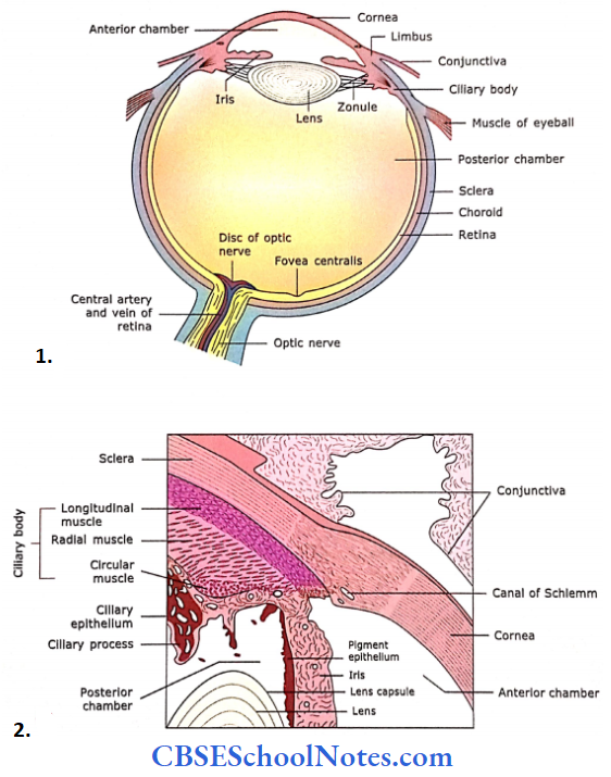
  1. The sagittal section of the eye shows Its gross structure.
  2. Schematic diagram showing sclerocorneal Junction, ciliary body and process, Iris and lens.

Fibrous Coat

Sclera

The sclera is a tough, fibrous connective tissue layer. It consists of irregularly arranged dense connective tissue, i.e., flat bundles of type 1 collagen fibres, a network of elastic fibres and fibroblasts. Its thickness ranges from 0.6 mm to 1 mm.

Cornea

The cornea is located at the front of the eye and is continuous with the sclera at the limbus. It is a transparent portion of the eye, hence, avascular.

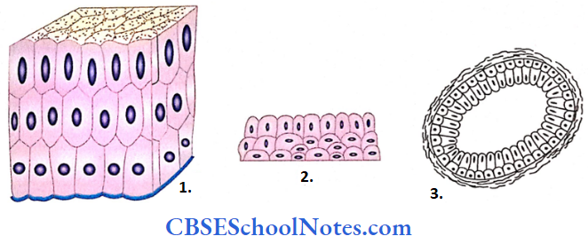
It is about 1 mm thick and consists of five layers. From superficial to deep these are:

Eye Eyelid Lacrimal Gland Structure Of Cornea

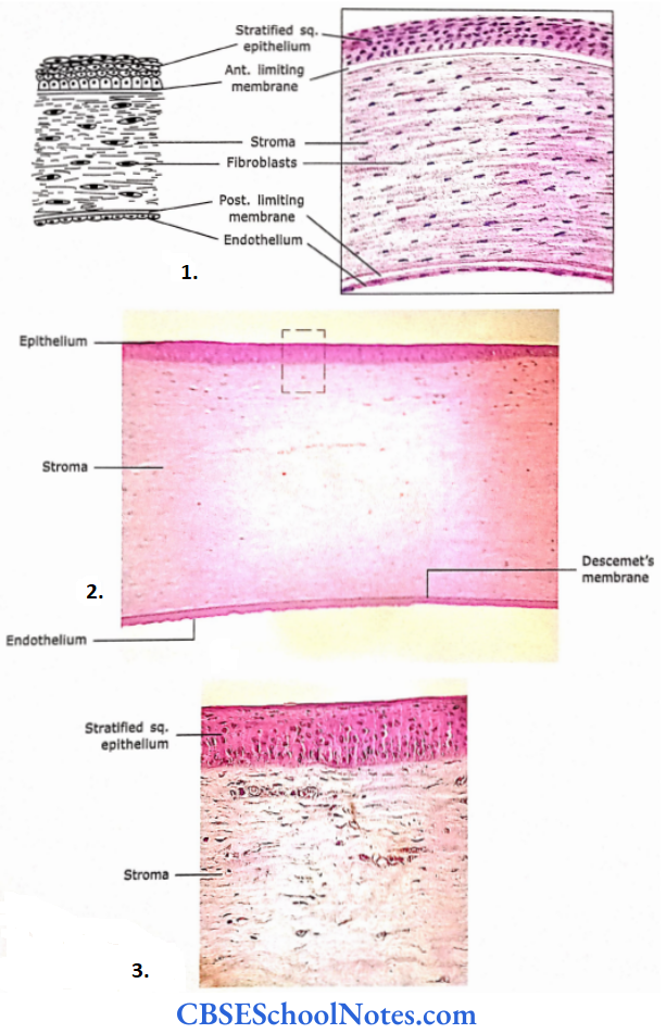
  1. Section of the human cornea showing various layers.
  2. Section of the cornea at low magnification.
  3. Photomicrograph at high magnification.

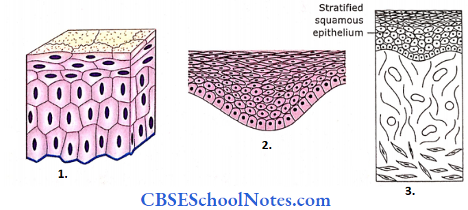
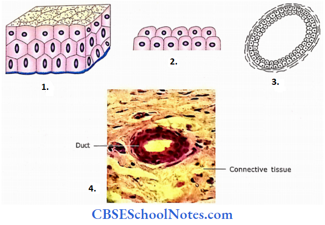
Corneal epithelium: It consists of stratified squamous, non-keratinized epithelium. Sensory nerves supply it. The cornea is very sensitive and any irritation or injury causes severe pain, excessive lacrimation, photophobia and blinking of eyelids.

Bowman’s membrane: It is about 10-12 mm thick, structureless, homogeneous lamina consisting of randomly oriented thin fibrils of collagen.

  • This layer is also known as an anterior limiting membrane. It acts as a barrier to the spread of infection. It is believed that Bowman’s membrane is synthesized by both the corneal epithelium and underlying stroma.
  • If damaged, it cannot regenerate. It heals by the formation opaque scar, which interferes with vision.

Corneal stroma: It constitutes the bulk of the cornea (90% of thickness). It is made up of thin lamellae of mostly type I collagen fibres, which are arranged in many layers.

  • The bundles of collagen fibres, within a lamella, are highly ordered. In the adjacent lamellae, the bundle of collagen fibrils are arranged at a right angle to each other.
  • The cornea is transparent because of the orderly arrangement of collagen fibrils. The flattened fibroblasts are located between the lamellae.
  • The canal of Schlemm is present at the sclerocomeal junction (limbus). It is the site of the outflow of the aqueous humour from the anterior chamber of the eye into the venous system.

Descentet’s membrane: This layer is also known as a posterior limiting membrane. It is an acellular, homogeneous basement membrane consisting of interwoven meshwork of collagen fibrils. It is a product of endothelium.

Endothelium: It is a single layer of squamous or low cuboidal cells on the posterior surface of the cornea, resting on Descemet’s membrane.

  • All the metabolic exchange between the cornea and aqueous humour takes place through the endothelium. It is responsible for the synthesis of proteins, which are necessary for maintaining Descemet’s membrane.
  • It also reabsorbs excessive fluid from the stroma to keep it relatively dehydrated, which helps in the refractive quality of the cornea.

Fibrous Coat Remember

The cornea is a transparent avascular membrane. It consists of two noncellular layers (Bowman’s membrane and Descemet’s membrane) and three cellular layers (corneal epithelium, corneal stroma and endothelium).

If Bowman’s membrane is damaged, it cannot regenerate. It heals by the formation of an opaque scar, which interferes with vision. Descemet’s membrane is an unusually thick basement membrane for a very thin endothelial lining.

Fibrous Coat Clinical Applications

Corneal Transplants

  • If a person has a defective cornea (presence of opacity), it interferes with normal vision. This cornea may be removed and a normal cornea obtained from a dead person (through eye donation) can be transplanted.
  • Corneal transplants are the most common and successful organ transplants. This is because the cornea is an avascular tissue.
  • Hence, antibodies that may cause rejection of transplanted cornea do not enter the transplanted tissue.

Glaucoma

The failure of drainage of aqueous humour from the anterior chamber of the eye leads to a prolonged increase in the intraocular pressure. The condition is known as glaucoma. It is an important cause of blindness.

Vascular Coat

The vascular coat consists of a choroid, ciliary body and iris.
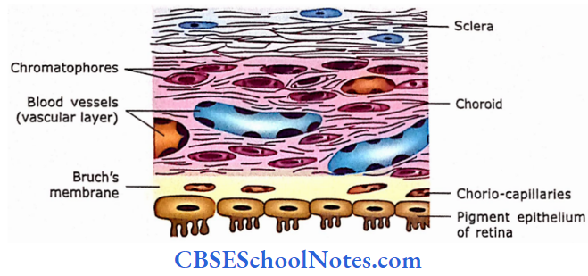
Choroid

The choroid is the posterior portion of the middle coat of the eyeball. It is the thin, highly vascular layer that lies between the sclera and retina. It consists of four layers:

  • Suprachoroid layer: This layer consists of fine collagenous fibres, elastic fibres and pigment cells (chromatophores).
  • Vascular layer: This layer consists of large blood vessels between loose connective tissue and pigment cells.
  • Chorio-capillary layer: This layer consists of a capillary network, which is essential for the nutrition of the retina.
  • Bruch’s membrane: This membrane separates the choroid from the retina. It is a thin (1-4 mm) refractile membrane. It also acts as the basal lamina for the pigment cell layer of the retina.

Vascular Coat Remember

The choroid is a pigmented vascular layer, which lies between the sclera and the retina. It is separated from the retina by Bruch’s membrane.

Eye Eyelid Lacrimal Gland Diagrammatic Representation Of Sclera, Choroid And Pigmented Epithelial Layer Of Retina

Ciliary Body

The ciliary body is made up of ciliary muscle (smooth muscle) and ciliary process. The smooth muscle of the ciliary body is oriented in longitudinal, radial and circular directions.

  • The ciliary muscle alters the shape of the lens for near and far vision. From the ciliary body, there come out 60-70 short ciliary processes with suspensory ligaments of the lens.
  • The ciliary processes are covered by bi-laminar epithelium and have a core of loose connective tissue and blood vessels.
  • The outer cell layer is the non-pigmented columnar epithelium, whereas the inner cell layer is composed of a pigmented simple columnar epithelium. The ciliary epithelium produces aqueous humour.

Ciliary Body Remember

The ciliary body is the anterior part of the vascular coat. It is located between the iris and the choroid. The ciliary body is made up of ciliary muscle (smooth muscle) and ciliary process.

The smooth muscle of the ciliary body is oriented in longitudinal, radial and circular directions.

Iris

The iris arises from the ciliary body and lies between the cornea and the lens. It consists of smooth muscle fibres in the connective tissue stroma (fine collagen fibres), melanocytes and blood vessels.

  • It has no epithelium on its anterior surface, but the posterior surface of the iris is lined by ciliary epithelium. This layer is heavily pigmented and the colour of the eye depends on its pigmentation.
  • The smooth muscle of the iris is arranged in two layers, i.e., dilator pupillae and sphincter pupillae. The dilator pupillae fibres are radially oriented.
  • While sphincter pupillae are oriented circumferentially near the pupil. Iris acts as a diaphragm, which controls the amount of light entering the eye.

Iris Remember

The iris is the anterior extension of the vascular coat. It acts as a diaphragm, which controls the amount of light entering the eye.

Nervous Coat

Retina

This is the inner coat of the eyeball and lines its posterior surface. The retina contains photoreceptors (rods and cones), which are essential for vision. The retina has a specialized area where vision is most acute, the fovea centralis or macula.

  • This area contains only cones, which are essentially bare (the overlying layers are pushed to the side). The retina also has a ‘blind spot,’ the optic disc.
  • Where the optic nerve leaves the eye and there are no photoreceptor cells. The retina has several layers from the outside in the following.


Eye Eyelid Lacrimal Gland Structure Of Retina

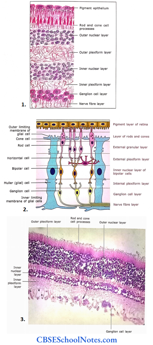
  1. The microscopic structure of the retina.
  2. Diagrammatic representation of principal cell types of the retina and their connections.
  3. Photomicrograph of the retina at high magnification.

Pigment Epithelium

It is the outermost layer of the retina, which is separated from the choroid by Bruch’s membrane. This epithelium consists of low cuboidal cells, which contain melanin granules.

  • Epithelial cells have long apical processes that occupy spaces between the outer segment of rods and cones.
  • The melanin pigment of epithelium absorbs light, thus preventing its reflection from the outer coats of the eye. The pigment epithelium also phagocytizes the tip of rod and cone cell processes.

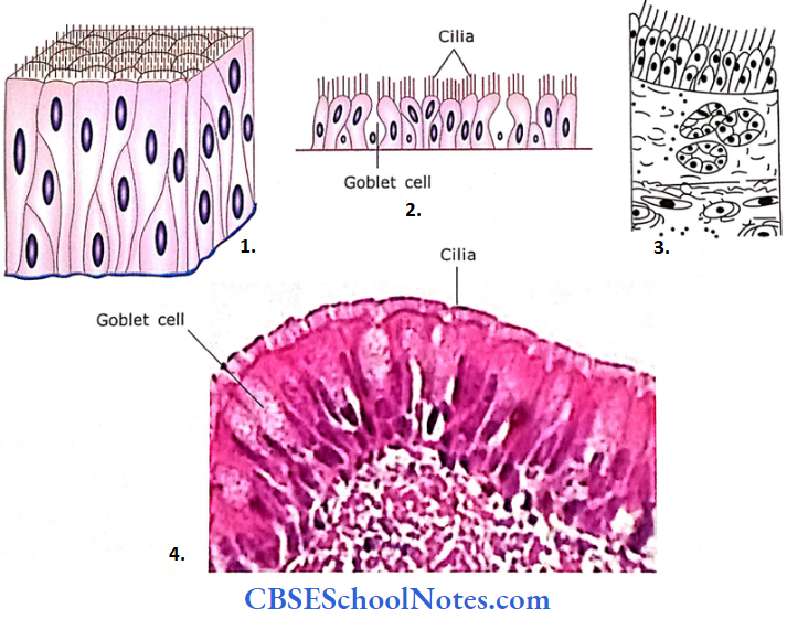
Layer of Rod and Cone Cell Processes

This layer consists of outer segments of rod and cone cells. The processes of rod cells are cylindrical and cones are thicker, cone-shaped. This layer is a photoreceptor in function.

  • Rods and cones are photoreceptor cells. They transfer the light energy into receptor potential. Both types of cells are long slender cells but the outer segments of rods are cylindrical or rodshaped
  • Whereas those of cones are tapered or cone-shaped. The rod cells are much more in number [120 million] compared to cone cells [6 to 7 million]. The parts of rod and cone cells are shown.
  • The rod consists of the outer segment, inner segment, inner fibre and spherule. The parts of the cone cell are almost the same except the terminal part is called the pedicle, instead of the spherule.

Outer Nuclear Layer

This layer consists of nuclei belonging to rods and cones. The nuclei of these cells are arranged in several layers. This layer is darkly stained.

  • Between the second and third layers, there is the presence of a pink linear marking called an outer limiting membrane or lamina.
  • This results because of zonula adherens of the glial cells (Muller cells) with the cell bodies of photoreceptor cells.
  • The Muller cells are supporting cells of the retina. They have long slender bodies that are radially oriented in the retina.

Outer Plexiform Layer

This layer stains lightly. The layer contains synapses between rods and cones with the dendritic processes of bipolar cells and horizontal cells.

Inner Nuclear Layer

It consists of cell bodies and nuclei of bipolar cells, horizontal cells, amacrine cells and Muller’s cells. The bipolar cells are oriented perpendicular to the layers of the retina.

  • They have synaptic contact with rod or cone cells at the outer (dendritic) end and with ganglion cells at the inner (axonal) end. The horizontal cells are oriented parallel to the layers of the retina.
  • Their processes form synaptic contact with rod and cone cells in the outer plexiform layer.
  • The amacrine cells are situated in the inner portion of this layer and have synaptic contact with axonal processes of bipolar cells and dendrites of ganglion cells in the inner plexiform layer.

Inner Plexiform Layer

In this layer, the axons of bipolar cells synapse with dendrites of ganglion cells and amacrine cells.

Ganglion Cell Layer

Mainly consists of the body and nuclei of large multipolar neurons (ganglion cells). They receive input from bipolar cells and their axons form optic nerve.

Nerve Fibre Layer

It consists of central processes (axons) of ganglion cells, which gather at the optic papilla (disc) and leave the eye as the optic nerve. As soon as these fibres leave the eyeball, they become myelinated.

The blood vessels of the retina are present only in the nerve fibre layer. However, sometimes they may reach as deep as the outer nuclear layer.

Nerve Fibre Layer Remember

The retina consists of ten layers of cells and their processes. Road and cone cells of the retina are specialized photoreceptors. Roads are specialized to perceive dim light, while cone cells perceive bright light and colour.

Nerve Fibre Layer Clinical Application

Detachment of Retina

As the apical ends of rods and cone cells are not firmly attached to the pigment epithelium, the retina may get separated from the pigment epithelium. This condition is called detachment of the retina.

  • This is a common but serious condition. It may be treated by laser surgery. If it remains untreated, rods and cones are damaged, leading to blindness.
  • This is because rod and cone cells get their nutrition from a chorio-capillary layer of choroid.

Lens

The lens is a transparent, flexible, biconvex disc. The lens is involved in near and distant vision, which is achieved by changing its curvature. The lens consists of a lens capsule, anterior epithelium and lens substance.

Lens Capsule

The lens is covered by a lens capsule, which is a homogeneous basal lamina coat. The capsule is made up of type 4 collagen and proteoglycans.

Anterior Epithelium

Beneath the capsule, simple cuboidal epithelium covers the anterior surface of the lens.

Lens Substance

The anterior epithelial cells toward the equator become columnar. The columnar cells of the equatorial region further elongate to form long fibres.

  • These long fibres are about 7-10 mm in length, 8-12 pm wide and 2 pm thick. In the cross-section, their shape is like a hexagon.
  • Although these fibres are called lens fibres, they are in a true sense modified elongated epithelial cells. The younger fibres are nucleated, whereas old fibres lose their nuclei.

Lens Substance Clinical Application

  • Cataract
    • The entire lens substance consists of transparent lens fibres. However, with increasing age or metabolic disorders (diabetes), the lens may become opaque.
    • This interferes with clear vision. This condition is called a cataract. In this condition, the lens is removed from its capsule and replaced with a plastic lens.
  • Presbyopia
    • The lens is highly elastic. However, the elasticity of the lens is gradually lost due to increasing age. This leads to difficulty in accommodation for near objects (reading, etc).
    • This is usually observed after 40-45 years of age. The condition (presbyopia) is corrected by wearing glasses with a convex lens.
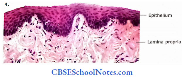
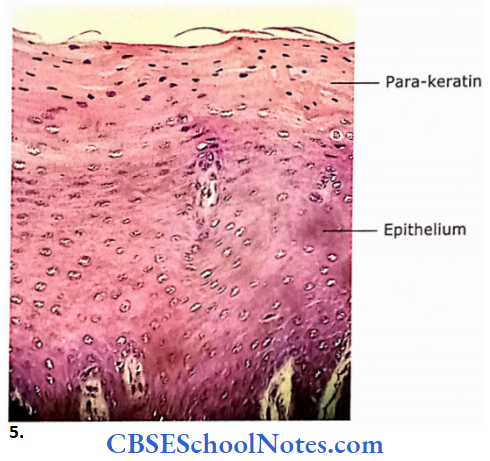
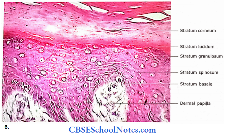
Eyelid

The eyelids protect the eye. From external to internal surface eyelid consists of many layers, i.e., skin, loose connective tissue, orbicularis oculi muscle, tarsal plate and palpebral conjunctiva.

  • The skin of the eyelid is thin, loose and elastic.
  • The loose connective tissue is present deep in the skin, which is devoid of fat.
  • The third layer of the eyelid is formed by bundles of skeletal muscle (orbicularis oculi). The tarsal plate is made up of dense connective tissue and forms the skeleton of the eyelid.
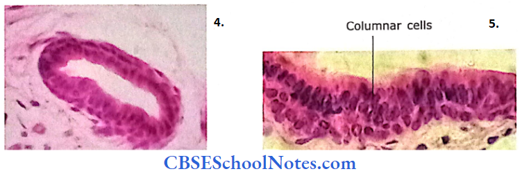
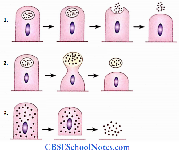
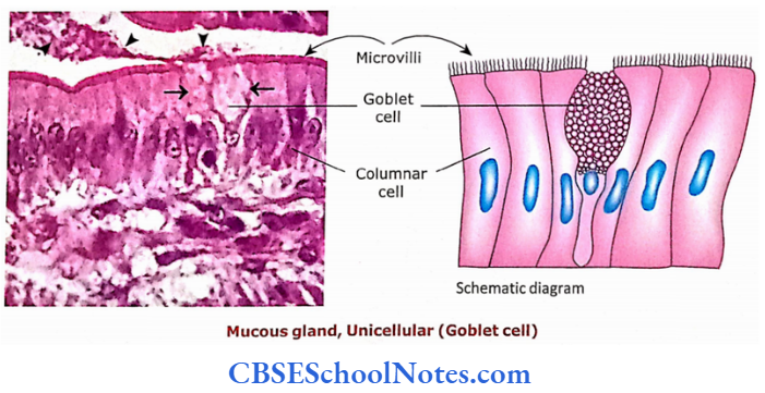
  • The inner surface of the lid is lined by palpebral conjunctiva. The palpebral conjunctiva consists of stratified columnar epithelium with goblet cells.

There are three types of glands present in eyelids.

  • Tarsal glands (Meibomian glands) are long sebaceous glands, which are present in the tarsal plate.
  • They produce sebaceous secretion, which forms an oily layer on the surface of the ear, thus preventing an early evaporation of tears.
  • Glands of Zeis (sebaceous gland of the eyelid) are small modified sebaceous glands, which are connected with the follicles of eyelashes.
  • Glands of Moll are small sweat glands that pour their secretion into eyelashes. They are simple spiral tubular glands.

Eye Eyelid Lacrimal Gland Histological Structure Of Eyelid

  1. Histological structure of eyelid.
  2. Section of eyelid.

Eyelid Remember

The main function of eyelid is to protect the eye. On its outer surface, it is covered by skin and its inner surface is covered by palpebral conjunctiva. Three types of glands are present in the eyelid.
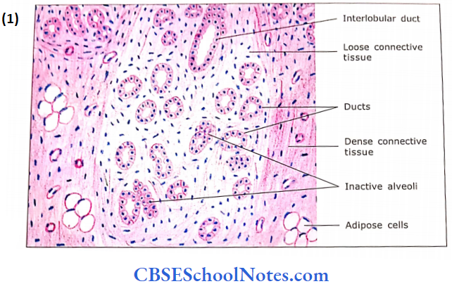
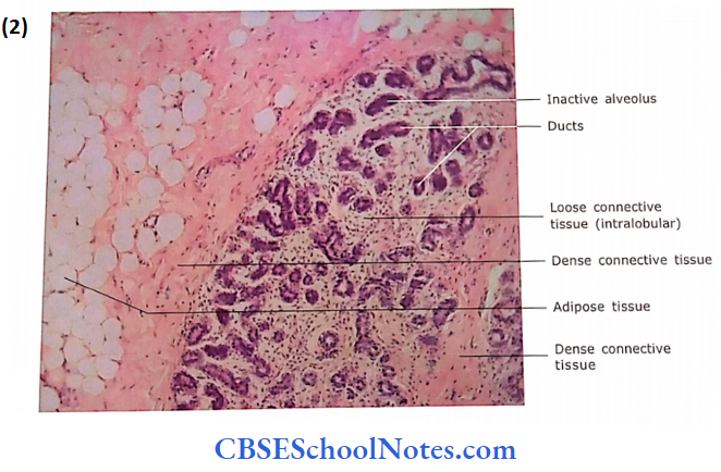
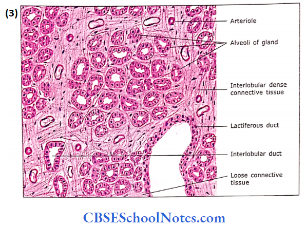
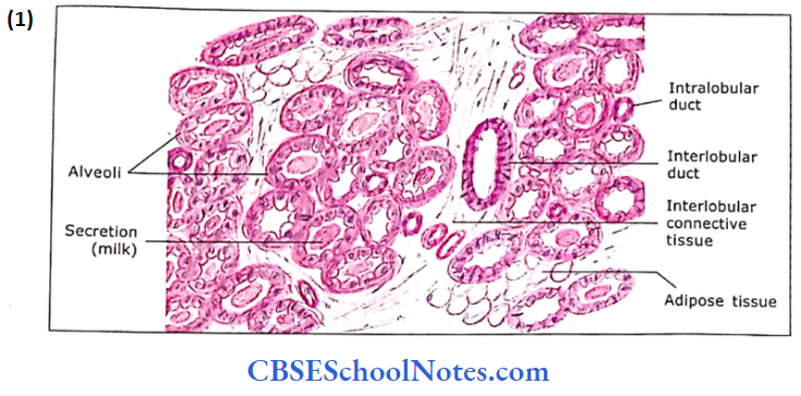
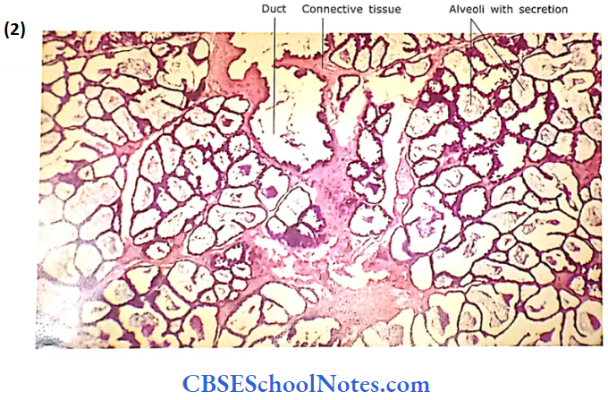
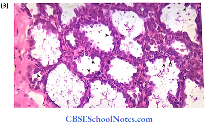
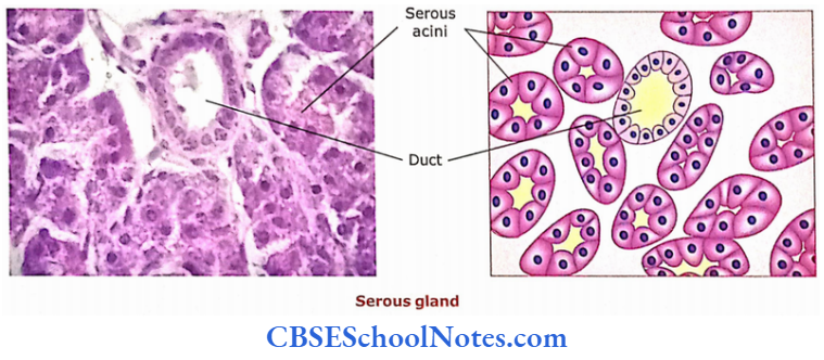
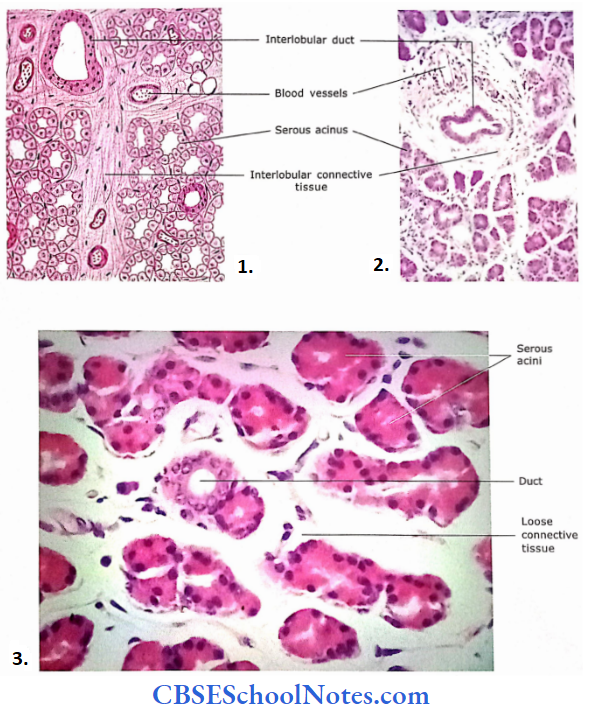
Lacrimal Gland

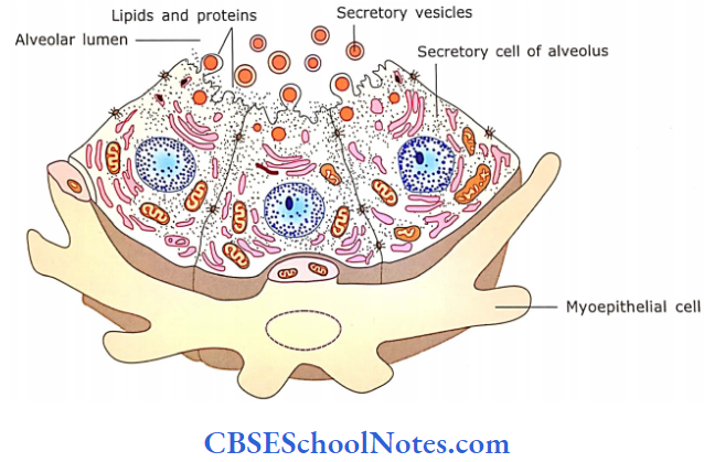
The lacrimal gland is a compound tubuloacinar serous gland. It consists of many separate lobes, which pour their secretion (tears) in the superior conjunctival fornix through small ducts.

  • The lacrimal gland acini has the features of a serous salivary gland. These acini are lined by light-staining low columnar cells.
  • The acini are large and have a wider lumen compared to the serous salivary gland.

Myoepithelial cells are located within the lamina of acini and help in the release of tears.

Eye Eyelid Lacrimal Gland Microscopic Structure Of Lacrimal Gland

  1. Section of lacrimal gland.
  2. Lacrimal gland as seen under low magnification.
  3. Magnified view of lacrimal gland

What Is Pineal Gland and Its Functions

Pineal Gland

The pineal gland (epiphysis cerebri) is a small endocrine gland (weighs 0.1-0.2 g). It arises as an outgrowth from the roof of the third ventricle of the brain. The gland is covered by the pia mater, which forms its capsule. Thin septa extend inward from this capsule to divide the gland into small irregular cords or lobules of cells.

What It Pineal Gland and Its Functions

The pineal gland consists of two main cell types:

  1. Pinealocytes and
  2. Interstitial cells

1. Pinealocytes:

Pinealocytes are the most numerous cell types, accounting for 95% of cells in the gland.

  • These are pale-staining epithelioid cells. Their nucleus is deeply indented and cytoplasm contains both rough and smooth endoplasmic reticulum.
  • The pinealocytes have a few long processes that have bulbous expansions at their terminal end. The pinealocytes secrete melatonin hormone.

2. Interstitial Cells:

These cells are far less numerous than pinealocytes (only 5%).

  • They have dark staining nuclei and resemble the astrocytes of the brain in having long cell processes containing many fine filaments.
  • Interstitial cells occur in proximity to pinealocytes and blood vessels.

The pineal gland also contains many mast cells, many blood vessels, and sympathetic nerve fibers. The extracellular calcified deposits of “brain sand” (corpora arenacea) are found in the pineal gland of older individuals. These concretions are found due to the deposition of calcium phosphates and carbonate on the substance secreted by pinealocytes.

Pineal gland Functions

  • The functions of the pineal gland are still unclear in humans.
  • The following functions can be attributed to lower animals
  • Melatonin appears to have a role in light-influenced reproductive cycles.
  • The release of melatonin is governed by the circadian (daily) dark-light cycle. The secretion of melatonin increases in the dark phase of the cycle.
  • Melatonin suppresses the release of gonadotrophic hormones of anterior pituitary. Daylight has the reverse effect.
  • The arginine vasotocin is another hormone secreted by pinealocytes. It also has antigonado-trophie activity.
  • The pineal gland, in humans, also plays an important role to adjust the sudden change in daylight in persons suffering from “jet lag.”

Pineal gland Remember:

The pineal gland contains two types of cells, i.e., pinealocytes and interstitial cells. The pinealocytes secrete melatonin, while interstitial cells are like astrocytes of the brain. The pineal gland is a photosensitive organ therefore thought to respond to dark and light periods.

Histology of Male Reproductive System Notes

Male Reproductive System

The testis is the main organ of reproduction in males. It is involved in the production of sperm and testosterone. The accessory organs are the epididymis, ductus deferens (which convey the sperm from the testis to the prostatic part of the urethra) and penis (a copulatory organ).

During the passage of sperm from the testis to the penis, accessory glands like the seminal vesicle, prostate and bulbourethral glands provide a fluid vehicle for sperm and make them more motile.

Male Reproductive System Of Reproductive System Of Testis And Epididymis

Testis

The testes lie outside the body cavity in the scrotum. They are ovoid and measure approximately about 5 cm in length and 2.5 cm in diameter. Each testis weighs 10-15 g. The testis has a thick white fibrous connective tissue capsule called the tunica albuginea.

The tunica vasculosa is a highly vascularized connective tissue, which underlies the tunica albuginea.

  • On the posterior border of the testis, the dense connective tissue of tunica albuginea projects into its interior and forms the mediastinum of the testis.
  • Through the mediastinum, blood vessels, nerves and ducts of the testis enter and leave the organ.
  • The connective tissue septa extend between mediastinum and tunica albuginea and divide the testis into about 250 compartments called as lobules
  • Each lobule contains one to three tightly coiled tubules, the seminiferous tubules.
  • Thus, each testis contains more than 500 seminiferous tubules. Each seminiferous tubule is about 30-70 cm long and about 200 cm in diameter.
  • These tubules are sites where sperm are produced. Sperms are produced in these tubules.
  • In between seminii- erous tubules, there is the presence of loose connective tissue (interstitial tissue) and blood vessels.
  • The interstitial tissue contains endocrine cells, Leydig cells or interstitial cells, which produce testosterone.
  • At the apex of the lobule, near the mediastinum, seminiferous tubules open into tubuli recti, which connect the open end of each seminiferous tubule to the rete testis.

Rete testis are epithelial-lined labyrinthine spaces within the mediastinum testis. The spermatozoa after passing through rete tesis travel through 10-20 short tubules known as efferent ducts. These efferent ducts fuse with epididymis.

Male Reproductive System Structure Of Testis Epididymis And Ducts Deferens

Testis Remember:

The main function of testes is to produce spermatozoa and synthesize the hormone testosterone.

Microscopic Structure Of Testis

The testis consists of seminiferous tubules, interstitial tissue and blood vessels.

Seminiferous Tubules

Each seminiferous tubule is long and convoluted. It is surrounded by a layer of connective tissue called lamina pro-pria. This layer consists of flattened or spindle-shaped myoid cells arranged in one or more layers. The myoid cells are contractile and help spermatozoon and testicular fluid to move through seminiferous tubules. Deep into lamina propria is basal lamina.

A dense capiUan network surrounds each seminiferous tubule- A dense capillary network surrounds each seminiferous tubule. Deep into lamina propria is basal lamina. On the basal lamina, seminiferous tubules are lined by a complex stratified epithelium.

Male Reproductive System Structure Of Testis At Low Magnification

Male Reproductive System Structure Of Testis Higher Magnification

Male Reproductive System Structure Of Testis Cross Section Of Seminiferous Tubule

Male Reproductive System Structure Of Testis Photomicrograph Showing Cross Section Of Seminiferous Tubule

Male Reproductive System Phortomicrograph Of Testis Sections Seminiferous Tubule At Low Magnification

Male Reproductive System Adjcent Parts Of Three Seminiferous Tubules

Which contains two major cell classes.  

  1. Supporting cells and
  2. Spermatogenic cells.

1. Supporting Cells (Sertoli Cells)

Sertoli cells are tall columnar cells, which extend from the basal lamina to the free surface of the epithelium (lumen of tubule). These cells have irregular outlines as they have lateral cell processes, which surround all spermatogenic cells except those resting on basal lamina. Sertoli cells are non-dividing cells in adults. They have ovoid euchromatic nuclei with one or more prominent nucleoli.

Sertoli cells Ultrastructure:

The Sertoli cells exhibit faint longitudinal striations.

  • The cell has a meshwork of thin actin filaments, bundles of intermediate filaments and microtubules arranged parallel to the cell axis.
  • These filaments and microtubules are involved in the change of shape of Sertoli cells that help in the movements of the germ cells toward the surface.

Sertoli cells Functions: 

Sertoli cells provide mechanical support to the spermatogenic cells.

  • They provide nutrition to the spermatogenic cells.
  • Sertoli cells form a blood-testis barrier, thus preventing the movement of extracellular molecules into the seminiferous epithelium.
  • They also secrete some components of testicular fluid.

Blood-Testis Barrier:

Complex intercellular tight junctions are present between lateral processes of Sertoli cells over spermatogonia. The inter-sertoli junctions comprise a “blood-testis barrier” to prevent sperm-related proteins from entering the circulation.

The blood testis barrier serves an important role in isolating developing sperm cells and spermatozoa from the immune system. This prevents the formation of sperm-specific antibodies.

  • These junctional complexes divide the seminiferous epithelium into two compartments, i.e., basal and luminal.
  • The basal compartment is present between basement mem¬brane and junctional complexes. It contains spermatogonia and primary spermatocytes.
  • In the luminal compartment, secondary spermatocytes and spermatids are present.
  • The movement of primary spermatocytes from the basal com¬partment to the luminal compartment takes place by the formation of new junctional complexes beneath the primary spermatocytes.
  • Once the secondary spermatocyte is formed then the junctional complex situated above the dividing pri¬mary spermatocytes break down and spermatocytes move to the luminal compartment.
  • Thus, the junctional complexes separate the antigenic haploid germ cells (secondary spermatocytes, spermatids and sperms), which are present in the luminal compartment form the immune system of adults. This prevents the formation of sperm-specific antibodies.

Male Reproductive System Representation Of Sertoli Cell

Sertoli cells  Remember:

The Sertoli cells serve many functions, i.e., form the blood testis barrier; secrete androgen-binding proteins and hormones; provide support and protection to spermatoge¬nic cells, and provide nourishment to developing sperm. Sertoli cells also phagocytose cytoplasmic remnants of spermatids

2. Spermatogenic Cells

Besides Sertoli cells, the seminiferous tubules are also lined with spermatogenic cells. The spermatogenic cells are arranged as complex stratified epithelium and consist of stem cells (spermatogonia) at the base of the epithelium.

  • The other cells at successively higher levels are primary spermatocytes, secondary spermatocytes, spermatids and spermatozoa. These cells are in different stages of differentiation of sperm.
  • Thus seminiferous tubule consists of two different cell populations, i.e., Sertoli cells and a population of germ cells.
  • The germ cells (spermatogonia) divide at the base of the epithelium and slowly move upward while they differentiate into spermatozoa.
  • The spermatozoa are then released into the lumen of the seminiferous tubule.
  • The spermatogenesis is under the control of pituitary hormones, luteinizing hormone (LH, ICSH) and follicle-stimulating hormone (FSH).

Interstitial Tissue and Blood Vessels:

In between the seminiferous tubules are many fenestrated capillaries, lymphatics, loose connective tissue and Leydig (interstitial) cells. The blood vessels apart from the usual function of blood supply, supply cooler blood to the testes.

  • The heat of the testicular artery is partly dissipated by its proximity to the pampiniform plexus of veins, which carries cooler venous blood from the testis and surrounds the artery in the spermatic cord.
  • The extra-abdominal scrotal position and dartos muscle also regulate the temperature of the testis.
  • Leydig cells are 3-8 pm in diameter. They are acidophilic, polyhedral in shape and found in clusters. They secrete testosterone.
  • Testosterone stimulates spermatogenesis by influencing the Sertoli cells. It also supports the structure and functions of the accessory sex organs and ducts.
  • Testosterone is also responsible for male secondary sex characteristics.

Seminiferous tubules Remember:

Seminiferous tubules consist of complex stratified epithelium surrounded by a thin connective tissue layer, i.e., lamina propria. The epithelium is composed of two different types of cells, i.e., Sertoli cells and spermatogenic cells.

Mature Spermatozoon:

The mature spermatozoon is commonly considered to have a head, neck and tail

  1. Head: The nucleus having condensed chromatin is covered in front by the acrosome. The acrosome is later released at the time of fertilization to disperse the corona radiata and digest the zona pellucida of the ovum.
  2. Neck: It is a short segment containing the centriole that gives rise to the core of the flagellum (9+2). or axoneme.
  3. Tail: The tail has 3 pieces, i.e., midpiece, principal piece and end piece. The mid piece is 5-7 urn in length. Here. 9 coarse fibres form a sheath around the flagellar core. Mitochondria become helically condensed around the sheath, which provides energy for sperm motility. The principal piece is about 45 pm in length. The axoneme and 9 coarse fibres are enclosed in a fibrous sheath. The end piece, 5-7 urn long, consists of axoneme enclosed by plasmalemma.

Male Reproductive System Structure Of Spermatozoon Steps Of Spermigenesis And Enlarged View And Internal Structure

Spermatozoon Remember:

The spermatozoon consists of the head, neck and tail. The head consists of a nucleus while the tail is divided into three regions, i.e., midpiece, principal piece and end piece. The head is about 5 μm long, while the tail is approximately 55 μm in length.

Sperm Clinical Application

  • Immotile Sperm: Sperms are highly motile in the female genital tract.
    • However, in a condition known as immotile cilia syndrome, sperm are unable to move from one place to another.
    • This results in infertility.
    • The immobility of sperm is due to the absence of protein (dynein) required for the motility of cilia and flagella. This also affects the cilia present throughout the body.
    • Thus, immotile cilia syndrome is also associated with chronic respiratory infections because of the presence of immotile cilia on the respiratory epithelium.
  • Sperm-specific Antibodies: The secondary spermatocytes, spermatids and spermatozoa possess specific proteins, which are recognized as foreign by the body.
    • Usually, these proteins are isolated by the blood testis barrier.
    • However, in case of breakage of the immune barrier there occurs the formation of sperm-specific antibodies.
    • These antibodies agglutinate the sperms, thus preventing their movements. This leads to infertility.
    • This kind of infertility can be detected by estimating the level of anti-sperm antibodies in the blood serum.

Duct System

The duct system of the male reproductive organ consists of the following tubules or ducts.

1. Straight Tubule (Tubuli Recti)

  • Straight tubules are the final portions of seminiferous tubules. The proximal part of the tubules is lined with simple columnar cells and Sertoli cells. The distal part is lined by simple cuboidal cells with microvilli.

2. Rete Testis

It consists of a system of flattened anastomosing channels in the dense connective tissue of the mediastinum that drains the straight tubules. The rete testis is lined by low cuboidal epithelium with microvilli and a single flagellum.

3. Efferent Ducts

  • They collect the sperm from the testis. Efferent ducts consist of 12-15 coiled tubes that coalesce to form the head of the epididymis.
  • The lumens of ducts are wavy in appearance.
  • This is because these ducts are lined by alternating groups of simple ciliated columnar and groups of cuboidal cells. The cilia of columnar cells propel the still non-motile sperm.
  • Cuboidal cells are probably absorptive.
  • Beneath the base membrane, in lamina propria, a thin layer of circular muscle is present.

Duct system  Remember:

Tubuli recti (straight tubule) and rete testis are present within the testis and both are lined by low cuboidal cells with microvilli. The efferent ducts are interposed between the rete testis and epididymis.

4. Epididymis

Efferent ducts fuse to form this 20-foot-long, highly coiled tube, which can be divided into head, body and tail.

  • The epididymis is placed at the posterior border of the testis. The whole organ and individual tubes are surrounded by vascular connective tissue.
  • The lining of the tubule is the pseudostratified epithelium. It consists of low basal cells and tall columnar cells. The tall columnar cells are with long stereocilia.
  • The function of epithelium is not well understood as stereocilia are non-motile. Probably, the epithelium is involved, both in secretion and absorption.
  • The lumen of the tubule may show the collected sperms. Beneath the distinct basement membrane, lamina propria contains circularly arranged smooth muscle fibres.
  • The smooth muscle helps to push the sperm along, especially in the proximal segment.

Epididymis  Functions

The epididymis is so long that it may take a month for sperm to make the journey.

  • The distal or tail segment stores the sperms, where they mature and lose the last bit of cytoplasm attached to their head and middle piece and become motile, thereby acquiring the capacity to fertilize an ovum.
  • Smooth muscle in the wall contracts rhythmically during ejaculation to move the sperm along.
  • It also contributes a viscid nutritive substance.
  • The epithelial cells of epididymis also phagocytose the degenerated sperms and residual bodies.

Epididymis Remember:

Epididymis is formed by the fusion of efferent ducts. It is a highly coiled tubule divided into head, body and tail. The tail of the epididymis is continuous with ductus deferens. Within the epididymis sperms are stored, mature and thereby acquire the capacity to fertilize an ovum.

Male Reproductive System Microscopic Structure Of Epididymis Of Convoluted Tubule

Male Reproductive System Microscopic Structure Of Epididymis Of Tubules Of Epididymis Lumen Filled With Spermatozoa

Male Reproductive System Microscopic Structure Of Epididymis Of Medium Magnification

Male Reproductive System Microscopic Structure Of Epididymis Of High Magnification

5. Ductus Deferens

It is a thick muscular tube extending from the tail of the epididymis to the prostatic part of the urethra. It drains the epididymis.

The ductus deferens consists of the following layers in its wall:

  • Mucosa: The lumen of the duct is irregularly star-shaped. The epithe¬lium is pseudostratified columnar with stereocilia and resembles that of epididymis. The lamina propria underlying the epithelium contains collagenous and elastic fibres.
  • Muscle Layer: It consists of three layers of smooth muscle, i.e., outer and inner thin layers of longitudinal muscle and a well-devel¬oped thick middle layer of circular muscle. The muscle layer is very thick (1-1.5 mm) compared to the thickness of the mucosa.
  • Adventitia: The adventitia is made up of loose areolar tissue, which contains many blood vessels and nerves. The terminal part of the ductus deferens is dilated to form an ampulla. Here, the mucosa is thrown into tall branching folds covered by a low columnar epithelium.

Ductus deferens Functions

  1. The ductus deferens does not store sperm.
  2. The duct is involved in rapid propulsion during ejaculation because of its strong musculature.

Male Reproductive System Ducts Deferens

Male Reproductive System Ducts Deferens

Ductus deferens Remember

The wall of the vas deferens consists of three layers of smooth muscle. This strong musculature helps in the propulsion of sperms from the tail of the epididymis to the ejaculatory duct during ejaculation.

Accessory Sex Glands

1. Seminal Vesicle

The seminal vesicles are elongated sac-like structures with a highly convoluted irregular lumen. Each gland consists of. single tube, about 3-1 mm in diameter, 12-15 cm m light,

Which is folded upon itself to measure about 5 cm in length. It joins with the ductus deferens to form an ejaculatory duct  The wall of the seminal vesicle is composed of the following three layers

  • Mucosa: The mucosa is thrown into highly complex folds.
    • These folds join with each other to form many crypts and cavities.
    • The core of these folds is formed by connective tissue derived from lamina propria. The lamina propria is rich is elastic fibres.
    • The epithelium is pseudostratified low columnar or cuboidal. In some places, epithelium is simply columnar or cuboidal.
    • These cells are secretory. The epithelium varies greatly in height and appearance with activity, blood testosterone level and age.
  • Muscle Layer: The muscle layer is made up of smooth muscle, which is thinner than that of ductus deferens. The muscle is arranged in two layers, i.e., inner circular and outer longitudinal. The contraction of the muscle at the time of ejaculation expels the secretion of the gland into the ejaculatory duct.
  • Adventitia: A thin layer of loose connective tissue surrounds the muscle layer.

Seminal Vesicle Functions

  • The gland secretes seminal fluid, which is a yellow viscous fluid containing fructose (an energy source for sperms) and prostaglandin.
  • The seminal fluid is alkaline in nature.
  • The pale yellow colour of semen is due to lipochrome pigment released by seminal vesicles.
  • The gland is under the control of androgen. It helps to flush the sperm out of the urethra.

Male Reproductive System Seminal Vesicle

Male Reproductive System Seminal Vesicle At Low Magnification

Male Reproductive System Seminal Vesicle At High Magnification

Seminal Vesicle Remember:

The seminal vesicle is not involved in the storage of sperm. It secretes a yellow viscous fluid that contains fructose and prostaglandin and constitutes about 70% of ejaculate.

2. Prostate Gland

The prostate is the largest of the accessory glands. It is about the size of the chestnut. It surrounds the first part of the male urethra after it emerges from the bladder.

  • It is comprised of 20-50 tubulo-alveolar glands, which open by 15-25 ducts into the prostatic urethra. The stroma of the gland consists of fibromuscular tissue in which glandular tissue is embedded.
  • Thus, the prostate is known as a fibro¬muscular glandular organ. The prostate gland is surrounded by a thick capsule.
  • Three groups of glands surround the prostatic urethra concentrically, i.e., mucosal, sub-mucosal and main prostatic gland. The mucosal glands are small tubular glands situated in the mucosa, which open directly into the prostatic urethra.
  • The submucosal glands are situated deep in the mucosa and are tubulo-alveolar type.
  • The prostatic glands are situated in the outer zone of the prostate. Both submucosal and main prostatic glands open through long ducts into the prostatic urethra.
  • The alveoli of the glands are surrounded by the fibromuscular stroma. The stroma consists of smooth muscle, collagenous and elastic fibres.
  • The fibro-muscular stroma runs in different directions and contains blood vessels, lymph vessels and nerves. The glandular alveoli are of variable sizes and irregular lumens.

The epithelium lining the alveoli is secretory. It is either a simple columnar or a pseudostratified columnar.

  • However, in some places, the epithelium may be low cuboidal. The variation in the epithelium (low cuboidal to pseudostratified co¬lumnar) is due to its functional state.
  • In old people, the lumen of some of the gland alveoli may show the presence of prostatic concretions (corpora amylacea), which are oval-dense bodies of glycoproteins
  • This results from to condensation of secretory products, which may become calcified. The significance of these bodies is not known.
  • The prostatic urethra, above the opening of ejaculatory ducts, is lined by transitional epithelium. The lower part of the prostatic urethra is lined by stratified columnar epithelium
  • . The epithelial lining is surrounded by lamina propria and outside by smooth muscle.

Male Reproductive System Postate Gland

Male Reproductive System Prostate Gland At Low Magnification

Male Reproductive System Alveoli Of Prostate Gland At High Magnification

Prostate Gland Functions

  • The prostate secretes 10-30% of final ejaculate.
  • The fluid is thin and contains acid phosphatase, citric acid, amylase, fibrinolysin and prostate-specific antigen (PSA). Fibrinolysin helps in liquefication of semen.
  • Prostatic secretion is facilitated by the contraction of smooth muscles of the stroma at the time of ejaculation.
  • Prostatic secretion promotes the mobility of sperm.

Prostate Gland Remember:

Three groups of glands surround the prostatic urethra concentrically, i.e., mucosal, sub-mucosal and main prostatic gland. The prostatic secretion contains acid phosphatase, citric acid, amylase, fibrinolysin and prostate-specific antigen (PSA). Fibrinolysin helps in liquefication of semen.

Prostate Gland Clinical Application

  • Enlargement of Prostate:
    • The glandular tissue of the prostate starts proliferating after 40-45 years of age in almost 50% of men. However, 80% of males are affected by 70 years of age.
    • The enlarged prostatic tissue compresses the prostatic urethra, which leads to difficulty in passing urine.
    • The disease is called benign prostatic hypertrophy. It can be treated by surgical removal of a part of the gland.
    • The malignant prostatic tumour is the second most common cancer in men. The tumour arises from glandular tissue of the prostate gland, hence called as adenocarcinoma of the prostate.
    • The level of PSA increases in cancer of the prostate and is used for early detection of cancer. In this case, complete removal of prostate is required.

Bulbourethral Gland

The bulbourethral glands (Cowper’s gland) lie in the urogenital diaphragm and empty into the proximal portion of the penile urethra  The gland is about the size of a pea. It discharges a mucus-like lubricant. It is a compound tubuloalveolar gland. The epithelium of the secretory part varies from simple cuboidal to simple columnar.

3. Bulbourethral Gland Clinical Application

  • Semen: The semen contains spermatozoa and secretion of acces¬sory sex glands. The volume of the ejaculate is about 3 mL, 95% of which is secretions from accessory glands. The sperm concentration varies from 50 to 250 million/mL. A male whose sperm count is less than 20 million/mL of ejaculate is considered as sterile.
  • The following glands contribute to the formation of semen:
    • Bulbourethral Gland: The secretion is mucus-like fluid, which acts as a lubricant. Secretion starts appearing much before ejaculation begins.
    • Prostate Gland: The secretion of the prostate coagulates the semen, which is later liquefied by fibrinolysin.
    • Seminal Vesicle: The secretion is rich in fructose, which provides energy to sperms.
  • Impotence:
    • The inability to achieve an erection is called as impotence.
    • I Impotence may be temporary or permanent. The temporary impotence may be due to drugs or psychological factors.
    • The permanent impotence is due to lesions in the brain, hypothalamus, and spinal cord and injury to autonomic nerves.
    • It may be also due to various systemic diseases such as multiple sclerosis, Parkinson’s disease and diabetes

Bulbourethral Gland Remember:

Secretion of the bulbourethral gland acts as a lubricant and its release in the urethra is due to sexual stimulation.

Penis

The penis is an erectile copulatory organ. It is a common organ through which both semen and urine are discharged. The penis is made up of three cylindrical bodies of erectile tissue

The corpora cavernosa are placed dorsally while a single corpus spongiosum is placed ventrally.

  • The urethra passes within the corpus spongiosum and opens at the tip of dilated part of the penis called as glans penis. The glans is covered with a fold of skin called a prepuce.
  • In the shaft of the penis, each of the three erectile bodies is covered by a thick connective tissue sheath called the tunica albuginea.
  • The tunica albuginea also fonus an incomplete partition between two corpora cavernosa. The tunica albuginea is covered with a layer of loose connective tissue and skin.
  • A cross-section of the penis shows the following structures from superficial to deep. The most superficial structure is thin skin, which is devoid of any hair.
  • Deep to the skin is the presence of a loose connective tissue layer, which is devoid of fat (adipose tissue). This layer of loose connective tissue is also called Buck’s fascia.
  • It binds the tunica albuginea of all three erectile tissues with each other. Deep to this, the tunica albuginea covers all three erectile cylindrical bodies, i.e., two dorsal corpora cavernosa and one ventral corpus spongiosum.

Male Reproductive System A Cross Section Of Through Penis

The erectile tissue of the corpora is a sponge-like mass of endothelial-lined vascular spaces. The walls of these spaces are formed by numerous.

  • Trabeculae consist of collagen fibres, elastic fibres and smooth muscle. These spaces are supplied by afferent arteries, which are branches of central deep arteries. T
  • these spaces are drained to veins on the inner aspect of the tunica albuginea. They penetrate the tunica obliquely to join the deep dorsal vein of the penis.
  • During the erection of the penis, blood fills the cavernous vascular spaces because of the vasodilation of arteries due to psychic and afferent sensory input.
  • These spaces expand as they are filled with blood under pressure.

The peripheral veins are compressed against the inner surface of tunica albuginea, hence blood outflow diminishes considerably. This causes the erection of the penis. Erection is controlled by the parasympathetic nervous system, while ejaculation is controlled by the sympathetic nervous system

The corpus spongiosum is traversed by the penile urethra throughout its length. The urethra is lined by stratified columnar or pseudostate-tied columnar epithelium. The tip of the urethra, at the glans penis, is lined by stratified squamous non-keratinized epithelium. There are many small mucous glands of Eittre, which are scattered along the length of the urethra. They secrete mucus and have e lubricating function.

Penis Remember:

Filling of cavernous spaces (corpora cavernosa and cor¬pora spongiosum) with blood causes the erection of the penis.

Central and Peripheral Nervous Systems Notes

The Central Nervous System

The central nervous system consists of the brain and spinal cord. The cut surface of any part of the central nervous system consists of grey matter and white matter.

  • The cerebrum and cerebellum have a cortical layer of grey matter on the surface deep to which is white matter. However, in the spinal cord, grey matter is inside, surrounded by white matter.
  • Out of the various parts of the brain, we should study only the cerebral cortex and cerebellar cortex which are of histological significance.

Some Important Definitions

  • Grey matter: It is a collection of neurons, neuroglial cells, the processes of these cells lying adjacent to the cell body, and blood vessels inside the central nervous system (brain and spinal cord).
  • White matter: It consists of bundles of nerve fibers; such as sociated neuroglial cells and blood vessels. The Myelin sheath that surrounds the nerve fibers gives it a white appearance.
  • Nuclei: These are islands of grey matter situated deep inside the cerebrum, cerebellum brain stem, and spinal cord For Example., the thalamus, basal nuclei, etc.
  • Ganglion: These are collections of nerve cells (grey matter) outside the brain and spinal cord For Example., dorsal root ganglia and sympathetic trunk ganglia.

Cerebral Cortex

Most of the cerebral cortex (except the cortex of hippocampal formation and piriform lobe) is described as having six layers. However, the distinction between these layers is not well-marked.

  • These layers are distinguished based on the predominance of cell type and arrangement of fibers.
  • The arrangement of fibers can be visualized only by special stains and not by H and E. From superficial to deep, the following are the layers of the cerebral cortex.

Nervous System Structure Of Cerebral Cortex

  1. In a section of the cerebral cortex, it is difficult to distinguish various layers.
  2. Schematic diagram to show neurons in various layers of the cerebral cortex.
  3. Section of the cerebral cortex. Six layers of the cerebral cortex are ill-defined as they merge.

Molecularor Plexiform Layer

This layer is situated just beneath the pia mater. It consists predominantly of fibers and neuroglial cells. A few horizontal cells of Cajal are also present.

  • The peripheral portion consists largely of fibers, which travel parallel to the surface. In its deeper part lie the horizontal cells of Cajal.
  • Their cell body and processes are disposed of horizontally. This layer shows the blood capillaries and many nuclei of neuroglial cells.

External Granular Layer

This layer consists of two types of neurons, i.e., small pyramidal and granular or stellate cells. Their apical dendrites extend in the first layer and the axon ends in the deeper layer.

External Granular Layer Further Details

Pyramidal Neurons

These are triangular and their size ranges from 10 to 120 μm. Their apical dendritic end faces the surface of the cerebral cortex.

  • Dendrites take origin from all three angles and synapse with the fibers in other layers. The axon is given off from the base of the cell and extends to deeper layers.
  • The large pyramidal cells (Betz cells) are seen in the motor cortex and their axons form pyramidal (corticospinal) fibers.
  • The stellate or granular cells are star-shaped small neurons (8 μm). Their processes extend only into neighboring areas.

External Pyramidal Layer

The layer consists of medium pyramidal cells. This layer also consists of a few stellate cells and cells of Martinotti. The axons ofpyramidal cells form association and commissural fibres.

The Martinotti cells are small, triangular, or polygonal cells seen almost in all the layers of the cortex. Their axons travel toward the surface of the cortex.

Internal Granular Layer

This layer consists of densely packed granular cells with a white horizontal fiber layer called the external band of Baillarger. This layer provides connections between neurons of different layers.

Internal Pyramidalor Ganglionic Layer

This layer consists of large pyramidal cells (cells of Betz) and a few cells of Martinotti. The horizontal fibers in the deeper part are called the internal band of Baillarger.

Fusiform Layeror Layer of Polymorphic Cells

This layer predominantly contains spindle-shaped fusiform cells and a few stellate and Martinotti cells. The fusiform cells are located in the deeper layer of the cerebral cortex.

These cells lie perpendicular to the surface with axons coming out from the center of the cell and dendrites from both ends. Deep in the sixth layer of the cerebral cortex lies white matter.

Fusiform Layeror Layer of Polymorphic Cells Remember

The cerebral cortex is composed of 14-16 billion nerve cells approximately. The cortex consists of 6 layers of cells. The two principal neurons are pyramidal cells and granular (stellate) cells. Large-sized pyramidal cells may measure up to 120 μm.

Fusiform Layeror Layer of Polymorphic Cells Clinical Application

Alzheimer’s Disease

The disease is of unknown cause and occurs in old people. To begin with, they suffer from loss of memory, but later all their intellectual capabilities are also lost (dementia).

In this disease, the neurons of the cerebral cortex accumulate late tangled masses of filaments in the cytoplasm and later degenerate. The motor system remains unaffected.

Fusiform Layeror Layer of Polymorphic Cells Further Details

Histologically, one can differentiate motor and sensory cortex. The motor cortex consists predominantly of pyramidal cells in layers 3 and 5. These pyramidal cells are densely packed and large.

On the other hand, the sensory cortex shows very few pyramidal cells in layers 3 and 5 and most of the layers contain small granular cells.

Cerebellar Cortex

The histological structure of the cerebellar cortex is uniform throughout the cerebellum. It consists of three layers, i.e., molecular layer, Purkinje cell layer, and granular layer. The molecular layer is situated just beneath the pia mater.

Nervous System Structure Of Cerebellum

Nervous System Photomicrograph Of Cerebellar Cortex At Low Magnification Cortex Is Folded And Present Fissures And Folla

  1. The structure of two adjacent cerebellar folia shows outer grey and inner white matter.
  2. The grey matter of the cerebellar cortex consists of three layers, i.e., molecular, Purkinje, and granular layer.
  3. Photomicrograph of cerebellar cortex at low magnification, cerebellar cortex is folded and presents fissures and folia.
  4. Photomicrograph of cerebellar cortex showing three layers, i.e., molecular layer, Purkinje cell layer, and granular cell layer.

Molecular Layer

It stains lightly with eosin and is featureless as it consists of a few cells and more myelinated and unmyelinated fibers.

  • This layer consists of a few scattered stellate cells in the superficial part and a few basket cells in the deeper part.
  • However, the dendritic processes of Purkinje cells and the axonal processes of granular cells (that run parallel to the surface of the cortex) occupy most of the molecular layer.
  • The axons of granular cells come in synaptic contact with the dendrites of several Purkinje cells and basket cells. The molecular layer also contains the terminal part of climbing fibers.

Purkinje Cell Layer

These cells are arranged in a single row between molecular and granular layers. Purkinje cell (Golgi type F) is the large pyriform or flask-shaped neuron, that sends numerous dendrites into the molecular layer.

These dendrites synapse with axons of granular cells and climbing fibers. Purkinje cells give a single thin axon, which passes through a granular layer to end in deeper nuclei of the cerebellum.

Granular Layer

The granular layer stains deeply with the hematoxylin because it is densely packed with granular cells. Granular cells are small neurons with round nuclei surrounded by a thin rim of cytoplasm.

  • These cells receive impulses from various parts of the CNS through Mossy fibers. The Mossy fibers in the granular layer end as the dilated terminal.
  • On which the dendrites of granule cells and axons of Golgi cells (type 2) synapse to form light-stained areas called glomeruli.
  • Granular cells send their axons into the molecular layer where they branch in the form of T and come in synaptic contact with dendrites of various Purkinje cells and basket cells.
  • At the junction of molecular and granular layers, Golgi type 2 cells are found. Their vesicular nuclei are larger than granule cells. They contain chromophil substances present in the molecular layer.
  • However, their axons form synaptic contact with glomeruli in the granular layer. Deep to the granular layer the cerebellar cortex lies in contact with white matter.

Granular Layer Remember

The cerebellar cortex consists of three layers, i.e., the molecular layer, the Purkinje cell layer, and the granular layer. The histological structure of the cerebellar cortex is uniform throughout the cerebellum.

Purkinje cells are large pyriform or flask-shaped neurons arranged in a single row between the molecular and granular layers.

Granular Layer Further Details

Neuronal Circuit of Cerebellum

  • The input to the cerebellum is from different parts of CNS and is in the form of Mossy and climbing fibers
  • The climbing fibers ascend to the molecular layer where they form synaptic contact with the dendritic arborization of Purkinje cells.
  • The Mossy fibers end in glomeruli and form synaptic contact with granular cells in the granular layer.
  • The granular cells convey the impulse received by Mossy fibers to the dendrites of Purkinje cells.
  • The output of the cerebellum is in the form of axons of Purkinje cells, which synapse with the intracerebellar nuclei.
  • The other neurons like Golgi cells, basket cells, and stellate cells interconnect the intracortical circuit to modify the outgoing impulses.

Nervous System Various Types Of Neurons And Afferent And Efferent Fibers Of Cerebellar Cortex

Spinal Cord

The human spinal cord is about 45 cm long and has cervical, thoracic, and lumbosacral parts. A cross-section of the spinal cord shows a central canal lined by ependyma made up of simple ciliated columnar cells.

  • The central canal is surrounded by grey matter, which contains neurons, nerve fibers, neuroglial cells, and blood vessels.
  • The grey matter is surrounded by white matter, which consists of bundles of nerve fibers and neuroglial cells. The surface of the spinal cord is covered with pia mater.

Grey Matter

The grey matter of the spinal cord appears roughly in the form of an H. It has anterior and posterior grey columns or horns. The anterior grey column contains large-size multipolar motor neurons.

  • The nucleus of the motor cell is a large, spherical, light-staining structure with intensely staining nucleolus. The cytoplasm contains clumps of dark staining basophilic Nissl substance.
  • The Nissl substance extends into the dendritic processes neuron but not into the axon. The axons of motor cells form the ventral spinal root.
  • The neurons of the posterior grey column are much smaller than the anterior horn cells. Within the grey matter, besides the sensory and motor nerve cells, there are numerous neuroglial cells and blood vessels.

Nervous System Structure Of Spinal Cord

  1. Drawing to show the arrangement of the grey and white matter of the thoracic spinal cord (transverse section).
  2. The section of a part of the spinal cord shows the motor neurons in the anterior grey column and fibers in adjacent white matter.
  3. Section of spinal cord (at low magnification) showing centrally placed grey matter and peripherally placed white matter.

White Matter

The white matter is composed primarily of myelinated nerve fibers, but also neuroglial cells and blood vessels. The nerve fibers are comprised of ascending and descending tracts.

  • Nerve fibers are surrounded by a myelin sheath, which in turn is surrounded by a fine connective tissue sheath called epineurium. As the myelin sheath gets dissolved during the preparation of H and E sections.
  • The dark-staining axons (in a transverse section of the spinal cord) are surrounded by a clear space, which had been occupied by myelin.

White Matter Remember

A cross-section of the spinal cord shows a central canal surrounded by grey matter. The grey matter is surrounded by white matter. The grey matter of the spinal cord is in the form of an H and has anterior and posterior grey columns or horns.

  • The anterior grey column contains large-size multipolar motor neurons, while neurons of the posterior grey column are much smaller than anterior horn cells and are sensory neurons.
  • The white matter of the spinal cord consists of ascending and descending nerve fibers, which are mostly myelinated.

White Matter Clinical Application

Multiple Sclerosis

It is the most common disorder of the nervous system affecting young adults. In this condition, myelinated nerve fibers of the brain and spinal cord are progressively damaged due to the destruction of the myelin sheath.

  • This affects the sensation, movements, body functions, and balance. Damage to the optic nerve may cause blurred vision. If nerve fibers in the spinal cord are affected.
  • It may cause weakness or heaviness in the limbs. Damage to the fibers in the brain stem may affect balance. The demyelination is thought to result from an autoimmune disease with inflammatory features.

Guillain-barre syndrome

ln this disease, there occurs the demyelination of peripheral nerves and motor nerves arising from the ventral roots. The person suffers from muscle weakness and difficulty in respiration.

Peripheral Nervous System

All nervous tissues other than the brain and spinal cord are classified as the peripheral nervous system. The peripheral nervous system consists of nerves (made up of bundles of nerve fibers) and ganglia (collection of neurons and nerve fibers outside CNS).

Nerve

A nerve is defined as the collection of nerve fibers, which may be myelinated and or unmyelinated, and held together by connective tissue.

  • A nerve consists of nerve fibers (axons or dendrites), supporting neuroglial cells (Schwann cells) and connective tissue.
  • A nerve fiber is first surrounded by Schwann cells (which may form the myelin sheath around it), then it is surrounded by a thin layer of connective tissue called endoneurium.
  • The endoneurium is made up of delicate collagen fibers and a few fibroblasts.
  • At the light microscopic level, the endoneurium shows the nuclei of fibroblasts, which are difficult to distinguish from the nuclei of Schwann cells.
  • Numerous fibers are held together to form a bundle of nerve fibers, which is surrounded by a sheath, called perineurium.

The perineurium is a cellular sheath made up of 3-4 layers of squamous-shaped cells and extracellular material. These cells show basal lamina and are contractile.

Nervous System Transverse Section Of A Pheripheral Nerve

  1. A part of a transverse section of a nerve showing nerve fibers (axons) arranged in bundles and covered by a connective tissue sheath (perineurium).
  2. A drawing of the transverse section of a few nerve fibers is shown in an enlarged view.
  3. Under microscope.

Nervous System Longitudinal Section Of Nerve

Nervous System Photograph Of Longitudinal Section Of Nerve At High Magnification

  1. At low magnification.
  2. At medium magnification
  3. Photograph of a longitudinal section of nerve at high magnification.
  4. Micrograph of a longitudinal section of nerve fibers (Sliver Stain).

Differences between dorsal root ganglion and sympathetic ganglion

Nervous System Difference Between Dorsal Root Ganglion And Sympathetic Ganglion

The perineurium forms a semi-permeable barrier. Thus, the cells of perineurium are not in true sense connective tissue cells, which comprise epineurium and endoneurium.

  • These cells are more like an epithelioid tissue. Many bundles of nerve fibers are finally covered by a dense connective tissue sheath called epineurium.
  • The epineurium not only forms the outermost covering of a nerve, but it also goes inside between perineurial bundles, to bind them together. The epineurium contains blood vessels and adipose tissue.
  • The fine branches of blood vessels after penetrating the perineurium reach the endoneurium to supply it and nerve fibers.

Nerve Remember

Peripheral nerves are covered by three different connective tissue sheathes, i.e., epineurium, perineurium, and endoneurium.

Dorsal Root Ganglion

It is also known as sensory ganglion. This ganglion is the collection of sensory neurons on the dorsal root of the spinal nerves.

  • Each ganglion is surrounded by a connective tissue capsule, which is the epineurium of the dorsal root. Beneath the capsule, the ganglion contains large cell bodies arranged in groups.
  • Also, between and around the groups of neurons there are bundles of myelinated nerve fibers. Most nerve fiber bundles are seen in the central part of the ganglion and groups of nerve cells are seen in the peripheral part.
  • The neurons of the dorsal root ganglion have large, spherical bodies with large pale-staining euchromatic nuclei and dark-staining nucleoli. Each neuron is surrounded by the satellite cells.
  • The satellite cells, which form a sheath around the neuron, are much smaller compared to neurons. They are flattened or low cuboidal neuroglial cells and form an inner capsule around each neuron.
  • The fibroblast and fibers form an outer capsule surrounding the inner capsule formed by satellite cells. The satellite cells prevent unwanted depolarization of sensory neurons.

Nervous System Relationship Of Sympathetic Trunk With The Spinal Cord

Dorsal Root Ganglion Remember

The dorsal root (sensory) ganglion is a collection of pseudo-unipolar, rounded sensory neurons on the dorsal root of the spinal nerve.

Nervous System Structure Of Dorsal Root Ganglion

Nervous System Structure Of Dorsal Root Ganglion.

  1. Section of dorsal root ganglion (spinal ganglion) showing spherical nerve cells with euchromatic nuclei and prominent nucleolus.
  2. Section of dorsal root ganglion (at low magnification) consisting of large-size pseudo-unipolar neurons.
  3. Photomicrograph at high magnification.

Sympathetic Trunk Ganglion

The sympathetic ganglion is the ganglion of the autonomic nervous system and lies along the sympathetic trunk. They contain cell bodies of postsynaptic motor neurons of the autonomic nervous system.

  • The ganglion is covered with a thin capsule of connective tissue. It consists of small, irregular neurons dispersed between the nerve fibers. Cells are multipolar and, therefore appear irregular in shape.
  • They contain eccentrically placed nuclei with prominent nucleoli. The cytoplasm contains small Nissl bodies. The satellite cells are less in number than in dorsal root ganglion cells.
  • In between nerve cells, there is supportive connective tissue, blood vessels, and bundles of nerve fibers (both myelinated preganglionic and unmyelinated postganglionic).

Sympathetic Trunk Ganglion Remember

Autonomic ganglion houses cell bodies of postganglionic autonomic nerves.

Nervous System Section Of Sympathetic Ganglion Showing Small Irregular Nerve Cells

  1. Section of sympathetic ganglion showing small irregular (multipolar) nerve cells
  2. Photomicrograph of sympathetic ganglion showing multipolar neurons, scattered between nerve fibers.

Development of the Urinary System: Kidney and Bladder Notes

Urinary System

The urinary system consists of two kidneys, two ureters, one urinary bladder and one urethra. Two kidneys produce urine, while ureters conduct urine from the kidneys to the urinary bladder, which stores the urine. The urethra drains the urine from the urinary bladder to the exterior.

Kidney Functions:

  1. Excretion: The major function of the kidney is excretory, i.e., it eliminates the waste material (urea) and excess metabolites (electrolytes, water, glucose and amino acids). However, when these metabolites are not in excess it retains them by reabsorption.
  2. Endocrine function: Kidney also has an endocrine function, i.e., it secretes renin (involved in the regulation of blood pressure and retention of sodium) and erythropoietin (regulates RBC production) in embryonic life.
  3. Conversion of vitamin I) into calcitriol: Proximal convoluted tubules of the kidney are involved in the conversion of vitamin D into active hormone [calcitriol, l,25-(OH)2
    vitamin DJ that regulates plasma calcium level.

To have a comprehensive understanding of the histological structure of the kidney, readers are advised to learn the gross structure of the kidney structure of nephron and the blood supply of the kidney.

Gross Structure Of Kidney

The naked-eye view of a hemisected kidney shows the outer cortex and inner medulla.

  • The cortex lies just beneath the connective tissue capsule. The medulla is made up of 8-12 conical structures called pyramids.
  • The cut face of the pyramid displays a striated appearance it consists of numerous parallel tubules and blood vessels.
  • The broad base of each pyramid is directed toward the cortex and apex (renal papilla) facing into a minor calyx.
  • There are about 8-12 minor calyces, which join to form two or three large extensions called major calyces.
  • Major calyces unite to form a funnel-shaped renal pelvis, which is present at the hilus of the kidney. The renal pelvis becomes narrow and forms the ureter.

Urinary System Outer Cortex Ad Inner Medulla

The cortex at the margin of each pyramid extends inward between the pyramids as renal columns. Some of the striated patterns from the base of the pyramid may extend into the cortex, which are called medullary rays. Each medullary ray is the collection of straight tubules and col¬lecting ducts extending in the cortex. A lobe of the kidney is defined as a renal pyramid with its overlying cortex and part of its laterally associated renal columns 

A kidney lobule is defined as a medullary ray with its laterally associated cortical tissue. Thus, a lobule consists of a col¬lecting duct and all the nephrons draining to it. As there are about 20,000 medullary rays in the cortex, approximately the same number of lobules are estimated to be present. The cortex of the kidney consists of about 2-3 million tubular structures called nephrons.

Urinary System Lobe Of Kidney Includes Renal Pyramid

Kidney Remember:

The lobe of the kidney includes the renal pyramid with its overly¬ing cortex and parts of its later ally-associated renal columns, While on the other hand, the kidney lobule consists of the centrally placed medullary ray with its laterally placed cortical areas. Thus, the number of kidney lobes is equal to number of pyramids present in the kidney (i.e., 8-12). While, the number of kidney lobules is equal to the number of medullary rays present in the cortex (i.e., about 20,000).

General Structure Of Nephron

The functional unit of the kidney is (lie nephron. Bach nephron has several parts, i.e.,

  1. Renal corpuscle
  2. Proximal convoluted tubules
  3. The loop of Henle and
  4. Distal convoluted tubule.

1. Renal Corpuscle:

  • The blind end of each nephron is expanded in the cortical region into a double-walled cup called Bowman’s capsule made up of an outer parietal epithelium and an inner visceral epithelium. The cup encloses a tuft of capillaries called a glomerulus.
  • Bowman’s capsule and glomerulus together constitute the renal corpuscle. The renal corpuscle has two poles, i.e., the vascular pole (where arterioles arc present) and the urinary pole, from where proximal convoluted tubules take origin.
  • An ultrafiltrate of the glomerular blood enters the space between two layers of Bowman’s capsule. The filtrate then passes to the proximal convoluted tubules.

2. Proximal Convoluted Tubule:

The proximal convoluted tubule is quite tortuous and begins at the Bowman’s capsule. It makes many convolutions in the cortex near the Bowman’s capsule from which it arises, it then enters the medullary ray and continues as the descend¬ing thick segment of the loop of Henle.

3. Loop of Henle:

The loop of Henle consists of a descending limb (both thick and thin segments), a hairpin turn and an ascending limb (thin and thick segments).

  • The upper part of the descending limb is thick and is in continuation of the proximal convoluted tubule.
  • The lower part of the descending limb is a thin segment, which is continuous with the hairpin turn.
  • The ascending limb has a lower-thin and upper-thick segment. The thick segment ascends back into the cortex.

4. Distal Convoluted Tubule:

As the ascending thick segment of the loop of Henle comes close to the vascular pole of its originating renal corpuscle, it continues as distal convoluted tubules. After this, it makes many shorter loops in the cortex before it opens in the collecting tubule.

Urinary System Nephrons Showing Its Various Parts Relations Of Sigment

Two different types of nephrons are located in the cortex.

A nephron whose glomerulus is located adjacent to the base of the pyramid is called a juxtamedullary nephron. All other nephrons are called cortical nephrons.

  • The collecting tubules are present in cortical tissue and drain into increasingly larger tubules called collecting ducts.
  • A collecting duct travels first in the medullary ray and then in the pyramid to its apex.
  • At the apex of the pyramid, several large collecting ducts open, which are called as ducts of Bellini. 
  • A nephron, its collecting tubule and its collecting duct together form a unit called a uriniferous tubule.
  • The nephrons are embryologically derived from metanephros while collecting tubules and ducts from the ureteric bud.

Nephron Remember:

The nephron is the functional and structural unit of the kidney. It consists of renal corpuscle (glomerulus and Bowman’s capsule), proximal convoluted tubule, loop of Henle and distal convoluted tubule that opens in collecting tubule. Each kidney contains about 2 million nephrons.

Relationship between Parts of Nephron and Zones of Kidney:

  • Renal corpuscles are located at varying levels in the cortex and only in cortical tissue.
  • Renal corpuscles close to the capsule (cortical nephrons) send their tubules down to the outer zone of the medulla.
  • Renal corpuscles in the juxtamedullary area send their tubules deep into the inner zone of the medulla.
  • Descending and ascending thick limbs occupy the outer zone, while the thin limbs occupy the internal zone almost to the apex of the pyramid.
  • Thus, cortical tissue contains renal corpuscles, proximal and distal convoluted tubules and initial parts of collecting tubules.
  • The medullary ray contains thick segments of the loop of Henle and collecting ducts. The medulla contains a thick and thin segment of the loop of Henle, vasa recta and collecting ducts.

The cortex of the kidney Remember:

The cortex of the kidney contains renal corpuscles, proxi¬mal and distal convoluted tubules and initial parts of the collecting tubules, while the medulla contains thick and thin segments of the loop of Henle, vasa recta and collecting ducts.

Renal Blood Supply of Kidney

The renal artery after entering the hilus of the kidney divides into a few segmental arteries, which in turn form interlobar arteries. Each interlobar artery runs between two pyra¬mids through the renal column.

  • At the cortico-medullary junction (at the base of the pyramid), the interlobar arteries turn to arch over the base of the pyramid. These arteries are known as arcuate arteries.
  • The arcuate arteries give off branches, which ascend into the cortex between lobules as interlobular arteries. These arteries are located between medullary rays.
  • Many intralobular arteries arise from the interlobular artery and are called as afferent arterioles of glomeruli. They form the capillary network of glomeruli.
  • Blood from glomeruli is drained by efferent arterioles. The efferent arterioles give rise to a second network of capillaries, which are called as peritubular capillaries
  • The peritubular capillaries of cortical glomeruli surround the local uriniferous tubule and do not go into the medulla.
  • However, the peritubular capillaries of juxtamedullary glomeruli descend into the medulla along with loops of Henle. In the medulla, they form capillary loops called recta before returning to the cortex. The ascending limb of the vasa recta forms the arterial limb, while its ascending limb forms the venous limb.
  • The venous return from the peritubular capillary networks is via interlobular, arcuate, interlobar and renal veins.

Urinary System Renal Blood Supply

Blood Supply of Kidney Remember:

Both kidneys receive large volumes of circulating blood, i.e., approximately 1000 mL of blood enters two kidneys each minute, from which about 125 mL/min glomerular filtrate is formed. Both kidneys form about 180 L of glomerular filtrate out of which only 2 L is excreted as urine while the remaining 178 L is reabsorbed by the kidneys.

Microscopic Structure Of Kidney

Following is the histological structure of various parts of the nephron i.e., renal corpuscle, proximal convoluted tubules, loop of Henle and distal convoluted tubule

1. Renal Corpuscle

It is also known as the Malpighian corpuscle. It consists of Bowman’s capsule and glomerulus. The Bowman’s cap¬sule has an outer parietal layer and an inner visceral layer  The parietal layer is lined by simple squamous epithelium, while the visceral layer is lined by podocytes. The space between the parietal and visceral layer is the urinary space (Bowman’s space), which receives the ultrafiltrate of blood. At the urinary pole, the space between two layers is continuous with the lumen of the proximal convoluted tubule. The squamous epithelium of the parietal layer at this pole becomes continuous with the cuboidal epithelium of the proximal convoluted tubule. The glomerulus is the tuft of capillaries fed by an afferent arteriole and drained by an efferent arteriole. Both these arterioles are present at the vascular pole of the renal corpuscle.

Urinary System Renal Corpuscle And Juxtaglomerular Complex

The visceral epithelium of Bowman’s capsule is closely applied to the endothelial lining of capillaries. The cells of the visceral layer become modified and are called Propodocytes and have many radiating processes, which in turn contain secondary processes, called foot processes or pedicels. The foot processes of neighbouring podocytes interdigitate with each other. These foot processes are separated from each other by narrow intercellular spaces that are called filtration slits. The gap of the filtration slit is occupied with a thin membrane called a filtration slit membrane or slit membrane

Urinary System Relationship Of Podocyte To Glomercular Capillary

Ultrastructure of Filtration Barrier:

The endothelial cell layer and podocyte cell layer (visceral layer of Bowman’s capsule) share a common fused basal lamina. The foot processes of podocytes are closely applied to the common basal lamina.

The filtration barrier is made up of three components

  1. Fenestrated Endothelium: The pores of capillary endothelial cells are about 70-90 nm in diameter. These pores are not spanned by a pore diaphragm and allow the passage of molecules up to 70,000 molecular weight. Endothelial cells of glomerular capillaries possess a large number of aquaporin-1 (AQP-1) water channels, which allow fast filtration of water through the endothelium.
  2. (Glomerular basement Membrane: It consists of fused basal lamina of endothelium and visceral layers of Bowman’s capsule (podocytes). It is made up of type 4 and 8 collagens, proteoglycan and glycoproteins. It forms the major unit of barrier and serves to retain the necessary plasma proteins from leaking out.
  3.  Filtration Slit Membrane (diaphragm): It spans between adjacent foot processes of podocytes and measures 25 nm in width and 4-6 nm in thickness. The slit diaphragm is formed by a transmembrane protein called nephrin. Nephrin proteins emerge from opposite foot processes and form a central density, which has pores. Mutation in the nephrin gene leads to congenital nephritic syndrome which is characterized by proteinuria. This membrane shows the presence of small pores, which prevent the passage of albumin and large molecules from the blood to glomerular filtrate.

Urinary System Glomerular Filtrtion Barrier

Filtration Barrier Remember:

The glomerular filtration barrier is formed by the fenestrated endothelium of glomerular capillaries, glomerular basement membrane and visceral layer of Bowman’s capsule, which consists of epithelial cells that become modified and are called podocytes. The foot processes of neighbouring podocytes interdigitate with each other and contain gaps, which are known as filtration slits. The gap of the filtration slit is occupied with a thin membrane called a filtration slit membrane or slit membrane. This slit membrane acts as a part of the filtration barrier. Thus, the filtration barrier consists of endothelial cells, fused basal lamina and a filtration slit.

Process of Glomerular Ultrafiltration:

Fluid first passes through the pores of capillar}’ endothelial cells then it is filtered by the basal lamina.

  • Fluid containing small molecules, ions and macromolecules passes through lamina densa and pores in slit diaphragm of filtration slits.
  • If molecules are small (<1.8 nm) and are un¬charged then they pass easily through the slit diaphragm. However, large molecules cannot pass through the slit diaphragm.
  • The large molecules, which are unable to cross the barrier are rapidly removed by the intraglomerular mesangial cells, otherwise, the basal lamina will get clogged with these large molecules. The fluid, which after passing through bar¬riers reaches the Bowman’s space is called the glomerular ultrafiltrate.

Mesangial Cells:

The capillaries of the glomerulus are held together by mesangium (mesangium = between vessels). Mesangial cells are of two different types, i.e., extraglomeru lar and intraglomerular. Extraglomerular cells are located at the vascular pole, while intraglomerular cells are located within the renal corpuscle.

Mesangium is a connective tissue consisting of mesangial cells in an extracellular matrix. These cells are most numerous near the vascular pole of the renal corpuscle. Mesangial cells correspond to the pericyte and may be enclosed by the basal lamina of the glomerular capillaries.

They have many functions:

  1. Their phagocytic function helps to remove large protein and filtration residues from the glomerular basal lamina. Thus, the integrity of the filter is maintained.
  2. They participate in the turnover of basal lamina.
  3. Mesangial cells are contractile, thus regulating the glomerular filtration rates.
  4. Mesangial cells synthesize and secrete interleukin-1 and platelet-derived growth factor (PDGF).
  5. The} provides structural support to podocytes.
  6. Mesangial cells proliferate in certain types of nephropathy.

Urinary System Relationship Of Podocyte To Glomercular Capillary

Juxtaglomerular Apparatus:

This is an apparatus present near the vascular pole of a renal corpuscle and helps in maintaining blood pressure. The macula densa, Juxtaglomerular cells and extra mesangial cells constitute the juxtaglomerular apparatus

  • Macula Densa: These are specialized cells at the beginning of the distal convoluted tubule that lie adjacent to afferent and efferent arterioles at the vascular pole of the corpuscle.
    • These cells are narrow, columnar and crowded together.
    • Cells of macula densa can sense a low sodium concentration of urine in the distal convoluted tubules and help in the release of renin.
  • Juxtaglomerular Cells: The smooth muscle cells in the wall of afferent arteriole (and sometimes in efferent arterioles also), which lie close to macula densa, become modified to form juxtaglomerular cells.
    • These cells contain secretory granules and no myofilaments.
    • They secrete renin hormone, which increases blood pressure. (Renin breaks the angiotensinogen of blood plasma into angiotensin 1, which subsequently is broken down to angiotensin 2 in the lungs. Angiotensin 2 raises the blood pressure by its vasoconstriction activity and controls the glomerular filtration.)
    • It also stimulates the synthesis and release of aldosterone, which in turn acts on collecting ducts to increase the reabsorption of sodium and water.
    • This leads to a further rise in blood volume and blood pressure.
    • Thus, the juxtaglomerular apparatus regulates blood pressure by activating the rennin-angiotensin-aldosterone system.
  • Extraglomerular Mesangial Cells: These cells are present in the space between the distal tubule, and afferent and efferent arterioles at the vascular pole of the corpuscle.
    • These cells have receptors for angiotensin 2 and may regulate the glomerular filtration rate.
    • These cells connect the sensory cells of macula densa with the juxtaglomerular effector cells and transmit the signals through gap junctions.
    • They also send signals to the contractile mesangial cells for vasoconstriction within the glomerulus.

Urinary System Vascular Pole Of Renal Corpuscle

Juxtaglomerular apparatus Remember:

The juxtaglomerular apparatus consists of macula dense (which are specialized cells in the beginning of the distal convoluted tubule that lie adjacent to afferent glomerular arterioles), juxtaglomerular cells, (which are smooth muscle cells in the wall of afferent arteriole) and extraglomerular mesangial cells.

The cells of macula densa are specialized to detect the low concentration of sodium and volume of glomerular filtrate in the distal tubule. This leads to the release of renin by juxtaglomerular cells causing the conversion of angiotensinogen to angiotensin 1 which is subsequently converted to angiotensin 2. Angiotensin 2 is a potent vasoconstrictor and helps in the release of aldosterone.

Kidneys Clinical Application

Glomerulonephritis, Filtration Barrier and Proteinuria:

  • If kidneys are infected by bacteria, glomeruli are highly affected.
  • The urine of a healthy person does not contain protein because the molecules of protein are too large to pass through filtration barrier.
  • However, in diseases like glomerulonephritis, the filtration unit (basal lamina of capillaries) may get damaged and large amounts of protein and RBCs can leak into the urine from the blood.
  • The presence of protein in urine is known as proteinuria, while the appearance of blood (RBCs) in urine is called haematuria.
  • The leakage of protein results in low protein levels in the blood. This causes a collection of fluid in tissue and widespread swelling.
  • Proteinuria may also occur in diseases like diabetes mellitus due to damage to the filtration unit of the kidney.

Kidney Failure and Dialysis:

Kidney failure results from the loss of normal function of both kidneys due to a variety of causes, i.e., fall in blood pressure, infection, glomerulonephritis, toxic chemicals, drugs, diabetes mellitus, etc.

  • In kidney failure, kidneys are unable to remove waste products and excess water from the blood, thus disrupting the chemical balance of the blood.
  • Methods of treatment of kidney failure may include drugs, dialysis or kidney transplant.
  • Dialysis is the procedure in which the functions of the kidney (removal of wastes and excess water from the blood) are performed by a machine.
  • Each dialysis treatment takes about 3-4 hrs and has to be repeated 3 times a week.

2. Proximal Convoluted Tubule

If starts from the urinary pole of a renal corpuscle and extends up to a thick portion of the descending limb of Henle. This part of the tube is 60 m in diameter and complexly coiled (convoluted). The length of the proximal convoluted tubule is almost double that of the distal convoluted tubule. It is present in the cortex only.

The tube is lined with simple cuboidal or low columnar epithelial cells. These tubules have small uneven lumen (Fig. 17.9 and 17.12). There is the presence of a brush border formed by tall microvilli on the apical surface of cells. Nuclei are round and centrally placed. The cytoplasm stains deeply with eosin. The basal part of the cell may show vertical striations, due to the presence of mitochondria.

Proximal Convoluted Tubule Ultrastructure:

  • The electron micrograph of cells of proximal convoluted tubules shows the features that indicate that these cells are involved in absorption and transport.
  • The presence of microvilli lateral and basal infoldings of plasma membrane increases the surface area of cell for absorption and transport.
  • The presence of mitochondria between basal folds provides for the high-energy requirements needed for active transport.
  • Fluid and absorbed substances return to the fenestrated blood capillaries present adjacent to proximal convoluted tubules.

Proximal tubules Functions:

  1. In proximal tubules, there occurs reabsorption of 80% of salts (Na and Cl), water (85%)
  2. Most amino acids, ascorbic and lactic acid (100%), filtered proteins, glucose and bicarbonate
  3. The remaining molecules and fluids are removed in the other portions of the nephron.

Urinary System Microscopic Structure Of Cortex Of Kidney Glomeruli And Proximal And Distal Convluted

Urinary System Microscopic Structure Of Cortex Of Kidney Glomeruli And Proximal And Distal Convluted Under Microscope

3. Loop of Henle

The proximal convoluted tubule continues downward into the medullary ray and medulla as the loop of Henle.

  • The histological structure of the thick descending limb of the loop of Henle is similar to that of the proximal convoluted tubule.
  • The descending and ascending thin limbs of the loop are about 15 m in diameter and are lined with squamous epithelial cells bearing few microvilli. The cytoplasm is pale staining, nuclei bulge into small lumen.
  • The thin limb resembles a venule in cross-section. The histological structure of the thick ascending limb of the loop of Henle is similar to that of the distal convoluted tubule (see below).

Loop of Henle Ultrastructure:

The thin limb of the loop of Henle is lined by squamous epithelium bearing a few’ short microvilli. The presence of very few organelles (including mitochondria) and very few infoldings of plasma membrane indicates that these cells are only involved in the passive transport of fluid and salts.

Loop of Henle Functions:

  • The loop of Henle is the essential element in the production of hypertonic urine.
  • The thin descending limb is permeable in both water and salt.
  • In contrast to this the thin ascending limb is permeable to salt but not to water.

Urinary System Microscopic Structure Of Medulla Of Kidney

Urinary System Microscopic Structure Of Medulla Of Kidney Under Microscope

Urinary System Microscope Structure Of Medulla Of Kidney In Logitudinal Section

Urinary System Kidney Medulla Showing Collecting Ducts

4. Distal Convoluted Tubule

As the distal convoluted tubule is half the length of the proximal convoluted tubule few distal tubules are seen in a microscopic field. The diameter of the distal tubule is less as compared to proximal tubules (15-30 μm). The tubules are lined by cuboidal epithelium.

The cytoplasm of cells stains light eosinophilic. The brush border is not present and the height of cuboidal cells is short (5 μm). These two facts are responsible for the large regular lumen of distal tubules.

Urinary System Renal Cortex As Seen At Low Magnification

Urinary System Renal Cortex Of A Renal Corpuscle Proximal And Distal Convoluted Tubules

Differences between proximal and distal convoluted tubules:

Urinary System Differences Between Proximal And Distal Convoluted Tubules

The differences between proximal and distal convoluted tubules are presented in Table

Distal Convoluted Tubule Ultrastructure:

  • The cells of distal convoluted tubules show very few short microvilli, but lateral and basal infoldings of the plasma membrane are very prominent.
  • The mitochondria are oriented parallel to the long axis of the cell.
  • All these features indicate that cells are involved in the active transport of ions.

Urinary System Structure Of Epithelal Cells Lining The Convoluted Tubules Proximal Convouted

Distal Convoluted Tubule Functions:

  • It is involved in the reabsorption of salt, water and bicarbonate. The distal tubule also secretes potassium and hydrogen ions.
  • The distal convoluted tubule is under the control of an antidiuretic hormone, which promotes the reabsorption of water and salts

4. Collecting Tubules

Collecting tubules begin in the cortex and proceed to the medullary ray where they join the larger collecting tubules called as collecting ducts. These ducts in the medulla run toward the apex of the pyramid and join each other to form the duct of Bellini. Collecting tubules are about 40 pm in diameter while ducts are much wider.

Both tubules and ducts are lined by cuboidal to low columnar epithelium. The brush border is not present and cells are lightly stained with eosin. The cell outline is clear and both, tubules and ducts, have a much larger lumen

 Collecting Tubules Ultra Structure:

Collecting tubules and ducts are lined by two kinds of cells, i.e., principal and intercalated cells. Most of the lining cells are principal cells, which are wide, low columnar. They have few organelles, lateral and basal infoldings of the plasma membrane and several mitochondria. Intercalated cells are few and they have microvilli and basal infoldings.

Collecting Tubules Functions:

The function is the concentration of urine by sail-free water re-absorption that occurs under the influence of ADH. The result is hypertonic urine.

  • The light microscopic structure of the cortex of the kidney: A section from the cortex of the kidney shows the glomeruli, proxi¬mal and distal convoluted tubules, blood vessels and col¬lecting tubules.
  • The light microscopic structure of the medulla of the kidney: A section from the medulla of the kidney shows the thick and thin segment of the loop of Henle, collecting ducts and blood ves¬sels (capillaries).

Ureter

The ureter is a tube with a star-shaped lumen varying in length from 25 to 35 cm. It conducts urine from the renal pelvis to the urinary bladder. The following three layers comprise the wall of the ureter

  1. Mucosa
  2. Muscle layer
  3. Adventitia

The mucosa consists of lining epithelium and lamina pro-pria. The epithelium is transitional and is 4-5 cells thick. The lamina propria is wide and made up of loose connective tissue. Blood vessels and lymphatics are present in it.

The muscle layer consists of an inner longitudinal and outer circular layer of smooth muscle fibres. In the middle and lower partial ureter, a third outer layer of longitudinal smooth muscle is also present.  these three layers of muscle arc are not well defined and are difficult to mark off front of each other. The outermost layer (adventitia) is made up of loose connective tissue and contains many blood vessels, nerves and fat cells.

Urinary System Structure Of Ureter At Higher Magnification Of Ureteric Wall

Urinary System Structure Of Ureter Under Microscope

Urinary System Structure Of Ureter Transverse Section Of Ureter

Urinary System Structure Of Ureter High Magnification View Of Transitional Epithelium Superficial Layer

Kidney Stones Clinical Application

Calcium salts and uric acid are excreted In the glomerular filtrate. These salts are less soluble in water The water is reabsorbed from the glomerular filtrate to concentrate the urine Kidney stones occur when urine is saturated with these salts. These salts tryst into stone-like structures. Kidney stones can take years to form These stones are usually formed in the renal pelvis. A small stone may dislodge from the kidney and may pass to the ureter where it may cause severe pain.

Urinary Bladder

Following are the layers in the wall of the urinary bladder

  1. Mucosa
  2. Muscle layer
  3. Serosa/Adventitia

The mucosa is made up of transitional epithelium and lamina propria. The empty bladder shows many mucosal folds and epithelium increases in thickness up to eight cell layers.

  • The superficial cell layer takes dark eosinophilic stains due to the presence of plaques. Plaques are modified areas of the plasma membrane.
  • These plaques are more rigid and thicker than the rest of the apical plasma membrane (interplaque region). Plaques give attachment to actin filaments on their inner surface. The functional significance of these plaques is not known.
  • Probably, these plaques act as osmotic bar¬rier to water and salts.
  • When the bladder is filled, the mucosal folds disap¬pear and the epithelium becomes thin to about 3-4 cells. The lamina propria is made up of moderately dense con¬nective tissue. It may occasionally show small lymphatic nodules among the blood vessels and lymphatics.
  • The thick muscle coat is made up of smooth muscle fibres running in all directions. Between the bundles of muscle fibres is loose connective tissue.
  • Although the three muscle coats, i.e., transverse, longitudinal and oblique are described, these layers are difficult to distinguish. In the region of trigone, the mucosa is thin and directly applied to the muscle layer.
  • The superior surface of the bladder is covered by serosa (peritoneum) while all other surfaces are covered with tu¬nica adventitia.

Urinary Bladder  Clinical Application 

Bladder Tumours and Bladder Stones

  • Tumours in the bladder may be either non-cancerous or cancerous.
  • The tumour starts growing from the epithelial lining of the bladder and projects into the cavity of the bladder.
  • The bladder is also a very common site for the formation of stones.
  • Stones are formed because of the crystallization of waste products present in urine.

Urinary System Structure Of Urinary Bladder

Urinary System High Section Of Urinary Baldder

Urinary System Photomicrograph Of Urinary Baldder

Urethra

The male urethra is long and consists of prostatic and penile urethra. The male urethra is described along with prostate and penis (see male reproductive system).

The female urethra is short (about 3 cm long) and near the bladder, it is lined by transitional and in the middle portion by pseudostratified columnar epithelium.

  • Near the external opening, it is lined by stratified squamous epithelium.
  • The submucosa consists of loose connective tissue, which contains many venous plexuses and elastic fibres.
  • The muscle coat consists of an inner longitudinal and an outer circular coat of smooth muscle fibres.
  • At the terminal end, the urethra is surrounded by skeletal muscle fibres, which constitute the external sphincter.

Histology Of Female Reproductive System Notes

Female Reproductive System

The female reproductive system comprises the external genitalia and internal organs. The external genitalia consists of labia majora, labia minora, vestibule and clitoris. As the functions of mammary glands are closely associated with the reproductive system, it is considered an accessory reproductive organ.

The internal reproductive organs are listed below :

  • Ovaries: These are exocrine organs. They produce maturing ova (secondary oocytes), which are discharged and passed into the uterine tubes where they may be fertilized. The ovaries are also endocrine organs because they produce hormones like progesterone and estrogen.
  • Uterine Tubes: The uterine tubes or oviducts transport secondary oocytes where they may be fertilized. The fertilized or unfertilized ova are then transported to the uterus.
  • The Uterus: The development of embryo and fetus occurs in the uterine cavity.
  • Vagina: It is a fibromuscular organ, which gives passage to the fetus at the time of birth.
  • Placenta and Umbilical Cord: These are accessory reproductive organs through which a mother can nurture a fetus until the time of parturition. Placenta is also a major endocrine organ, which produces hormones, i.e… chorionic gonadotrophin and progesterone.
  • Mammary Glands: These are also considered to be part of the accessory female reproductive system.

Female Reproductive System The Components Of Human Femal Reproductive Organs

 Female reproductive organs Remember:

The female reproductive organs, under the influence of hormones, undergo regular cyclic changes from puberty to menopause.

Ovary

The ovaries are almond-shaped paired structures, each attached to a broad ligament on either side of the uterus. Each ovary measures about 3 cm in length, 1.5 cm in width, and 1 cm in thickness.

Female Reproductive System The Ovary Of Germinal Epithelium Tunica albuginea And Cortex And Medulla

Each ovary consists of the following parts:

  • Germinal epithelium: The surface of the ovary is covered with a single layer of low cuboidal or squamous epithelium that is called germinal epithelium. The term germinal epithelium is a misnomer because it does not give rise to germ cells (ova).
  • Tunica albuginea: A connective tissue layer lies beneath the germinal epithelium, i.e., the tunica albuginea. A cross¬section of the ovary shows an outer cortex and inner medulla
  • Cortex: It is the peripheral portion of the ovary, which lies beneath the tunica albuginea. It contains germ cells (oocytes) in ovarian follicles.
    • The ovarian follicles arc in various stages of development in highly cellular connective tissue (stroma).
    • The connective tissue cells are known as stromal (interstitial) cells.
    • Their structure is like fibroblasts. The primordial follicles are found in large numbers deep into tunica albuginea.
    • The growing follicles (primary and secondary follicles) show stratified follicular cells. Few mature follicles with follicular fluid are also present in the cortex.
    • Theca externa and theca interna surround large-sized follicles.
    • At certain places in the cortex, atretic follicles and corpus luteum can be seen.

Female Reproductive System The Cortex Of Ovary Showing Primary And Growing Follicles

Female Reproductive System The Cortical Region Of The Ovary A Low Magnification

  • Medulla: It is present deep in the cortex and consists of loose fibroblastic connective tissue, lymphocytes, blood vessels, and nerves. The demarcation between cortex and medulla is indistinct.

Ovary Remember:

The ovary, deep to tunica albuginea, shows an outer cortex and inner medulla. The cortex contains highly cellular connective tissue (stroma) and germ cells (oocytes) in ovarian follicles. The ovarian follicles are in various stages of development.

Ovarian Follicles

A section passing through the cortex of the ovary shows the ovarian follicles in different stages of development. An ovarian follicle consists of centrally placed oocyte and peripherally placed surrounding cells. When an oocyte is surrounded by a single layer of cells, these cells are called as follicular cells. When these cells multiply to form several layers, they are called granulosa cells. The following developmental stages of ovarian follicles are seen in the ovarian cortex of an adult reproductive female.

1. Primordial Follicle:

The primordial follicle consists of a developing ovum (primary oocyte) surrounded by a single layer of flattened epi-thelium (follicular cells). A large number of primordial follicles are found in the stroma of the cortex just beneath the tunica albuginea. The oocyte measures about 25-30 m in size and its plasma membrane is in close contact with follicular cells.

2. Primary Follicle:

After puberty, a few primordial follicles start to grow during each menstrual cycle. The oocyte enlarges and measures about 50 to 80 m.

  • The surrounding single layer of lint-tened cells changes to low cuboidal.
  • Oocyte and follicle cells now secrete a gel-like glycoprotein layer surrounding the oocyte.
  • This is called zona pellucida.
  • These single-layer cuboidal-shaped follicular cells divide rapidly to form six to seven layers of cuboidal cells called granulosa cells.
  • The outermost cells rest on a well-defined basement membrane, which is separated from the ovarian stroma.

The surrounding stroma now differentiates into two layers:

  1. Theca interna, a highly vascular layer of secretory cells and
  2. Theca externa is the outer layer of connective tissue cells. It mainly contains smooth muscle cells and collagen fibers.

The follicle is now called as primary follicle

3. Secondary Follicle:

The granulosa cells begin to secrete follicular fluid, thus few small fluid-filled spaces appear between follicular cells. Now diameter of the follicle measures about 0.2 mm and the oocyte measures 125 m. These spaces now coalesce into a single large space (antrum) surrounded by follicular cells. The antrum is filled with a fluid. The follicle is now called a secondary follicle.

4. Graafian Follicle (Mature Follicle):

The follicle now increases in size and its antrum also enlarges. It measures about 10 mm or more. The primary oocyte completes its first meiotic division and becomes a secondary oocyte. The secondary oocyte starts its second meiotic division and reaches the metaphase stage at about the time when the follicle bursts and releases its secondary oocyte. This process is called ovulation. The theca interna further develops and produces estrogen. Similarly, granulosa cells are also involved in the production of ovarian hormones.

The follicle in which the above events are taking place is called as Graafian follicle or mature follicle.

Female Reproductive System Primordial Follicle Primary Follicle And Secondary Follicle

Female Reproductive System Primary Follicle With A Primary Oocyte

Primordial follicle Remember:

A primordial follicle consists of a developing ovum (pri¬mary oocyte) surrounded by a single layer of flattened epithelium (follicular cells).

  • In the development of primary follicles, follicular cells divide rapidly to form 6-7 layers of cuboidal cells called granulosa cells.
  • The surrounding stroma now differentiates into two layers, i.e., theca interna and theca externa.
  • In the secondary follicle, there occurs the accumula¬tion of liquor folliculi among the granulose cells.
  • A mature follicle or Graafian follicle is formed by the continued proliferation of granulosa cells and continued formation of liquor follicle until its size just before ovulation reaches about 1 cm or more. This follicle contains the secondary oocyte.

Corpus Luteum

After ovulation, the wall of the follicle collapses and becomes infolded. The blood vessels and stromal cells now invade the granulosa cells. The granulosa cells and theca interna cells enlarge, accumulate lipids,s and become pale-staining luteal cells. The structure is now called as corpus luteum, which is now a spherical body.

Two kinds of lutein cells are seen in the corpus luteum:

  1. Those arising from granulosa cells are called granulosa lutein cells and they form the bulk of corpus luteum and form progesterone.
  2. Those arising from theca interna cells are called theca lutein cells.

They are much smaller, less in number, and deeply staining and are found at the periphery. They secrete estradiol. Cells of theca externa form a capsule. If fertilization takes place then the corpus luteum will survive for the next few months. But if fertilization does not take place then corpus luteum will last for only 9 days.

When the corpus luteum degenerates, the lutein cell becomes swollen, thin, and pyknotic and a scar of connective tissue replaces the dead lutein cells. This white scar is called corpus albicans. The corpus albicans persist in the cortex for several months.

Female Reproductive System Corpus Luteum Showing Large And Light Staining Polyhedral Lutein And Granulosa Lutein Cells

Atretic Follicles

For each menstrual cycle usually only one follicle reaches maturity and ovulates. The other maturing follicles, in various stages of development, start to degenerate. This process of regression and ultimate degeneration and disappearance of follicles is called follicular atresia.

After ovulation, the wall of the collapsed follicle undergoes reorganization to form the corpus luteum.

Two kinds of lutein cells are seen in the corpus luteum, i.e.,

  1. Granulosa lutein cells (they form progesterone) and
  2. Theca lutein cells (they form estradiol).

If fertilization takes place then the corpus luteum will survive for the next few months.

  • However, if fertilization does not take place then corpus luteum will last for only 9 days.
  • In the process of follicular atresia, the vascular connective tissue from theca invades the membrane granulosa and antrum, the granulosa cells and oocyte degenerate, and wrinkled zona pellucida remains for some time.
  • The base membrane under the membrane granulosa and cells of the theca interna enlarge.
  • The basement membrane becomes a distinct glossy membrane and theca cells look like theca lutein cells.
  • The follicle eventually disappears as the ovarian stroma invades the degenerating follicle.

The primary oocyte is arrested for 12-50 years in the prophase stage of the first meiotic Division. Just before ovulation, the secondary oocyte is also arrested at metaphase in the second meiotic division which is completed only if the oocyte is penetrated by a spermatozoon.

Ovary Clinical Application

  • Polycystic Ovary:
    • In this condition, both the ovaries consist of fluid-filled follicular cysts and atrophic secondary follicles that lie beneath the thick tunica albuginea.
    • This condition may result due to excessive production of estrogens; failure of ovulation; and absence of progesterone production due to failure of the follicle to transform into corpus luteum.
    • Females suffering from polycystic ovaries are infertile and have scanty menstruation. These patients can be treated by hormones.

Uterine Tube (Fallopian Tube)

Passing from the open end to the uterine cavity, there are four different regions of the uterine tube: the infundibulum, ampulla, isthmus, and interstitial portion in the wall of the uterus. These regions differ according to the size of their lumina and the relative thickness of their wall. The ovum is received by the uterine tube for fertilization in its ampullary part. From here, it is transported to the uterine cavity.

The uterine tube consists of the following layers :

1. Mucosa

The mucosa of all regions is lined by simple ciliated colum¬nar epithelium and peg-shaped noil-ciliated secretory’ cells. The size and activity of these two types of cells vary depending on the level of estrogen and progesterone (stage of the menstrual cycle).

  • The lamina propria is made up of richly vascularized loose connective tissue. Peg cells have secretion functions.
  • Their secretion provides nutrition and a protective environment for spermatogonia. The secretion also helps incapacitation of spermatozoa. a process by which spermatozoa become fully mature and capable of fertilizing the ovum.
  • It also provides nutrition to fertilize the egg as it travels through the uterine tube, and the cilia of columnar ciliated cells beat towards Lucius. This helps in the movement of the zygote toward the uterus.
  • Because of the presence of branching mucosal folds (leaf-like structure). the lumen of the uterine tube is highly irregular. The mucosal foldings are maximum in the ampullary nan and minimal in the interstitial part of the tube.

2. Muscle layer:

This is present in two distinct layers, i.e., inner circular and outer longitudinal. The thickness of the muscle coat increases from the lateral end to the medial end of the tube (from the infundibulum to the interstitial portion).

3. Serosa: It is the peritoneal covering of the broad ligament.

Female Reproductive System Structure Of Uterine Tube

Female Reproductive System Photomicrograph Of Uterine Tube Of Branching Mucosal Folds

Female Reproductive System Epithelium Of Mucosal Folds Uterine Tube At High Magnification

Uterine tube Remember:

The wall of the uterine tube consists of three layers, i.e., mucosa, muscle coat, and serosa. The mucosa is lined by simple ciliated columnar epithelium and peg-shaped non-ciliated secretory cells.

Uterus

The uterus is a pear-shaped organ divided into three parts, i.e.. fundus, body, and cervix. The nulliparous uterus measures 7.5 cm in length, 5 cm in width, and 2.5 cm in thickness. During pregnancy, it increases tremendously in size. The uterine wall of the fundus and body consists of three layers, i.e., perimetrium, myometrium, and endometrium . The histology of the cervical part of the uterus is different and will be described separately.

1. Perimetrium:

This consists of two layers a mesothelial lining and a connective tissue layer rich in blood vessels and elastic fibers. This is the continuation of the peritoneum of the broad ligament.

2. Myometrium: It is the thickest layer of the uterus. It consists of compactly arranged smooth muscle bundles, which are arranged in three ill-defined layers.

  • The inner and outer layers of muscle fibers are arranged longitudinally.
  • The middle layer is a thick layer of circularly or spirally arranged muscle fibers. This layer contains large blood vessels and interstitial connect-live (issue.
  • The myometrium undergoes considerable enlargement of during pregnancy.
  • This is due to the hypertrophy of existing muscle lilacs and the addition of new smooth muscle fibers.
  • New smooth muscle fibers are produced by the division of existing muscle cells and differentiation of mesenchymal cells.
  • These changes occur under the influence of estrogen.

3. Endometrium:

  • This is the mucosal lining of the uterine cavity.
  • It consists of simple columnar secretory epithelium overlying thick lamina propria.
  • Simple tubular glands are present in lamina propria, which open directly to the surface.
  • These glands are usually coiled in deep portions. Hence, many cross-sections of glands are seen in the deep part (near the myometrium).
  • Coiled (spiral) arteries are present in between the glands.

The endometrium can be divided into two zones:

A narrow 1/3 deep layer is called as basal stratum (stratum basalis) and a wide 2/3 superficial layer called as functional stratum (stratum functionalis).

Uterus Remember:

The uterine wall of the fundus and body consists of three layers, i.e., perimetrium, myometrium, and endometrium. The endometrium is the mucosal lining of the uterus.

  • It consists of two layers:  A narrow 1/3 deep layer is called stratum basalis and a wide 2/3 superficial layer is called stratum functionalis. The stratum functionals of endometrium proliferate and then degenerate during each menstrual cycle

Cyclic Changes in Endometrium:

The endometrium undergoes monthly cyclic changes in its thickness and histological appearance. These changes are under the control of ovarian hormones. The cyclic changes of the endometrium are divided into three phases.

  • Follicular Phase (Proliferative Phase): It coincides with the secretion of estrogen from developing follicles in the ovary. It extends from day 4 to day 14 of menstrual cycle. It is also known as the pre-ovulatory phase as ovulation occurs on day 14 of the menstrual cycle.
  • Secretory Phase: It coincides with the secretion of progesterone by corpus luteum. It correlates with day 15 to day 28 of the menstrual cycle.
  • Menstrual Phase (Menses): If the ovum is not fertilized the shedding of the superficial endometrium (functional stratum) occurs along with loss of blood.
    • The stratum basale remains intact.
    • This phase occurs because of the cessation of the selection of progesterone by corpus lutcum.
    • This phase lasts for roughly the first 5 days of the cycle. The first day of menstruation is considered the first day of a new cycle.

Histological Structure of Endometrium in Different Phases

1. The Endometrium of the Proliferative Phase:

This phase begins at the end of the menstrual phase (on about 511 days of the cycle). In this phase, there occurs the repair of damaged endometrium by the proliferation of cells in the stratum basale. There appear new surface epithelium and stroma, and blood vessels and glands begin to grow by numerous mitotic divisions.

At the end of this phase:

  • The thickness of the endometrium is about 3-4 mm.
  • The stroma is abundant and highly cellular. It consists of fibroblast cells, a few collagen fibers, and a network of reticular fibers.
  • The endometrial glands are straight and have narrow lumen with a slightly wavy’ appearance.
  • Spiral arteries are now long reaching up to the middle of the endometrium. They are slightly coiled.

2. The Endometrium of the Secretory Phase:

The endometrium now comes under the influence of progesterone secreted by the corpus luteum. The endometrium becomes thicker and measures about 6-7 mm. The increase in thickness is due to the collection of fluid (edema) in the stroma.

  • The endometrial glands show increased secretory activities and because of this they become more tortuous and acquire lateral sacculation.
  • Thus, in a section, the lumen of glands is dilated because of the accumulation of large quantities of secretory products.
  • The spiral arteries are highly coiled and now extend throughout the endometrium, i.e., up to the superficial part.
  • The above changes are seen in stratum functionale. Very little change takes place in the stratum basale.

3. The Endometrium of the Menstrual Phase

This phase results because of necrosis of the endometrium secondary to constriction of coiled arteries. These changes occur due to a decline in the ovarian secretion of estrogen and progesterone.

  • The epithelium and underlying tissue are lost.
  • The fragments of necrotic stroma, spiral arteries, and glands are sloughed off.
  • The eroded surface is covered with blood clots.
  • The vaginal discharge consists of blood, uterine fluid, and fragments of necrotic endometrial tissue of stratum functional,

Female Reproductive System The Structure Of Uterus Of Secretory Phase And Proliferative Phase Of Endometrium

Female Reproductive System Photomicrograph Of Proliferative Phase Of Endometrium

Cortex

The cervix is the narrow lower part of the uterus. ‘The lumen of the cervix is narrow and known as a cervical canal. The upper end of the canal communicates with the cavity of the body of the uterus and the lower end with the vagina. The upper and lower openings are referred to as internal and external os, respectively. The lower portion cervix projects into the vagina and is called as portion vaginalis.

The histology of the cervix is different than the fundus and the body of the uterus. The cervix is not lined by endometrium, hence does not show cyclic changes similar to endometrium. The surface epithelium of cervical mucosa is mucus-secreting and lamina propria contains a large branched gland.

There are no spiral arteries in the cervical mucosa.

  • The cervical canal is lined by tall columnar mucus-secreting epithelium.
  • This type of epithelium also lines the branched tubular cervical glands in lamina propria. They secrete mucus, which is rich in the enzyme lysozyme.
  • The secretion of mucus increases many folds during mid-cycle (at the time of ovulation), which helps in the migration of sperms into the uterus.
  • The secretion of mucus is under cyclic control of ovarian hormones.
  • The lamina propria is made up of loose connective tissue where cells predominate.
  • Deep to lamina propria is a muscle layer consisting of smooth muscle and intervening connective tissue.
  • The portion of the cervix, which projects in the vagina, is lined by stratified squamous epithelium. At the externals, there is a sudden change from columnar to stratified squamous epithelium.

Cervical canal Remember:

The cervical canal is lined by tall columnar mucous-secreting epithelium. The branched tubular cervical glands are present in lamina propria, which also secrete mucus. At the external os, there is a sudden change from columnar to stratified squamous epithelium.

Cervical and Vaginal Smear Clinical Applications

Examination of Cervical and Vaginal Smear (Pap Smears or Papanicolaou Test)

  • As the epithelial cells of the vagina and cervix are constantly shed off. the cervical and vaginal smear is examined to study the characteristics of these cells (cytology).
  • This examination gives information of clinical importance. The pap smear can tell us whether the lady is in the first or second half of the menstrual cycle (estrogen or progesterone phase).
  • The pap smear is also useful in the detection of early cervical cancer (carcinoma in situ).
  • The cancer of cervical (cervical carcinoma) is the most common in males. It is derived from the stratified squamous epithelium of the cervix.

Vagina

The vagina is a fibromuscular tube. It has the following layers

  1. Mucosa
  2. Muscular Layer
  3. Adventitia

1. Mucosa:

The lining epithelium is stratified squamous, which is non-keratinized. although some of the superficial cells may contain keratohyaline.

  • The epithelium accumulates glycogen under the influence of estrogen (follicular phase) but diminishes later in the cycle.
  • Bacteria act on glycogen to produce lactic acid, which lowers the pH of the vagina and helps in controlling the infection.
  • The surface cells are continuously shed off. The vagina has no glands but is kept moist by the secretion of cervical glands.
  • The lamina propria is broad and contains many elastic fibers in moderately dense connective tissue.
  • Large numbers of small vessels are present throughout the lamina propria.

2. Muscular Layer:

It is made up predominantly of longitudinally and obliquely arranged bundles of smooth muscle fibers. In between the muscle bundles are connective tissue and blood vessels.

3. Adventitia: This layer is made up of connective tissue and contains blood vessels.

Female Reproductive System Microscopic Structure Of Vagina Of Squamous Epithelium

Female Reproductive System Structure Of Vagina The Longitudinal Section Of Vagina

Placenta

Readers are advised to read the embryological development of the placenta before reading

The following description of the placenta:

  • The placenta is involved in providing the nutrition, hormones, and oxygen to the fetus and removes the metabolic wastes of the fetus. The placenta is formed by both fetal and maternal tissue. The fetal part of the placenta is formed by chorion and the maternal by decidua basalis.  is a diagrammatic representation of various parts of a fully formed placenta.
  • From the chorionic plate, there are many stem villi, which branch repeatedly. The core of these villi contains fetal blood vessels and capillaries. The other side of the placenta has a decidual plate. It sends incomplete septa toward the chorionic plate, which divides the placenta into 15- 20 cotyledons.
  • The maternal blood comes to intervillous space from the spiral arteries of decidua. It bathes the chorionic villi. The exchange of gases and metabolic products occurs between the blood flowing in the capillaries of the villi and the maternal blood, which bathes these villi.
  • A slide of the placenta will show the cross-section of many villi. A villus is lined with the inner layer of cytotrophoblasts and the outer layer of syncytiotrophoblasts. The cytotrophoblasts are cuboidal in shape and lie on the basement membrane.
  • The syncytiotrophoblast layer is the layer of multinucleated cytoplasm with indistinct cell margins. The core of the villi contains umbilical blood capillaries.

Female Reproductive System Mature Of Palcenta Chorinic Plate And Decidua Basalis And One Stem villi Between Intervillus Septae

Placenta Remember:

The placenta is involved in providing the nutrition, hormones, and oxygen to the fetus and removes the metabolic wastes of the fetus. In the third trimester of. pregnancy, the placental barrier is formed by thin syncytiotrophoblastic cells, the basement membrane of the fetal capillary’ and the endothelial cell of the fetal capillary.

Embedded in a thin layer of legal conned tissue. The cross-sections of villi nix’ are surrounded by maternal blood (RBCs)

Female Reproductive System Placenta Showing Several Chorionic Villi Cut Transversely

In earlier stages of pregnancy (3-5 months), the placental barrier between fetal blood (in fetal capillaries) and maternal blood (in intervillous space) consists of:

  • Endothelium of fetal capillaries. The endothelium is non-fenestrated.
  • The basal lamina of fetal capillaries.
  • Fetal connective tissue
  • The basal lamina of cytotrophoblasts
  • Cvtotrophoblast
  • Syncytial trophoblast

As the placenta becomes older, the chorionic villi at full term show the disappearance of the cytotrophoblastic layer. The syncytial trophoblast becomes thin, and fetal capillaries increase in number and abut closely to the syncytial trophoblast. The thickness of the placental barrier reduces from 0.025 mm at the beginning to 0.002 mm at full term.

Female Reproductive System Stem Villi Branch At Low Magnification

Female Reproductive System Placenta At High Magnification

Now it has the following layers:

  • Thin syncytiotrophoblastic cells
  • The basement membrane of the fetal capillary
  • The endothelial cell of the fetal capillary

The syncytial Imphobliisl produces progesterone, estrogen, human chorionic gonadotrophin, and other hormones.

Female Reproductive System Placental Barrier

Female Reproductive System Placental Barrier Full term

Umbilical Cord

The umbilical cord extends between the placenta and the fetus. It brings the oxygenated blood from the placenta to the fetus through a single umbilical vein and carries deoxygenated blood to the placenta through two umbilical arteries. A cross-section of the umbilical cord shows the following structures

  • The amniotic membrane covers the umbilical cord. Thus, the cord is lined by flattened amniotic epithelial cells.
  • Deep to epithelial lining umbilical cord contains mucoid connective tissue (Wharton’s jelly).
  • Wharton’s jelly consists of highly branched fibroblasts, collagen fibers, and ground substance.
  • The fibroblasts are widely separated from each other because of the ground substance.
  • In the connective tissue, there is the presence of two umbili¬cal arteries and one umbilical vein. The umbilical arteries are thick-walled and show wavy internal elastic lamina and narrow lumen. The vein is thin-walled with a wide lumen.

Female Reproductive System Umbilical Cord

Female Reproductive System Nuclei Of Mucoid Connective Tissue

Mammary Glands (Breast)

Two mammary glands are modified sweat glands of the skin that have evolved in mammals to produce milk to nourish the offspring. Breast starts developing in females at puberty under the influence of hormones.

  • They reach their greatest development at about age 20. The striking changes in size and functional activity occur during pregnancy and lactation. Atrophic changes start at the age of 40 and increase after menopause.
  • Each breast consists of 15-20 individual, radially arranged mammary glands called lobes. A single large lactiferous duct drains each lobe. Therefore, 15-20 large lactiferous ducts converge upon the nipple to open as milk pores.
  • Each lobe (in a breast) is surrounded by a dense fi¬brous connective tissue capsule. The capsule in turn is surrounded by abundant adipose tissue. Each lobe consists of several smaller compartments called lobules.
  • Each lobule is composed of grapelike clusters of milk-secreting glands termed alveoli, which are embedded in connective tissue.

From alveoli, ducts may be traced as follows:

  1. Alveolus to Alveolar Ductule: The ductule drains an alveolus and is lined by low cuboidal epithelium.
  2. Intralobular Duct: Many ductules join to form this duct, which is lined by cuboidal epithelium. Both alveolar ductules and intralobu¬lar ducts are present within lobule, in between alveoli.
  3. Interlobular Ducts: These are lined by cuboidal to low columnar epithelium and are present in connective tissue septa between lobules.

Female Reproductive System Structure Of Breast Of Human Female Breast

Female Reproductive System Structure Of Breast Of Various Kinds Of Ducts In A Lobe

Lactiferous (Lobar Duct):

It is a large duct with columnar epithelium, terminating at the nipple. The lobar ducts ha ve a dilatation. the lactiferous sinus, under the areola, which may be lined by two layers of cuboidal or pseudostratified columnar epithelium.

Histology of Inactive (Non-lactating) Gland

The inactive mammary gland consists mainly of duets and their branches embedded in connective tissue stroma and fat cells.

  • The stroma of the gland is lobulated, although lobules in an inactive mammary gland are poorly defined. Each lobule consists of intralobular ducts and inactive alveoli in the form of solid epithelial spherical masses or cords. The intralobular connective tissue consists of loose vascular connective tissue with numerous fibroblasts.
  • Lobules are surrounded by interlobular connective tissue, which is made up of dense collagen fibers, adipose tissue, blood vessels, and interlobular ducts.
  • The glandular elements (alveoli) are minimal or absent. The glandular elements may be present in the form of small spherical masses of epithelial cells.
  • These masses are present at the terminal end of the smallest branches of the duct system.
  • These solid masses of cells do not have a lumen, but under the influence of hormones may develop into functional acini.

Female Reproductive System Mammary Gland Of Inactive Mammary Gland

Female Reproductive System Mammary Gland Of Photomicrograph Of Inactive Mammary Gland

Female Reproductive System MAmmary Gland Early Pregnency

Mammary gland Remember:

In an inactive mammary gland, there is the absence of alveoli, and only ducts and their branches are embedded in connective tissue stroma.

Histology of Active (Lactating) Gland

During pregnancy, the increased level of estrogen and progesterone influences the rapid growth and branching of the duct system. There occurs the formation of new acini (alveoli) at the terminal tip of ducts. The pre-existing sphere and cords of glandular cells start proliferating and form true alveoli

Female Reproductive System Non Pregent Brest And Pregnency And Lactation

  • In a lactating breast, the lobule of the gland is full of acini with a minimal amount of connective tissue.
  • There is a marked reduction in adipose tissue. New stroma is infiltrated by lymphocytes, plasma cells, and eosinophils.
  • Plasma cells secrete antibodies in the milk, which may be some degree of passive immunity to the newborn. Many alveoli me lined with low columnar cells with narrow lumen.
  • They lake acidophilic slain, Bill also shows some basophilia near the base. The apical pot lion (d cell shows the presence of lipid droplets.
  • The lipid droplets are also seen in the lumen of the alveoli. The secretion of lipids is apocrine, while the protein component of the milk is secreted through merocrine secretion.
  • Some alveoli may be in the resting phase, i.e… their epithelium is low cuboidal and their wide lumen is filled with lipid droplets (milk).
  • Myoepithelial cells are seen between the basement membrane and secretory cells.
  • The intralobular ducts are seen, but they are fewer in number compared to several alveoli.
  • They can be easily differentiated from alveoli because they take dark stains compared to alveoli.
  • The interlobular ducts may also show the presence of milk in their lumen. The bigger ducts now may be lined with stratified columnar cells.

Female Reproductive System Active Gland Of Connective Tissue

Female Reproductive System Active Mammary Gland

Female Reproductive System Low Columnar Cells

During pregnancy under the influence of hormones, there occurs the rapid growth and branching of the duct system and the development of secretory units known as alveoli. While in a lactating breast, the lobule of the gland is full of alveoli with a minimal amount of connective tissue.


Female Reproductive System Myoepithelial cell With Alveolar Cells

Mammary Gland (Brest) Clinical Application

  1. Breast Cancer: Breast cancers (carcinomas) arise due to the malignant proliferation of epithelial cells lining lactiferous ducts. The early detection of breast cancer by self-examination may reduce the mortality rate.
  2. Milk Ejection Reflex: As soon as a child suckles the breast, the sensory receptors in the nipple of the mother get stimulated.
    • This results: In the secretion of oxytocin from the posterior pituitary gland.
    • This hormone causes the contraction of myoepithelial cells 1 in alveoli and ducts, which results in the ejection of milk.
    • The sensory stimulation also inhibits the release of prolactin j inhibiting factor.
    • This leads to the release of prolactin from the anterior pituitary resulting in the secretion of milk from the breast.

Histology Notes

Types Of Epithelial Tissue Notes

Epithelium Tissue

What is Histology?

The subject of histology deals with the microscopic and ultramicroscopic structure of cells, tissues and organs of the body.

  • An aggregation of similar types of cells (and material surrounding them) is defined as tissue (For Example., epithelial tissue, connective tissue, muscular tissue and nervous tissue).
  • All the cells of a tissue work in a collective manner to perform a particular function. Various tissues combine to form an organ of the body (For Example., the intestine, kidney, liver, etc).
  • The intestine is formed by the combination of various types of tissues like epithelial (epithelium lines the mucous membrane), connective (forms lamina propria, submucosa and serosa), muscular (forms muscularis mucosae and muscle coats) and nervous (forms Meissner’s and myenteric nerve plexus).
  • Similarly, various organs of our body aggregate to form a system of the body (For Example., the urinary system is formed by the kidney, ureter, bladder and urethra).

A system of the body consists of related organs that have a common function. All the systems of the body function synchronously to form one living person.

Epithelium Tissue Histological Organization From Cell To Organ System

  1. Cells aggregate to form various kinds of tissues (epithelial and
    muscular tissue)
  2. Various tissues aggregate to form organs (kidney and ureter)
  3. Various organs form an organ system (urinary system).

Epithelium

The epithelium is the basic tissue of the body. It consists of cells arranged as continuous sheets, in either single or multiple layers. Cells forming epithelium, on their lateral surfaces, are in close apposition with one another and are held tightly together by various kinds of cell junctions.

One of the surfaces of epithelial cells is a free surface that is exposed to the body cavity, lumen of an internal organ or external surface of the body. The basal surface adheres to a thin, continuous, supporting layer called basal lamina or basement membrane.

Epithelium Remember

The epithelium is defined as a tightly bound continuous sheet of cells, covering the free surface (inner or outer surface) of the body.

What are epithelioid tissues?

At certain places in the body, cells are found aggregated in close apposition with one another like epithelial cells but do not present a free surface. These kinds of issues are called epithelioid tissues.

For Example., parenchyma of the adrenal gland, Leydig cells of the testis, islets of the Langerhans, pituitary gland, parathyroid gland and luteal cells in the ovary.

epithelioid tissues Remember

Epithelial cells when showing the absence of a free surface are called epithelioid tissue.

  • Epithelial tissue is avascular but derives its nutrition from capillaries present in the connective tissue layer beneath the basement membrane. Although epithelial tissue is devoid of blood supply, it is supplied by nerves.
  • Epithelial tissue is subjected to constant wear and tear and, hence has a high capability to regenerate. The secretory portion of glands and cells lining the ducts are epithelial as the glands are epithelial in origin.

Functions Of Epithelium

  • Protection: As epithelial tissue forms a boundary layer between the body and external environment (epithelium of skin), and lines the body cavities, it is protective.
  • Absorption: The epithelia of organs like kidneys and intestines are absorptive.
  • Acts as barrier: Epithelium may also act as a barrier, as in the skin and urinary bladder, which resists the absorption of water and toxic substances.
  • Excretion: Epithelium of a certain portion of the nephron of the kidney is involved in the excretion of harmful metabolites.
  • Secretion: Epithelium may also act as secretory as in intestine, For Example., goblet cells, and gastric and intestinal glands.
  • Detection of sensations: Epithelial tissue may combine with nervous tissue to form special organs of sense, For Example., organs of hearing, smell and vision.

Classification Of Epithelia

Epithelia are classified according to the number of cell layers, the shape of cells, the arrangement and the specialization of their free surface.

  • Epithelium with a single layer of cells is called as simple. While epithelium consisting of multiple layers of cells is called stratified.
  • A third type of epithelium that has the appearance of being stratified is classified as pseudo-stratified.
  • The fourth class of epithelium is called transitional because it can change its shape when stretched.

Epithelia Remember

Classification of epithelia is based on the shape and arrangement of the epithelial cells.

Classification Of Epithelia

  • Simple (single layer of cells)
    • Squamous
    • Cuboidal
    • Columnar
  • stratified (multiple layers of cells)
    • St. squamous
    • St. cuboidal
    • St. columnar
  • Pseudostratified (epithelium appears as if stratified)
  • Transitional (shape of epithelium is not fixed)

Based on the shape of the cells, simple epithelium is further classified as simple squamous, simple cuboidal and simple columnar.

Similarly, the stratified epithelium is further classified as stratified squamous, stratified cuboidal and stratified columnar depending on the shape of the cells on its uppermost layer (free surface).

Simple Epithelium

Simple Epithelium Squamous

Squamous Description

  • Consists of a single layer of fat cells.
  • The nucleus is oval or flat, situated in the centre of the cell.
  • On the surface view, cells appear to be arranged like floor tiles.

Squamous Location

  • It lines the heart, blood vessels and lymphatics. Here, it is called the endothelium.
  • It also lines the serous membranes of body cavities and the surface of viscera. Here, it is called mesothelioma.
  • It is present in lung alveoli, a parietal layer of Bowman’s capsule, certain tubules of the kidney and at certain places on the inner aspect of the tympanic membrane.

Squamous Functions

It helps in rapid transport of substances, filtration of fluids, diffusion of gases and osmosis.

Epithelium Tissue Simple Squamous Epithelium

  1. Three-dimensional arrangement.
  2. Sectional view.
  3. Squamous epithelium lining the lumen of blood vessels.
  4. Section of a venule lined with simple squamous epithelium called endothelium (arrowheads). Few blood cells are seen in the lumen of the vessel (arrows).
  5. Endothelium of arteriole.
  6. Squamous epithelium lining the parietal layer of Bowman’s capsule.

Simple Epithelium Cuboidal

Cuboidal Description

  • In sectional view, cells appear cuboidal in shape.
  • Nuclei are round and centrally placed. All nuclei are arranged at the same level.
  • When viewed from the top, cells are hexagonal

Cuboidal Location

  • Epithelium lining the follicles of the thyroid gland
  • Ducts of exocrine glands
  • Pigmented epithelium of the retina
  • Lens capsule
  • Surface of ovary
  • Certain ducts and tubules of the kidney

Cuboidal Function

Secretion and absorption.

Epithelium Tissue Simple Cuboidal Epithelium

  1. Three-dimensional view.
  2. As seen in sectional view.
  3. This epithelium is present in the thyroid gland.
  4. Collecting ducts of the kidney lined by simple cuboidal epithelium.
  5. Lining the thyroid follicles.
  6. Lining the duct of a glands

Simple Epithelium Columnar

Columnar Description

  • Cells of the epithelium are much taller compared to their width.
  • Nuclei are elongated and located in the lower half of the cells. All nuclei are placed at the same level in neighbouring cells).
  • On their free surface, modifications like microvilli or cilia may be seen. When viewed from the top, cells look hexagonal.
  • It may also contain goblet cells.

Columnar Location

Epithelium lining gall bladder, ducts of glands, gastrointestinal tract (from the stomach to anus), uterine tube, uterine cavity, cervical canal and central canal of the spinal cord.

Columnar Function

  • Secretion and absorption.
  • Ciliary action moves mucus in the respiratory tract and ovum in the uterine tube.

Epithelium Tissue Simple Columnar Epithelium

  1. Three-dimensional view.
  2. Simple columnar (without any modification on their apical surface, Example., stomach).
  3. With microvilli, Example., small intestine.
  4. With cilia.
  5. The simple columnar epithelium with cilia is seen in the uterine tube.
  6. Simple columnar epithelium from the intestine.
  7. Photomicrograph showing simple columnar epithelium with microvilli taken from the lining of the gall bladder.

Simple Epithelium Remember

In cross-section, the cells of the simple cuboidal and simple columnar epithelia are shaped like hexagonal solids.

Stratified Epithelium

Stratified epithelium is named based on the shape of the cells lining the topmost layer (free surface).

Stratified Squamous (non-keratinized) Description

  • Cells are arranged in many layers.
  • The basal layer is attached to the basement membrane and is usually columnar, cuboidal or rounded in shape.
  • Intermediate cells are irregularly polyhedral in shape and become increasingly flattened as they move towards the superficial layer.
  • The superficial layer consists of thin squamous cells.
  • Basal cells replace surface cells as they are shed off.

StratifiedSquamous (non-keratinized) Location

  • Epithelium lining the oral cavity, tongue, part of epiglottis, oesophagus and vagina.
  • StratifiedSquamous (non-keratinized) Functions
  • Protection of deeper tissue.

Epithelium Tissue Stratified Squamous Non-Keratinized Epithelium Three-Dimenstional, Sectional Views And Muscosal Lining Of Vagina

Epithelium Tissue Stratified Squamous Non-Keratinized Epithelium Taken From The Linling Of Oesophagus

Epithelium Tissue Para-Keratinized Epithelium From Tongue

Epithelium Tissue The Stratified Squamous Keratinized Epithelium From The Epidermis Of The Skin

  1. Three-dimensional view.
  2. Sectional view.
  3. As seen in the mucosal lining of the vagina.
  4. Stratified squamous non-keratinized epithelium taken from the lining of the oesophagus.
  5. Para-keratinized epithelium from the tongue. Parakeratin is an intermediate stage between non-keratinized to keratinized epithelium.
  6. The photomicrograph shows the stratified squamous keratinized epithelium from the epidermis of the skin.

Stratified Squamous (keratinized) Description

  • In this type of epithelium, superficial cells become dead, dehydrated andnon-nucleated like scales.
  • These dead cells become hard (comified) as they are filled with keratin.
  • Stratified Squamous (keratinized) Location
  • Epithelium of the skin

Stratified Squamous (keratinized) Functions

  • Protection of deeper structure.
  • It prevents the absorption of water.
  • Keratin prevents dehydration of underlying tissue.

Stratified Squamous (keratinized) Remember

In keratinized epithelium, cells lining the free surface are dead and filled with keratin. Nuclei are also absent in these superficial cells.

Stratified Cuboidal Description

  • The epithelium consists of two or more layers of cells.
  • Cells of the superficial layer are cuboidal in shape

Stratified Cuboidal Location

  • Ducts of sweat glands.

Stratified Cuboidal Functions

  • Provides passage to the secretion and acts as a barrier.

Epithelium Tissue Stratified Cuboidal Epithelium

  1. Three-dimensional view.
  2. Sectional view.
  3. As seen in the duct of the sweat gland.
  4. The stratified cuboidal epithelium is present in the excretory duct of an exocrine gland.

Stratified Columnar Description

  • Two or more layers of cells.
  • Cells of the superficial layer are columnar

Stratified Columnar Location

Epithelium lining large ducts of some glands, fornix of the conjunctiva and cavernous urethra.

Stratified Columnar Functions

Provides passage to the secretion and acts as a barrier.

Stratified Columnar Remember

The stratified epithelium is composed of more than one layer. It is classified based on the shape of cells forming the uppermost layer (surface layer).

Epithelium Tissue Stratified Columnar Epithelium

Epithelium Tissue Duct Of Pancreas And Epithelium Lining The Duct Of Serious Salivary Gland

  1. Three-dimensional view.
  2. Sectional view.
  3. As seen in the duct of the salivary gland.
  4. image
  5. The Duct of the pancreas,
  6. Epithelium lining the duct of the serous salivary gland.

Pseudostratified Epithelium

Columnar Description

  • It is not a true stratified epithelium but appears to be stratified.
  • All cells are attached to the basement membrane but are of different heights. Hence, not all reach the apical surface. Because of this, the nuclei of cells are at different levels.
  • The epithelium may be ciliated or non-ciliated and may contain goblet cells.

Columnar Location

  • The non-ciliated epithelium is found in the large excretory ducts, auditory tube and male urethra.
  • The ciliated epithelium is found in the upper respiratory tract.

Columnar Function

  • Protection of underlying tissue.
  • Ciliary movements remove mucus, while goblet cells secrete mucus.
  • Pseudostratified Epithelium Remember
  • Although pseudostratified epithelium looks as if cells are arranged in many layers all the cells of this epithelium are attached to basal lamina.

Epithelium Tissue Psudostratified Ciliated Columnar Epithelium

  1. Three-dimensional view
  2. Sectional view
  3. Trachea
  4. The Pseudostratified Ciliated Columnar epithelium is taken from the lining of an intrapulmonary bronchus.

Transitional Epithelium (urothelium) Description

  • The appearance of epithelium varies during stretched and relaxed conditions. When this epithelium is stretched then it looks like stratified squamous epithelium.
  • But when the epithelium is in a relaxed condition, it appears stratified cuboidal. Due to this apparent change in the shape this epithelium is called transitional epithelium.
  • This stratified epithelium is made up of 2-3 layers of cells as seen in a distended urinary bladder. However, when the bladder is relaxed many layers of cells are seen due to the folding of epithelial cells.
  • These changes in shape are confined to the urinary bladder only and not observed at any other place in the urinary tract. The deeper cells are cuboidal or polyhedral, while superficial cells are large and have rounded free surfaces (domeshaped or umbrella-shaped cells).
  • The apical surface of dome-shaped cells is thickened and more eosinophilic. This is due to the presence of plaques of intramembranous glycoprotein particles. Some cells of superficial cells may show two nuclei.

Transitional Epithelium (urothelium) Location

Epithelium lining the urinary tract.

Transitional Epithelium (urothelium) Functions

  • The presence of occluding junctions and intramembranous plaques forms an effective barrier, i.e., prevents the absorption of toxic substances in urine.
  • Distention

Transitional Epithelium (urothelium) Remember

Transitional and pseudostratified epithelia are a special class of epithelium.

Epithelium Tissue Transitional Epithelium

  1. Transitional epithelium
  2. Sectional view.
  3. Urinary bladder.
  4. The transitional epithelium is taken from the urinary bladder.

The Basement Membrane

The basement membrane is the thin supporting layer placed between the basal surface of the epithelium and underlying connective tissue.

  • This membrane is not seen very clearly in most of the organs in H and E (haematoxylin and eosin) preparations. However, when stained with the PAS (periodic acid Schiff) technique.
  • It appears as a well-defined magenta(pink) layer. This colour reaction is due to the presence of carbohydrates (sugar)in the basement membrane.

The Basement Membrane Structure

When seen under an electron microscope, the basement membrane appears to be made up of two layers, i.e., basal lamina and reticular lamina.

The basal lamina is associated closely with the basal layer of the cell surface and the reticular lamina is near the connective tissue layer. The reticular lamina consists of reticular fibres.

Basal Lamina

The basal lamina is further divided into lamina lucida and lamina densa. The lamina lucida is a layer of very low density and lies immediately beneath the epithelium.

  • The lamina densa is the outer layer of greater density, facing the underlying reticular lamina. Lamina lucida is almost featureless while lamina densa contains a network of extremely fine (3-4 nm) filaments (type 4 collagen) in an amorphous matrix.
  • Anchoring fibrils are type 7 collagen, which links basal lamina to reticular lamina. Chemically the basal lamina consists of proteoglycans, laminin and typeIV collagen.
  • Lamina lucida consists of molecules of glycoproteins and aminin while a network of type 4 collagen is Present in lamina densa. The type 4 collagen of basal lamina is produced by epithelial cells.
  • The basal lamina and basement membrane are sometimes used as interchangeable terms. However, the term basement membrane is mostly used in light microscopy and basal lamina in electron microscopy. Basal lamina in non-epithelial cells is known as external lamina.

Reticular Lamina

It is placed between the basal lamina and underlying connective tissue. It is manufactured by fibroblasts and is composed of type 1 and type 3 collagen.

These collagen fibres are bound to anchoring fibrils (type 7 collagen) of the lamina reticularis.

Epithelium Tissue Basement Membrane

The Basement Membrane Remember

Basement membrane, as seen by a light microscope, is made up of two layers, i.e., basal lamina and lamina reticularis.

The Basement Membrane Functions

  • It provides support to the epithelium.
  • The epithelium is attached to the underlying connective tissue with the help of basal lamina.
  • It provides support to the epithelium.
  • The epithelium is attached to the underlying connective tissue with the help of basal lamina.
  • It acts as a mechanical barrier preventing malignant cells from invading deeper tissues.
  • It provides a selective filtration barrier. Molecules of certain shapes, sizes and electrostatic charges only are allowed to pass through the basal lamina.
  • In some diseases of the kidney, the basal lamina of glomerular capillaries is thickened.

Intercellular Contacts

The cells of epithelia on their lateral surfaces are in contact with each other. These contacts provide adhesion and communication between adjoining cells.

  • The plasma membranes of two adjacent cells are usually separated from each other by a 15-20 nm gap. This gap is occupied by cell adhesion molecules (CAM), which are glycoproteins in nature.
  • Besides these adhesion molecules, opposing membranes also show some specialization for intercellular contacts that are called junctional complexes (cell junction). The following four junctional complexes are described.

Epithelium Tissue A Schematic Diagram To Show Various Types Of Intercellular Contacts

Zonula Occludens (Tight Junction)

This junction is located near the apical part of cells of tissues that line the body cavities. It is in the form of a circumferential (belt-like) band or ring that encircles the entire circumference of the cell.

In this junction, surrounding each cell near its apex, the outer surfaces of adjacent plasma membranes arc fused by a web-like strip of proteins. These transmembrane junctional proteins are called claudins and occludins.

Epithelium Tissue Zonula Occludens

Zonula Occludens (Tight Junction) Remember

The zonula occludens is present near the apical end of epithelial cells. This intercellular cell junction consists of localized sealing of the plasma membrane of adjacent epithelial cells.

Zonula Occludens (Tight Junction) Functions

It is a barrier device. As the adjacent plasma membranes are fused, it prevent the passage of large and small water-soluble molecules between cells.

  • It also prevents the leak of contents of organs into the blood or surrounding tissues, For Example., urine from the urinary bladder.
  • This kind of junctional complex is common in kidney tubules, intestines and urinary bladder.

Zonula Adherens

This junctional complex is present immediately below the zonula occludens. This complex also completely encircles the cell. There is a gap of 15-20 nm between opposing cell membranes.

  • In this complex, the electron-dense material is found along the cytoplasmic side of the membrane of each cell. The microfilaments (actin) are seen embedded in this electron-dense area.
  • These actin microfilaments are continuous with the filaments of the terminal web situated near the apex of the cell.
  • A specific glycoprotein i.e., the cell adhesion molecule (CAM) is present in the gap between two opposing membranes. This helps membranes to maintain their adherence to one another.

Zonula adherens Functions

It provides strong adhesion between adjacent cells. It gives stability to the terminal web of the cell. Terminal webs of adjacent cells are interconnected by zonula adherens.

Zonula Adherens Remember

Zonula adherens is present below occludentes and provides encircling band-like lateral adhesion between epithelial cells.

Epithelium Tissue Terminal Web As Seen In A Cell Enlarged View Of The Actin Filaments In Terminal Web And Microvilli

Terminal Web

The terminal web is a network of actin filaments present horizontally in the apical part of the cytoplasm just beneath the microvilli. The actin filaments of the terminal web are cross-linked and contractile.

  • On the periphery, this network is attached to the intracellular density of zonula adherens. The terminal web also gives attachment to the actin filaments of microvilli.
  • The contraction of the terminal web causes the microvilli to spread apart, thus increasing the space between microvilli thereby exposing more surface for absorption.

Desmosomes (Macula Adherens)

This type of junctional complex is not in the form of an entire cling band but is in the form of discs scattered over the lateral surface of cells. The opposing membranes are separated from each other by 30 nm distance.

  • This junction shows the presence of a dense disc (attachment plaque) on the cytoplasmic side of opposing membranes. The intermediate filaments of the cytoplasm converge and terminate on this dense disc.
  • The transmembrane linker proteins extend between opposing dense discs across the intercellular space. These linkerproteins are cadherins, For Example., desmogleins, desmocollins, etc.
  • Desmosomes are predominantly present in epithelia that are subjected to abrasion and physical stress (For Example., stratified squamous epithelium of epidermis).

Desmosomes (Macula Adherens) Functions

  • It provides stability to epithelium as a whole by linking cytoskeletons of adjacent cells.
  • It provides strong adhesion between cells.

Desmosomes (Macula Adherens) Remember

The desmosomes are spot-like (disc-like) junctions between epithelial cells, while hemidesmosomes are cell junctions between the basal cell membrane and underlying basal lamina.

Epithelium Tissue The Desmosome Or Macula Adherens Is A Disc-Like Junction Between Adjoining Cells

Hemidesmosomes

It looks like half a desmosome and connects cells to the basal lamina. Thus, they are found at the basal surface of cells. The basal lamina that faces the hemidesmosome is usually thick and connecting strands (integrins) extend between it and the plasma membrane.

Gap Junction (nexus)

Gap junctions allow ions and small molecules to pass from the cytosol of one cell to another. The two adjacent plasma membranes come close to each other and a gap of 3 nm is observed between the two.

  • These intercellular gaps are bridged by transmembrane protein channels called connexons. These six rod-like protein subunits (connexins) are so arranged around a central pore that they form a minute fluid-filled tunnel.
  • The diameter of this tunnel (central pore) is about 1.5-2 nm. The connexons of opposite membranes face each other and project 1.5 nm into the intercellular gap where they are linked end to end. Channels thus formed connect the cytoplasm of neighbouring cells.
  • The opening and closing of gap junction is regulated. An increase in calcium concentration or decrease in cytoplasmic pH closes gap junctions. While channels are open when there is a decrease in Ca+ concentration or an increase in pH in the cytoplasm.

Gap Junction Functions

  1. It coordinates the activities of cells in the epithelium, heart and smooth muscles.
  2. Gap junctions enable nerve or muscle impulses to spread rapidly between cells.
  3. It coordinates the activities in embryonic cells by distributing signalling molecules throughout the cell mass.

Gap Junction Remember

The nexus or gap junctions allow communication between adjacent epithelial cells by allowing the passage of small molecules, ions, hormones, amino acids and vitamins

Epithelium Tissue Enlarged View Of Gap Junction

Gap Junction Clinical Applications

Tumours of Epithelial Cells

  • A tumour is a swelling that results due to excessive proliferation of cells. Epithelial cells can give origin to both benign (harmless, non-cancerous) and malignant (cancerous) tumours.
  • Epithelial cells are a common site of origin of malignant tumours (cancers) and are called carcinomas. Cancers derived from glandular epithelial tissues are called adenocarcinomas.

Specialization Of The Free Surface Of Cell

Following modifications may occur on the free surface of epithelial cells to perform duties related to tissue function:

  • Microvilli
  • Stereocilia
  • Cilia

Microvilli

Under the light microscope, the epithelia lining the tubules of the kidney and intestine show fine vertical striations near their free surface.

  • This surface modification in intestinal absorptive cells is called a striated border and brush border for kidney tubule cells.
  • Under an electron microscope, this border is found to consist of cytoplasmic finger-like protrusions from the apical cell surface.
  • These cell processes arc closely packed and measure about 1-2 pm in length and about 80-90 ran in diameter. These processes are called microvilli. Microvilli are covered by a fuzzy coat called glycocalyx.
  • Each microvillus contains a bundle of 25-35 actin filaments. These actin filaments at one end arc attached to the terminal web in the apical cytoplasm and at the other end to the membrane at the tip of the microvillus.
  • In epithelia not involved in absorption, microvilli are few and lack actin filament.

Specialization Of The Free Surface Of Cell Functions

The formation of microvilli is a special modification of the cell surface to increase the surface area of the membrane. It achieves a 15-30-fold increase in the area.

  • Increased surface area is an adaptation to increase the capability for absorption. Microvilli are non-motile processes.
  • However, in some microvilli oscillatory, contractile movements may be seen due to the presence of actin filaments. This helps in the process of absorption.
  • Microvilli are small finger-like cytoplasmic projections from the free cell surface. It contains the core of actin filaments.

Epithelium Tissue Microvilli On The Apical Surface Of Cells.

  1. Seen as a brush or striated border under a light microscope.
  2. Seen as small projections in an enlarged view.
  3. Simple columnar epithelium showing striated border (microvilli).
  4. Electron micrograph of microvi li from the apical surface of cell lining intestinal villi. The microvilli are finger-like protrusions measuring about 1 to 2 pm.

Stereocilia

Stereocilia are found on the apical surface of the epithelium lining epididymis, ductus deferens and on the sensory hair cells of the cochlea of the inner ear.

  • Stereocilia are long, non-motile cytoplasmic processes measuring about 100-120 pm in length. Their structure resembles that of microvilli and is therefore called a large microvilli.
  • Their function is not known exactly. It is believed that they increase cell surface area and serve as an absorptive device. In the hair cells of the ear, they probably function in signal generation.

Stereocilia Remember

Stereocilia are very long non-motile microvilli. Their core consists of actin filaments.

Epithelium Tissue Stereocilia On The Epithelium Lining The Ductus Deferens

Cilia

Cilia are structural modifications of the cell surface that are capable of rapid, regular and synchronous to-and-fro movements. Cilia covering the epithelium move as waves in a grass field.

  • Cilia are short (5-10 pm in length, 0.3 pm in diameter), fine, hair-like structures arranged in rows on the apical cell surface. The shaft of each cilium is covered by the cell membrane.
  • The core of it consists of a central pair of microtubules (which are separated from each other) with nine pairs of evenly spaced microtubules around them, The outer pairs of tubules are connected to the central pair.
  • At the base of cilia are basal bodies that are believed to have originated from the centriole. Each basal body is similar in structure to a centriole and is made up of nine triplet microtubules around the periphery.
  • The 9+2 microtubules of cilia extend between the tip of the cilium to its base where the basal body is situated. Each of the paired microtubules of the cilium is continuous with the two inner microtubules of the triplet of the basal body.

Epithelium Tissue Cilia

  1. Under the light microscope, cilia are seen as hair-like structures.
  2. In an enlarged view, they are seen as finger-like projections.
  3. Photomicrograph shows the ciliated cells of the respiratory epithelium (trachea). A single cell may have up to 300 cilia.

Cilia Remember

Cilia are hair-like motile structures arranged on the api¬ cal surface of the cell. The core of cilium contains longitudinal microtubules arranged in a 9+2 organization called the axoneme.

Cilia Functions

  • The wave-like (rapid back-and-forth) movements of cilia on the surface of bronchial and tracheal epithelium help to move the mucus in one direction (towards the pharynx).
  • Cilia are responsible for the movements of ova in the oviduct.

Epithelium Tissue Cross Section Of A Cilium

  1. Schematic diagram showing cross-section of a cilium.
  2. To one of the peripheral microtubules, protein dynein is attached.
  3. Electron micrograph showing a cross-section of the cilium.

Glandular Epithelia

A gland is an organ that consists of specialized secretory cells. The material secreted by the gland is usually a liquid (enzyme, hormone, mucus or fat).

Glands are epithelial in origin. They may be unicellular and multicellular. The unicellular gland consists of a single cell distributed among non-secretory cells, For Example., the goblet cells.

The multicellular glands are formed by the invagination of surface epithelium in deeper tissue. These are classified into the following two types depending on how their secretion is released.

Exocrine glands – These types of glands remain in contact with the surface epithelium by the ducts and pour their secretions on its surface.

Endocrine glands – On the other hand, some glands lose their epithelial contact because of the disappearance of ducts. These types of glands pour their secretion directly into the blood.

They secrete hormones that act on target cells, which are usually situated some distance away from the gland.

Glandular Epithelia Remember

Exocrine glands secrete their product through a duct. Endocrine glands are ductless and secrete directly into the blood bloodstream or lymphatic system. Paracrine gland secretion remains locally and affects the surrounding cells.

Epithelium Tissue Formation Of Exocrine Gland

  1. Formation of exocrine gland.
  2. Formation of the endocrine gland.

Exocrine Glands

Classification of Exocrine Glands

1. Classification based on the shape and branching pattern of the duct

The classification of exocrine glands is based on the shape of secretory units (tubular or alveolar) and the branching pattern of their ducts. If the ductofa gland is unbranched, it is called as simple while glands with branched ducts are called compound.

  • Simple Glands: Simple tubular (For Example., crypt of Lieberkuhn); simple coiled tubular(For Example., sweat glands); simple branched tubular (where only the secretory part is branched,
    • For Example., fundic glands stomach); simple alveolar (mucous glands of the urethra); simple branched alveolar (where only secretory units are branched, For Example., meibomian gland).
  • Compound Glands: Compound tubular (Brunner glands); compound alveolar (For Example., mammary gland); compound tubulo-alveolar (submandibular gland).

Epithelium Tissue Types Of Glands

  1. Various types of simple glands were duct unbranched.
  2. Various types of compound glands

2. Classification based on the mode of release of their product

Exocrine glands secrete their products by three different methods.

  • Merocrine: Secretion is released by the exocytosis of secretory granules (For Example., pancreas and parotid gland. Here, neither cell membrane nor cytoplasm becomes part of the secretion).
  • Apocrine: In this process of secretion, the apical portion of the cell along with the secretory product is pinched off (For Example., the lipid component of milk from the mammary gland is secreted by this method. However, the protein component of milk is secreted by merocrine method).
  • Holocrine: In this process of secretion, the whole cell is shed along with secretory products (For Example., the sebaceous gland). As the secretory cell matures, it dies and becomes the secretory product.

Epithelium Tissue Methods Of Secretion Of Exocrine Glands

  1. Merocrine (secretion through exocytosis).
  2. Apocrine (an apical portion of the cell is pinched off).
  3. Holocrine (cell dies and becomes secretory product)

3. Classification based on the nature of their secretion

  • Mucous Glands: Mucous glands secrete mucus.
  • Serous Glands: They secrete thin watery secretion rich in enzymes.
  • Mixed Glands: They contain both mucous and serous secretory units. Sometimes, most of the secretory units are mucous acini and serous cells form crescentic caps on the acini. These crescentic caps are called serous demilunes.

Epithelium Tissue Mucous Secreting Unicellular Hland

Epithelium Tissue Mucous Secreting Salivary Gland

Epithelium Tissue Serous Gland-Acini Are Small And Dark Stained

Epithelium Tissue Mixed Salivary Gland

Epithelium Tissue The Theoretical And Realistic View Of Various Types Of Epithelia

  1. Simple squamous
  2. Simple cuboidal
  3. Simple columnar
  4. Pseudostratified columnar
  5. Stratified squamous and
  6. Transitional.

Cartilage: Histology, Types and Functions Notes

Cartilage

we have seen that cartilage is classified as a specialized connective tissue. It consists of all the three components of a connective tissue, i.e., ground substance, fibres and cells.

Cartilage differs from other connective tissues by the presence of substances in the intercellular matrix that give firmness to it.

The firm pliable matrix is capable of resisting mechanical stresses. Three types of cartilage are found in the body:

  • Hyaline cartilage
  • Elastic cartilage
  • Fibrocartilage

Although all the above cartilages consist of cells (chondrocytes), ground substance and fibres, they differ from each other because of the type and amount of fibres present in them.

  • Cartilage is not supplied with the blood vessels(it is avascular). However, it is usually surrounded by a membrane called as perichondrium, which is rich in blood vessels.
  • The chondrocytes receive nutritive substances from blood vessels in the perichondrium by diffusion through the ground substance. P
  • Erichondrium is present in most of the hyaline cartilage, and elastic cartilage but absent in fibro cartilage

Cartilage Remember

Cartilage is a special kind of connective tissue consisting 1 of chondrocyte cells, connective tissue fibres and specialized ground substances. The firm pliable matrix is capable of resisting mechanical stresses.

Hyaline Cartilage

Hyaline Cartilage Location

It is the most abundant type of cartilage in the human body. This
cartilage is found in the fetal skeleton, ends of adult bones(articular cartilage), nose, costal cartilage, trachea, bronchi and larynx.

Hyaline Cartilage Description

It consists of a homogeneous, transparent and amorphous intercellular matrix. The matrix consists of collagen fibres and ground substance.

Throughout the matrix, cartilage cells (chondrocytes) are present in small spaces called lacunae. Hyaline cartilage is surrounded by perichondrium.

Cartilage Microscopic Structure Of Hyaline Cartilage

  1. Schematic diagram showing features of hyaline cartilage
  2. As seen under a microscope.
  3. Photomicrograph of hyaline cartilage perichondrium in the upper part chondrocytes are present in lacunae
  4. Photomicrograph of hyaline cartilage perichondrium in the lower part chondrocytes are present in lacunae

1. Fibres

The ground substance of hyaline cartilage contains fine type II collagen fibres that are about 15-40 nm in diameter. The thin fibres do not show cross striations and form a three-dimensional network in the ground substance.

  • The thicker fibres show cross-striations and are arranged in a direction that can resist the strain to which cartilage is subjected. Collagen fibres provide stability and strength to the cartilage.
  • These fibres constitute about 40% of the dry weight of cartilage. These fibres are not seen in the histological section because they have the same refractive index as that of ground substance.
  • Some other types of collagen fibres(types 6,9,10 and 11) are also present in this cartilage in small amounts.

2. Ground Substance

It is a featureless(homogeneous) gel-like substance that stains blue with a basic dye(haematoxylin). The main constituent of ground substance is sulphated proteoglycans (aggrecan).

  1. The basophilia of the matrix is due to the presence of chondroitin and keratan sulfate which are acidic. The highest concentration of these substances is around the lacuna as it is newly formed.
  2. This dark blue staining around the lacuna is called a capsule or territorial matrix. As we go away from the lacuna the concentration of sulphated proteoglycans becomes less and less.
  3. Thus the matrix does not show that intense blue staining that is seen in a capsule. This matrix is called an inter-territorial matrix.

Hyaline Cartilage Remember

The matrix of hyaline cartilage consists of type 2 collagen fibres and sulfated proteoglycans, glycoproteins and water.

The matrix is homogenous as fibres are not seen in the histological section because they have the same refractive index as that of ground substance.

Cartilage A Portion Of Hyaline Cartilage

  1. A portion of hyaline cartilage showing lacunae containing shrunken chondrocytes.
  2. Photomicrograph of a portion of hyaline cartilage

Hyaline Cartilage Further Details

Chemical Composition of Ground Substance

The principal constituent of ground substance is proteoglycan which is present in high concentration and is responsible for the firmness of matrix.

  • The proteoglycan molecule consists of a core protein from which many glycosaminoglycan molecules radiate in a bottlebrush configuration. There are two types of glycosaminoglycans in the proteoglycans of hyaline cartilage.
  • These are chondroitin sulphate and keratan sulphate. About 80-100 molecules of proteoglycans are joined to a long molecule of hyaluronic acid to form large hyaluronate proteoglycan aggregates.
  • These aggregates are then bound to the thin collagen fibres present in the matrix. The glycosaminoglycan side chains form electrostatic bonds with the collagen.

Thus, the ground substance and collagen form a cross-linked molecular framework that resists tensile forces.

  • The ground substance also contains other types of proteoglycans that do not form aggregates. It also contains various other types of glycoproteins.
  • The ground substance also can collect large amounts of water molecules.
  • Because of a high degree of hydration hyaline cartilage solutes can diffuse easily through the matrix and it acts as an effective weight-bearing cartilage (For Example., articular cartilage).

Cartilage Diagrammatic Representation Of Molecular Organization Of Ground Substance of Cartilage

  1. A proteoglycan subunit is formed with a core protein to which about 100 glycosaminoglycan units(chains) are joined in bottlebrush configuration.
  2. About 80-100 molecules of proteoglycans are joined to a long molecule of hyaluronic acid with the help of a link protein.
  3. These hyaluronate proteoglycan aggregates bound to thin collagen fibres present in the matrix.

3. Cells

Only one type of cell(chondrocyte) is seen in the cartilage. These cells occupy lacunae in the matrix. In H and E preparation, these cells are usually shrunken and the gap is seen between the cell and the margin of a lacuna.

  • However, in elec tron microscopy cell appears elliptical and occupies a complete lacuna. Chondrocytes are responsible for the synthesis of collagen fibres and ground substances.
  • In young cartilage, chondrocytes show mitotic cell division and give rise to daughter cells. These newly formed chondrocytes produce fibres and ground substances around themselves.
  • Groups of daughter cells from one chondro cytes remain in close relationship and form cell nests. As newly divided chondrocytes produce a matrix, this surrounds them, and the chondrocytes move away from each other.

Cartilage The Hyaluronated Proteoglycan Aggregates Bounded To Two Thin Collagen Fibrils

  1. The hyaluronate proteoglycan aggregates bound to two thin collagen fibrils.
  2. Enlarged view of area enclosed in a rectangle. The chondroitin sulphate side chains of proteoglycan form an electrostatic bond with the collagen.

Perichondrium

Hyaline cartilage, on its free surface, is always covered with a fibrovascular membrane called perichondrium. The perichondrium is absent in the cartilage where it forms.

  • The free surface as in joint cavity articular cartilage) and where cartilage makes direct contact with bone (i.e., costal cartilage making direct contact with rib; epiphyseal cartilage making contact with metaphyses and epiphyses in a developing bone).
  • Perichondrium consists of two layers, i.e., an outer fibrous layer(made up of dense irregular fibrous connective tissue) and an inner cellular layer(made up predominantly of cells which may convert to chondrocytes when the cartilage is growing.
  • In adult cartilage, only a fibrous layer is present. Although cartilage itself is avascular, the fibrous layer of the perichondrium has blood capillaries.
  • These capillaries provide nutrition to cartilage cells by diffusion through the matrix. The hyaline cartilage displays both appositional and interstitial growth.

In the case of appositional growth, there is the addition of new cartilage at its surface, while in the case of interstitial growth new cartilage is formed within the existing cartilage by division and differentiation of chondrocytes.

Perichondrium Functions

  • Articular cartilage provides cushioning and a smooth surface for movements at joints.
  • Although it is flexible, it provides support because of its firmness (For Example., as in costal cartilage).
  • Firmness of cartilage keeps the lumen of the trachea and bronchi patent.
  • Most long bones of the fetus, to begin with, are cartilage models. Later this cartilaginous model is replaced by bone.

Elastic Cartilage

Elastic Cartilage Location

The elastic cartilage is present in the pinna of the ear, epiglottis, tips of arytenoids, corniculate and cuneiform cartilages of the larynx, external auditory meatus and auditory tube.

Elastic Cartilage Description

This cartilage is highly elastic. It looks yellow in the fresh state and is hence sometimes called yellow elastic cartilage. The elastic cartilage also consists of ground substance fibres, cells and perichondrium.

Fibres

Elastic cartilage contains a meshwork of branching and anastomosing elastic fibres that give it a yellow appearance. The elastic fibres are more heavily concentrated in the centre of cartilage than near the perichondrium.

This cartilage also contains a few type 2 collagen fibres. Elastic fibres are not seen in H and E stains but are visualized by special staining methods for elastic fibres(orcein stain).

Ground Substance

As in hyaline cartilage, it also contains proteoglycans.

Cells

  • Chondrocytes are present in lacunae. These cells are bigger than cells present in hyaline cartilage and are present singly or in groups of two.
  • Cells are closely placed as the intercellular ground substance is much less than in hyaline cartilage.
  • Although the matrix of hyaline cartilage calcifies in old age, the matrix of elastic cartilage does not calcify with age.

Perichondrium

It is the same as described in hyaline cartilage.

Elastic Cartilage Remember

Elastic cartilage is differentiated from hyaline cartilage as it contains elastic fibres in the matrix as well as in the perichondrium.

Elastic Cartilage Functions

It provides shape and support to the organ, with elasticity.

Cartilage Structure Of Elastic Cartilage

Cartilage Structure Of Elastic Cartilage.

  1. Schematic diagram of elastic cartilage.
  2. under microscope.
  3. Low power view of elastic cartilage from epiglottis.
  4. Photomicrograph of elastic cartilage

Fibrocartilage

It is also known as fibrocartilage it contains bundles of thick collagen fibres. The histological structure of fibro-cartilage resembles dense regular connective tissue. One may confuse it with the section of a tendon. However, it can be identified because chondrocytes are seen between collagen bundles.

Cartilage Structure Of Fibrocartilage

  1. Schematic diagram of fibrocartilage.
  2. Microscope.
  3. Section of fibrocartilage from intervertebral disc.

Fibrocartilage Location

Fibrocartilage is found in intervertebral discs (annulus fibrosus), pubic symphysis and manubriostemal joint.

  • The menisci of the knee joint and articular disc of tempo roman tabular and sternoclavicular joints are also fibrocartilage nous. Similarly, the glenoidal and acetabular labrum is also made up of fibrocartilage.
  • Sometimes, this cartilage is seen at the site of insertion of tendon in bone.

Fibrocartilage Description

Fibres

In fibrocartilage, all the collagen fibres are of type 1 and type 2 varieties. The proportion of type 1 and type 2 fibres varies in different types of fibrocartilage. In the intervertebral disc, both type 1 and type 2 are in equal proportion.

Ground Substance

A minimal amount of basophilic ground substance is seen around the chondrocyte.

cells

Very few chondrocytes are seen, which are oriented between large collagenous fibre bundles. Chondrocytes are either present in a row or scattered singly between bundles of fibres.

Perichondrium is absent in fibrocartilage.

Fibrocartilage Functions

Fibrocartilage is capable ofresisting compressive and shear forces, i.e., deformation, For Example., intervertebral disc.

Fibrocartilage Remember

Fibrocartilage consists of abundant type 1 collagen fibres and minimal ground substance. The perichondrium is absent in this type of cartilage.

Fibrocartilage Clinical Applications

  • Pseudoachondroplasia
    • It is due to a defect of cartilage oligomeric matrix protein (COMP) in the joints and is characterized by a more typical development of the head and face.
    • In this condition, there occurs the reduced proliferation of cartilage cells in the epiphyseal plate of long bones. This causes shortening of limbs, resulting in short stature.
    • However, the head and face are normal. The achondroplasia is inherited as an autosomal dominant disease. Shown in the accompanying photograph are seven pseudochondroplastic members of the Ovitz family.
    • A family of Romanian Jews who toured Eastern Europe as a musical troupe before World War 2 (their taller siblings working backstage), survived imprisonment at Auschwitz, and the family immigrated to Israel. They were photographed on arrival in Haifa in 1949.
  • Osteoarthritis
    • Osteoarthritis is a disease of old age. In this condition, the articular cartilage of interphalangeal joints of the hand, hip joint and knee joint progressively become thinner and ultimately break.
    • This exposes the bone beneath the cartilage. It is a highly painful condition and leads to a walking disability.

Comparison Between different types of cartilages

Cartilage Different Types Of Cartilages

Cartilage Pseudoachondroplasia In This Kind Of Achondroplasia, Head And Face Are Normal But Limbs Bones Are Short

Cartilage Compare The Healthy

  1. Healthy diseased
  2. Knee joints.

Histology of Bone Tissue Notes

Bone Tissue

Bone like cartilage, is also a specialized type of connective tissue. Similar to all other connective tissues, it also consists of ground substances, fibres and cells.

  • However, the bone is classified as specialized connective tissue because of the presence of minerals (calcium salts) in its intercellular matrix.
  • The presence of calcium salts makes bone tissue hard, which is suited for providing support and protection to the vital organs(lungs, heart and brain).
  • The hardness of bone is responsible for locomotion as it forms a skeletal framework and also provides attachment to muscles on its surface.
  • Although bone is the hardest tissue of the body, it constantly changes shape about stress is applied.
  • If pressure is applied to the bone, it leads to resorption, whereas tension applied to it results in the formation of new bone. These facts are used by orthodontists to treat malocclusion of teeth.

Before we study the histological structure of the bone, it would be useful to understand the gross structure and composition of bone.

Bone Tissue Remember

Bone is a specialized connective tissue. It is special because its intercellular matrix is mineralized making it hard. The hardness of the bone enables it to perform various functions associated with it.

The Macroscopic(Gross) Structure Of A Long Bone

A longitudinal section of a long bone consists of two knobby ends(epiphyses), joined by a long shaft(diaphyses). The expanded portion of the bone between epiphysis and diaphysis is called metaphysis.

  • Diaphysis consists of a thick wall of dense bone in which no spaces are visible to the naked eye exi amination; hence, called compact bone. Diaphysis encloses a central cavity known as the marrow cavity.
  • Both the ends of a long bone(epiphyses) are covered by a thin layer of compact bone and filled internally by the meshwork of thin and small rods and curved plates.
  • This meshwork looks like a sponge; therefore, this kind of bone is called spongy or trabecular bone. The articular areas of epiphyses(which are in contact with another bone to form a joint) are covered by hyaline cartilage(articular cartilage) in living bone.
  • Articular cartilage provides a smooth area to facilitate the movements between two bones forming a joint.

The entire outer surface of bone, except the area covered by articular cartilage, is covered by a connective tissue membrane called periosteum.

Similarly, the marrow cavity and spaces of spongy bone are also lined with a membranous layer called an endosteum. The marrow cavity and spaces of spongy bone are filled by bone marrow, which is highly vascular tissue.

  • In adults, bone marrow tissues are of two different kinds, i.e., yellow marrow and red marrow. The red marrow is present at the ends of the bone and is involved in the formation of blood cells, while yellow marrow is in the shaft of long bone and is predominantly made UP of adipose tissue.
  • From the above description, it is evident that bone tissue can be macroscopically classified into two distinct types, i.e., compact and spongy.
  • Students should note that the above description is of a living bone (bone present in a living person). The bones, which are handled in the classroom, are dry and devoid of many structural components of a living bone.
  • For example, a dry bone is not covered by hyaline cartilage at its epiphyseal ends. Similarly, it is also devoid of periosteum, endosteum, bone marrow, blood vessels and nerves.

Bone is not only a living tissue but it is also a dynamic tissue. It is continuously engaged in building new bone and breaking down old bone.

  • Each living bone is not just a bone tissue, but somewhat similar to an organ. It is evident by the fact that a bone consists of not only bone.
  • Tissue proper also consists of many other tissues like fibrous membranes(periosteum and endosteum), cartilage (articular cartilage), bone marrow (adi pose and haemopoietic tissues), nerves and blood vessels.
  • Similar to any other organ of the body, bone is also involved in various functional activities(locomotion, support and protection of delicate organs, formation of blood and storage of calcium).

Bone Tissue Longitudinal Section Of Upper End Of Tibia Showing Spongy End(Epiphysis) And Articular Cartilage.

Composition Of Bone Tissue

Like any other connective tissue, bone tissue is also comprised of the following three basic components: cells, fibres and ground substance. In addition to this, the intercellular matrix(fibres and ground substance) of bone tissue is mineralized.

1. Cells

There are four types of cells in bone tissue, i.e., osteogenic cells, osteoblasts, osteocytes and osteoclasts.

Osteogenic Cells

These cells are present in the cellular layer of the periosteum, endosteum and Haversian canals(see below). These are stem cells which, after cell division, give origin to osteoblasts. These cells are derived from embryonic mesenchymal cells.

Osteoblasts

These are bone-forming cells. They synthesize and secrete matrix(collagen fibres and ground substance). They are also responsible for the calcification of the matrix.

  • Thus, an osteoblast has a well-developed rough endoplasmic reticulum, Golgi complex and mitochondria that are needed for synthesis and secretion of matrix.
  • These cells also possess receptors for parathyroid hormones. During active bone formation, osteoblasts secrete a high level of alkaline phosphatase, thus its level increases in blood. By measuring blood alkaline phosphate levels, one can monitor bone formation.

Osteocytes

These are the main cells of bone tissue. They are formed from osteoblasts that have become entrapped in matrix secretion at the time of formation of new bone.

  • They occupy lacunae within the matrix and send cytoplasmic processes into the canaliculi, where they form gap junctions with the processes of adjacent osteocytes.
  • The cytoplasm and nucleus of an osteocyte show the features of a resting cell. Osteocytes not only play a role in the maintenance of the surrounding matrix.
  • But they also respond to various pressures and tensions applied to bone. They release osteocalcin and insulin-like growth factors, which help in the remodelling of bones.

Osteoclasts

These cells are involved in bone resorption. They are huge cells containing up to 50 nuclei and measuring about 50-150 pm in diameter.

  • The cytoplasm shows many lysosomes containing acid phosphatase. The osteoclast’s plasma membrane shows deep foldings(ruffled border) towards the site that comes in contact with bone.
  • Here, the cell releases powerful lysosomal enzymes and acids that help in the destruction of the mineralized matrix. As a result, a shallow depression (Howship’s lacuna) can be observed in the bone immediately below the osteoclast.
  • It is considered that osteoclasts arise from the fusion of many monocyte cells in the blood. In both origin and function, osteoclasts are closely related to macrophages.
  • However, according to new evidence, osteoclasts have bone marrow precursors in common with monocytes termed mononuclear phagocyte system.

Cells Remember

  • Bone Cells: Bone tissue consists of four different types of cells, i.e., osteogenic cells, osteoblasts, osteocytes and osteoclasts.
  • Osteocytes are mature bone cells derived from osteoblasts that have become entrapped in matrix secretion at the time of formation of new bone.
  • Osteoclasts are multinucleated large phagocytic cells that play a role in bone resorption.

Bone Tissue Schematic Diagram Of Various Types Of Bone Cells

  1. Osteogenic cell
  2. Osteoblast
  3. Osteocyte
  4. Osteoclast
  5. Photograph of a multinucleated osteoclast

2. Fibers

Bone consists of type 1 collagen fibres that are synthesized by osteoblasts. These fibres are embedded in the ground substance. The collagen fibres are responsible for providing tensile strength to the bone.

  • The collagen fibres within a lamella (see below) are oriented parallel to each other, but the fibres in one lamella are at an angle to those in an adjacent lamella.
  • Although the major structural component of bone matrix is type 1 collagen, to a lesser extent type 5 collagen is also found.

3. Ground Substance

It consists of a small amount of amorphous ground substance rich in proteoglycans. The proteoglycans of bone compared with the cartilage-have shorter core protein and fewer side chains.

  • The glycosaminoglycans are of three different types, i.e., hyaluronic acid, keratan sulphate and chondroitin sulphate.
  • Several multiadhesive glycoproteins are also present in the bone matrix. these are osteocalcin ( this captures calcium from circulation and stimulates osteoclasts in bone remodelling).
  • Osteonectin osteopontin and sialoprotein 1 and 2, are responsible for the attachment of collagen fibres to mineralized ground substances.

The ground substance and fibres form the organic part of the matrix, 90% of which is fibres and 10% ground substance. The inorganic component in the bone matrix consists of mineral salts.

Minerals

The principal constituent of the inorganic matrix is crystals of calcium phosphates. However, it also contains calcium carbonate, calcium fluoride, citrate, magnesium and sodium.

Most of the bone minerals are in the form of rod-like crystals that are arranged along the length of collagen fibres. A layer of water and ions surrounds each crystal.

Further Details

In bone, the matrix consists of about 25% water, 25% collagen fibres and 50% mineral salts. The hardness of bone is due to crystallized inorganic mineral salts which are responsible for the compressive strength of bone.

  • The flexibility of bone depends upon the presence of collagen fibres and is also responsible for its tensile strength.
  • If a long and thin bone-like fibula is treated with a weak acid the inorganic mineral salts will be removed from the bone but collagen fibres will remain.
  • The bone thus will lose its rigidity and will become so flexible that it can be tied into a knot.
  • On the other hand, burning fibula will destroy the organic matrix (collagen fibres) but mineral salts will remain in the bone and thus the shape of the bone will be maintained.
  • This bone will become as brittle as chalk. Thus bony hardness is due to inorganic salts while strength and flexibility are due to collagen fibers.

Ground Substance Remember

The predominant organic component of bone matrix is type 1 collagen, while inorganic components are crystals of calcium hydroxyapatite.

Microscopic Structure Of Bone

Bone tissue is made up of lamellae. It would be useful to understand the lamellar organization of bone before we study the histology of compact and spongy bone.

Lamellar Organization of Bone

As stated earlier, on naked eye examination, two different types of bones are observed, i.e., spongy and compact. Both these types of bones are made up of bone tissue layers or lamellae.

  • A lamella(layer) is the basic unit of adult bone. It would be useful to understand the lamellar organization of bone before we study the histology of compact and spongy bone.
  • A lamella is a thin plate of bone and is made up of collagen fibres and mineral salts embedded in a gelatinous ground substance. These lamellae are arranged one upon another.
  • The orientation of collagen fibres in each lamella is parallel to each other, but the collagen fibres in adjacent lamellae are oriented almost at a right angle to one another.

Small spaces are seen between adjacent lamellae that are called lacunae. Each lacuna is occupied by an osteocyte.

  • The adjacent lacunae are interconnected with one another and with a central canal with the help of numerous canaliculi that radiate from each lacuna.
  • Each lacuna is present between two adjacent lamellae, but the canaliculi travel through lamellae. Canaliculi arc minute canals that contain cytoplasmic processes of osteocytes and are filled with extracellular fluid.
  • This lamellar arrangement Of bone is found in both spongy and compact bones. Even a small spicule or trabeculae of spongy bone consists of several lamellae placed over one another.

Microscopic Structure Of Bone Remember

Bone tissue (both compact and spongy) is made up of lamellae. A lamella is a thin plate (layer) of bone made up of collagen fibres and mineral salts embedded in a gelatinous ground substance.

These lamellae are arranged one upon the other. A lamella (layer) is the basic unit of adult bone.

Bone Tissue Lamellar Organization Of Bone

  1. The bone lamellae are arranged one upon another. Osteocytes are present in lacunae and send their processes to canaliculi.
  2. Enlarged view of lacuna, osteocytes and canaliculi. The lacuna is present between two adjacent lamellae.

Structure of Spongy Bone

Spongy bone tissue is present at the epiphyses of long bones. A11 short flat and irregular bones also consist of spongy bones. Similarly, a thin rim around the marrow cavity of the diaphyses of long bone consists of spongy bone.

  • On naked eye observation, a spongy or cancellous bone is made of a three-dimensional meshwork of trabeculae or spicules. These trabeculae are made up of inter-anastomosing.
  • Thin, curved plates and rods. Between bone tissues (plates and rods) numerous interconnecting spaces are filled with bone marrow.
  • If we examine the cross-section of a trabecular rod or P’ate under a microscope, it appears to be made of several lamellae placed over one another.

Between these lamellae are small spaces(lacunae) containing osteocytes. Radiating from these lacunae are canaliculi, which are occupied by the processes of osteocytes.

  • The osteocytes situated in lacunae derive their nutrition through the canalicular system from blood vessels present in the bone marrow.
  • As trabecular rods and plates are not more than 0.4 mm in thickness, there is no need to have blood vessels within bone tissue. (However, in the case of compact bone, blood vessels are present within the bone tissue, i.e., in the Haversian canal.)

Structure of Spongy Bone Remember

Although the spongy bone also consists of lamellae, these lamellae are not organized in the form of Haversian canal systems (osteons).

Bone Tissue Structure Of Spongy Bone

  1. Spongy bone trabeculae.
  2. Section of a trabecula.
  3. A section of spongy bone.
  4. Section of spongy (cancellous) bone showing network of bony trabeculae.

Structure of Compact Bone

Compact bone forms the bulk of the diaphysis of long bones. It also forms a thin layer on the external surface of all other bones in which the core is made up of spongy bone.

  • For Example., short, flat and irregular bones. This bone is called as compact because no space is visible on naked eye examination.
  • On its outer and inner surface, it is covered by periosteum and endosteum, respectively.

The compact bone is also made up of lamellar bones. However, here lamellae are present in three different patterns:

  • Haversian system of lamellae
  • Interstitial lamellae
  • Circumferential lamellae

Haversian System of Lamellae

  • It is also known as osteon.
  • In this system, 4-15 concentric lamellae are arranged around a central canal that is called as Haversian canal. This canal contains a small amount of loose connective tissue, capillaries, nerves and lymphatics.
  • Between the concentric lamellae are lacunae that contain osteocytes.
  • The radiating canaliculi from lacunae connect the Haversian canal with all the lacunae present in an osteon.
  • This canalicular route facilitates the passage of nutrients and oxygen to reach the osteocytes from the blood capillaries in the Haversian canal.
  • The blood vessels and nerves from the periosteum go inside the compact bone through Volkmann’s canal and communicate with Haversian canals.
  • Volkmann’s canals not only communicate periosteal vessels with vessels in Haversian canals but also with vessels in adjacent Haversian canals and with vessels in the marrow cavities.
  • Volkmann’s canals are usually identified based on two features: firstly, they are horizontally or obliquely placed concerning the long axis of bone and secondly, are not surrounded by concentric lamellae.
  • The Haversian system is cylindrical with its long axis parallel to the long axis of bone. This is because osteons in a long bone are oriented in the direction of the line of stress.
  • Surrounding each osteon there is a thin layer of mineralized bone matrix called a cement line.
  • It is distinctly visible compared with surrounding lamellae due to the absence of collagen fibres in it.

Bone Tissue Diagram Of A Typical Haversian System (Osteon)

  1. Schematic diagram.
  2. As seen in the ground section of a dried compact bone.
  3. The Haversian canal is surrounded by concentric lamellae.

Interstitial Lamellae

As stated earlier, older bone tissue is constantly being replaced by new bone tissue. Because of this, the fragments of older osteons are seen in areas between osteons.

  • These areas also show the lamellar arrangement of bone, i.e., between lamellae are lacunae, which are occupied by osteocytes.
  • Radiating from lamellae are canaliculi. These kinds of lamel are called interstitial lamellae.

Bone Tissue Histological Structure Of Compact Bone

  1. Ground section of compact bone (transverse section)
  2. Ground section of compact bone (longitudinal section).

Circumferential Lamellae

The circumferential lamellae are of two different kinds. The outer circumferential lamellae are present on the outer surface of bone just beneath the periosteum.

They completely encircle the bone. Similarly, the inner circumferential lamellae encircle the marrow cavity.

Structure of Compact Bone Remember

The compact bone consists of four different types of lamellar systems, i.e., outer and inner circumferential, Hav Persian system (osteon) and interstitial lamellae.

Periosteum

Like perichondrium covering the cartilage, the bone on its external surface is also covered with a membrane called periosteum.

  • It consists of two layers: an outer fibrous layer made up of collagen fibres and an inner cellular layer made up of cells. These cells in a young developing bone are osteogenic.
  • In an adult(developed) bone, the cellular layer is not well developed and may have a few cells called periosteal cells. These cells may convert to osteoblasts when the need arises, i.e., in case of repair of bone after a fracture.
  • The blood vessels of the periosteum pass into compact bone, through Volkmann’s canals, to supply the nutrients to the outer layers of bone.
  • When a tendon is attached to bone, the collagenous fibres of the tendon after passing through the periosteum continue into the outer layers of bone. These fibres are called as fibres of Sharpey.

Endosteum

The endosteum is the thin lining of the bone that faces the marrow cavity and the spaces of spongy bone. This layer is mostly one cell thick and consists of cells that are concerned with bone formation.

Bone resorption or resting cells. The resting cells are flat or squamous in shape and may change to osteoblasts when the need arises.

Endosteum Remember

The periosteum is made up of two layers, i.e., outer fibrous and inner cellular. The inner cellular layer consists of osteogenic and osteoblast cells.

Endosteum Clinical Applications

Some Important Bone Diseases

Scurvy

As seen certain amino acids and vitamin C are necessary for collagen synthesis. Scurvy is caused by a dietary deficiency of vitamin C.

  • This leads to the synthesis of inadequate amounts of normal collagen and organic matrix.
  • In a patient suffering from scurvy, spongy bone consists of a reduced number of trabeculae. In the case of compact bone, the cortex of long bone is thinner than normal.

Rickets

This disease is due to inadequate mineralization of bone matrix in young individuals. This is due to an inadequate dietary supply of calcium and phosphorus.

  1. Vitamin D is needed for the absorption of calcium. Thus, if there is a deficiency of vitamin D, calcium absorption would be affected.
  2. Most cases of rickets are due to inadequate intake of vitamin D in infancy or childhood. Patients with rickets suffer from the bowing of long bones.
  3. This is because of loss of rigidity in weight-bearing long bones, as there is inadequate mineralization of bone matrix.

Osteomalacia

This disease is seen in adults and is due to an inadequate supply of minerals or vitamin D. The deformities due to osteomalacia are the same as seen in rickets.

Osteoporosis

Normally a balance is maintained between bone formation and bone resorption. When bone resorption becomes higher than bone formation, the bone becomes thin and unable to resist stress.

  • This leads to frequent fractures. This disease is due to poor calcium or phosphorus ratios, which is seen in persons above 50 years of age.
  • Osteoporosis is more common in women after menopause. After menopause, there is a reduction in estrogen levels which is considered the cause of osteoporosis.

Osteogenesis Imperfecta

This is a genetic disease. Here, there is a mutation of genes responsible for the synthesis of collagen. Due to inadequate and abnormal collagen, bones become weak and brittle.

  • Persons suffering from this disease are prone to frequent fractures. The formation of collagen begins as procollagen molecules.
  • Each rope-like procollagen molecule is made up of three chains, two alpha-1 chains and one alpha-2 chain.
  • Alpha 1 chain is produced by the gene COL1A1 and alpha 2 chains by COL1A2 gene. The COL1A1 gene is located on the long arm of chromosome 17 and the COL1A2 gene is located on chromosome number 7.
  • Mutation of these gene(s) leads to osteogenesis imperfecta. No genetic cure is available at present. Putting metal rods in long bones can prevent fractures of long bones.

Comparison Between Bone And Cartilage

Bone Tissue Comparison Between Bone And Cartilage

What is the ground section of a bone?

Ground sections of a bone are prepared from dry bones. Thin slices of dried bone are cut with the help of a saw and then further reduced in thickness on a grinding stone.

  • A ground specimen must be so thin that light can easily pass through it. These sections are then mounted on glass slides usually unstained.
  • As the ground sections of a bone are unstained, it is difficult to observe the details of a section under a microscope. These sections are best observed under minimal light.
  • For this, bring the condenser completely down and regulate the light passing through the diaphragm(from completely open to closed position) till you start seeing lacunae, canaliculi and lamellae.
  • You should also recollect that a dried bone is devoid of endosteum, periosteum, osteocytes in lacunae and blood vessels and nerves in Haversian canals. Hence, all the above structures will not be seen in a ground section.

All the spaces in a ground section(Haversian canals, lacunae and canaliculi) are filled with dust particles, during the preparation of the section, and therefore appear black.

Bone Tissue Photograph Of A Transverse Unstained Ground Section Of The Compact Bone

  1. Photomicrograph of a transverse unstained ground section of the compact bone.
  2. TS ground section of compact bone(magnified view).
  3. Longitudinal section of compact bone at low magnification.
  4. Canaliculi originating from lacunae.