CBSE Class 10 Science Chapter 3 Metals And Non-Metals
Elements

1. Metals
Physical Properties of Metals
- Strong, hard to cut except Na, K.
- Malleable, beaten into sheets.
- Ductile, can be drawn into wires.
- Sonorous, produce sound when we strike them.
- Good conductors of heat and electricity.
- High melting and boiling points except Na, K
Read and Learn More CBSE Class 10 Science Short Answer Questions
Chemical Properties of Metals
- React with oxygen to form oxides.
- Metals can lose electrons to form positive ions.
- Transition metals show variable valency
- Example: Cu+,Cu2+, Fe2+, Fe3+…………….
- Except few, metals react with cold water, some with hot, some with steam to form oxides or hydroxides and hydrogen gas.
- More reactive metals can displace less reactive metals from their salt solution.
- Metals react with dilute acids to form salt and hydrogen gas except with dil. HNO3 . They are good which is an oxidising agent.
Exceptional Properties
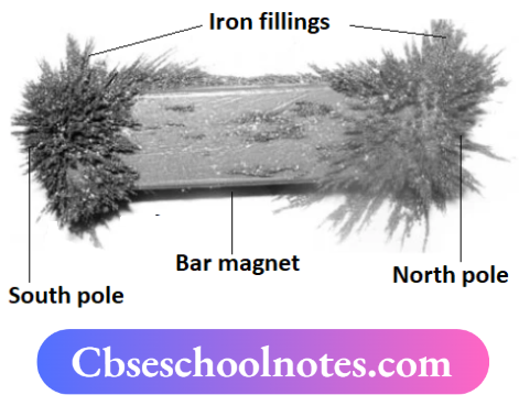
- Iron is malleable and strong. It easily rusts in moist air.
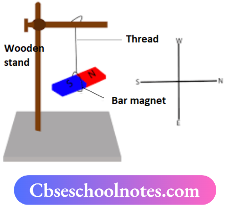
- Iron is magnetic, and melts at 1530°C.
- Sodium is so soft, can be cut by a knife. It floats on water, catches fire in the presence of water, and melts at 98°C.
- Gold is unreactive, highly malleable and ductile and looks attractive.
- Gold is used in jewellery, and melts at 1064°C.
Mg and Mn react with 5% HNO3 to produce Hz gas.
6. Al2O3. BeO and ZnO are amphoteric (acidic as well as basic) oxides.
7. NO, N2O, CO or neutral oxides.
2. Metalloids
Composition of Earth’s crust

Most of the metals occur as their compounds except gold, silver, and platinum which occur in free-state
3. Non-metals
Physical Properties of Non-Metals
- They are brittle.
- They exist as solids, liquids and gases.
- They are non-lustrous except for diamond and iodine.
- They are not conductors of heat and electricity except graphite.
- Low melting and boiling points except for diamond and graphite.
- Low density
Chemical Properties of Non-Metals
- They form acidic oxides.
- They gain electrons to form negative ions
- They are a good oxidising agent. oxidising agents
- Their oxide dissolves in water forming acids
- They do not react with dilute acids because they can’t lose electrons
- More reactive non-metals can displace less reactive non-metals from their salt solution.
CBSE Class 10 Science Chapter 3 Metals And Non-Metals Definitions
1. Metals: Metals are the element which can easily form positive ions by ionising electrons.
For example: Sodium, Potassium, Magnesium, Iron, Copper, etc.
2. Physical characteristics of metals:
- Conductors of heat and electricity
- Metals have shining surface
- Metals have a high tensile strength
- Metals have sonority
3. Sonority: It is the characteristic ofa metal to emit a deep resonant sound when struck.
4. Tensile strength: It is the resistance of a wire to pull action and is measured by the breaking stress in kilogram per square millimetre.
5. Reactivity of metals: The electropositive character of a metal determines its reactivity.
6. Metallichydrides: The compounds of metals with hydrogen are calledmetallichydrides.
These are usually obtained by passing hydrogen over heated metals.
7. Amphoteric oxides: Such oxides which react with both acids and bases to give salt and water are called amphoteric oxides. For example: A1203, ZnO.
8. Non-metals: Non-metals are the elements which form negative ions by gaining electrons.
9. Physical characteristics of non-metals:
- Non-metals are brittle.
- Non-metals are non-ductile.
- Non-metals have comparatively low melting and boiling points.
- Non-metals have low densities.
10. Minerals: The metals found in nature in a combined state are called minerals.
11. Ore: An ore is defined as a mineral from which a metal can be extracted economically.
12. Metallurgy: The extraction of a metal from a suitable ore and its refining for use is called metallurgy or metallurgical operations.
13. Calcination: It is the process of heating the concentrated ore in the absence of air
⇒ \(\underset{\text { Zinc carbonate }}{\mathrm{ZnCO}_3(s)} \longrightarrow \underset{\text { Zinc oxide }}{\mathrm{ZnO}(s)}+\underset{\text { Carbon dioxide }}{\mathrm{CO}_2(\mathrm{~g})}\)
14. Roasting: It is the process of heating the ore in the presence of air
⇒ \(2 \mathrm{ZnS}+3 \mathrm{O}_2 \longrightarrow 2 \mathrm{ZnO}+2 \mathrm{SO}_2\)
15. Slag: Some substances when heated with the ore combine with the earthy impurities and form an easily fusible mass. The easily fusible mass is called slag.
16. Gangue:
Ores are associated with earthy or rocky materials as impurities. These impurities are called gangue or matrix.
17. Smelting:
The roasted or calcined ore is mixed with coke and heated in a furnace to obtain free metal. The process of reducing the oxide with coke is called smelting.
18. Electrolysis:
Electrolysis is the process of conduction of electricity through molten or dissolved chemicals to decompose its components.
19. Corrosion: When the surface or metal is attacked by air, water and some other substance, it is said to corrode. The phenomenon is known as corrosion.
20. Galvanisation: The coating of iron with zinc is called galvanisation.
21. Anodising: It is the process of the formation of a thick layer of metal oxide over a metallic object.
22. Alloy: An alloy is a homogeneous mixture of two or more than two metals, which have nearly the same atomic size.
23. Amalgam: It is an alloy of metal with mercury.
24. Aqua regia: The mixture of cones. HCl and cone. HNO in the ratio 3:1.
25. Important chemical reactions of metals
Reaction with oxygen:
Metal + Oxygen Metal oxide
4 Na +O2 → 2Na2 O(s)
4K+ O2 → 2K2O(s)
2Cu +O2→ (heat) 2CuO(s)
4Al +3O2→(heat) 2AlO(s)
2 Zn +3O2→(heat) →2 ZnO(s)
4 Fe +3O→(heat)→2 Fe2O3(s)
2 Mg +O2 → (heat) 2MgO(s)
2 Ca +O2 →(heat)→ 2CaO(s)
Reaction with water
Metal + Water Metal Oxide + Hydrogen or hydroxide
2Na + 2H2O→(cold)→ 2NaOH + H2
2K + 2H2O→(cold) →2KOH + H2
Ca + 2H2O→(cold)→ Ca(OH)2 + H2
Mg + 2H2O→(hot)→ Mg(OH)2 + H2
2Al + 3H2O→(steam)→Al2O3 + 3H2
3Fe + 4H2O→(steam)→ Fe3O4 + H2
Reaction with dil acid
Metal + dil acid→ Salt + Hydrogen
Zn + 2HCl(dil)→ Salt + Hydrogen
Mg + H2SO4(dil)→ MgSO4 + H2
Mg + 2HNO3→(5%) →Mg(NO3)2 + H2
Mn + 2HNO3→ (5%)→ Mn(NO3)2 + H2
2Al + 6HCl → 2AlCl3 + 3H2
The metal reacts with salts
More reactive metal + salt → Salt + Less reactive metal
Fe(s) + CuSO4(aq) → FeSO4(aq) + Cu(s)
Mg(s)+FeSO(aq) → MgSO4(aq) + Fe(s)
Zn(s) + FeSO4(aq) → ZnSO4(aq) + Fe(s)
2Al(s) + 3CuSO4(aq) → Al2(SO4)3(aq) + 3Cu
Mg(s) + CuSO4(aq) → MgSO4(aq) + Cu(s)
Cu +2Ag NO3(aq)→ Ag +Cu(NO3)2
Ag + Cy (NO3)2 → No reaction
Metal oxide reaction with water
Metal oxide + water Metal hydroxide
K2O+H2O →2KOH
CaO+H2O→Ca(OH)2
MgO+H2O→Mg(OH)
NaO +HO → 2NaOH
Metal oxide reacts with acid
Metallic oxide + Acids → Salt + Water
Salt + Water MgCl +However
ZnO + 2HCl(dil) → ZnCl2 + H2O
CuO + H2SO4(dil) →CuSO4 + H2O
FeO + H2SO4(dil)→ FeSO4 + H2O
Al2O3 + 6HCl→ Al2Cl2 + 3H2O
26. Important chemical properties of metals
Reaction with oxygen
Non-metals + Oxygen Non-metal oxide (acidic oxide)
S + O2 → SO2
c + O2 →CO2
4P + 5O2 → 2P2O6, NO, CO and NO are neutral oxides
4B +3O2 → BO
Si +O2 → SiO2
Non-metallic oxides react with water
Non-metallic oxide + water→ Acids
CO2 + H2O H2CO3 (Carbonic acid)
SO2 + H2O→ H2CO3 (Carbonic acid)
SO3 + H2O→ H2SO4 (Sulphuric acid)
P2O5 + 3H2O→ 2H3PO4 (Phosphoric acid)
B2O3+ 3H2O → 2H3PO4 (Phosphoric acid)
SiO2 + H2O → H2SiO3 (Silicic acid)
H2SiO3(Silicic acid)
Acidic oxides + Base Salt + Water
CO2 + 2NaOH → Na2CO3+H2O
CO2 + Ca(OH)2 → Na2CO3+H2O
SO2 + 2NaOH → Na2SO3 + H2O
More reactive non-metals react with salt
More reactive non-metal + Salt → Salt+ Less reactive non-metal
More reactive non-metal + Salt
Cl2 + 2KBr→ 2KCl + Br2
Cl2 + 2KI → Cl2+ 2KI
Br2 + 2KI →2KBr + I2
Order of reactivity: Cl2 > Br2 > I2
Order of reactivity: Cl2 > Br2 > I2
Extraction of metals from ores

Alloys:
Homogeneous mixture of two or more metals which have better properties than metals. One of them can be non-metal also. Steel is an alloy of iron and carbon. It does not get rusted.
Important Alloys:
1. Stainless steel: It is an alloy of Fe. C. nickel and chromium which is hard, malleable and does not rust, It is used for utensils and surgical instruments.
2. Brass: It is an alloy of copper and zinc. It is used to make decorative articles
3. Bronze: It is an alloy of copper and tin. It’s used for medals and statues.
4. Solder is an alloy of an alloy of It is used for soldering purpose
5. Duralumin: Duralumin is an alloy of Alandmagnesium.It is used to make the body of the aeroplane
6. 24-carat gold is pure gold. It is very soft. It is alloyed with copper and silver to make it hard to make jewellery of 22-carat gold=\(\frac{22}{24} \times 100\)
= 91.6% pure
7. Al is used for transmission wires, cooking foil, drink cans, and coating CDs and DVDs. Aluminium powder is used In silver paints. Al is used as a reducing agent. Al Is used for making utensils and pressure cooker
Relative Reactivities of Metals

CBSE Class 10 Science Chapter 3 Metals And Non-Metals Short Question And Answers
Question 1. Give an example of a metal which
- Is a liquid at room temperature.
- Can be easily cut with a knife.
- Is the best conductor of heat.
- Is a poor conductor of heat.
Answer:
- Mercury is a liquid at room temperature.
- Sodium can be easily cut with a knife.
- Silver is the best conductor of heat.
- Lead is a poor conductor of heat.
Question 2. Explain the meanings of malleable and ductile.
Answer:
When a metal can be beaten into thin sheets with a hammer, the metal is called malleable and the property is called malleability. When a metal can be drawn into thin wires, the metal is called ductile and the property is called ductility.
Question 3. Write equations for the reactions of
- Iron with steam
- Calcium and potassium in water
Answer:
1. \(\underset{\text { Iron }}{3 \mathrm{Fe}(s)}+\underset{\text { Water }}{4 \mathrm{H}_2 \mathrm{O}(l)} \longrightarrow \underset{\text { oxide of iron }}{\mathrm{Fe}_3 \mathrm{O}_4(s)}+\underset{\text { Hydrogen }}{4 \mathrm{H}_2(g)}\)
2.

Question 4. Which gas is produced when dilute hydrochloric acid is added to a reactive metal? Write the chemical reaction when iron reacts with dilute H2SO4
Answer:
Hydrogen gas is produced when dilute hydrochloric acid is added to a reactive metal.
Reaction of iron with dilute sulphuric acid
⇒ \(\underset{\text { Iron }}{\mathrm{Fe}}+\underset{\text { Sulphuric acid }}{\mathrm{H}_2 \mathrm{SO}_4(\alpha q)} \longrightarrow \underset{\text { Ferrous sulphate }}{\mathrm{FeSO}_4(\alpha q)}+\underset{\text { Water }}{\mathrm{H}_2 \mathrm{O}}\)
Question 5. What would you observe when zinc is added to a solution of iron (2) sulphate? Write the chemical reaction that takes place.
Answer:
Zinc is more reactive than iron. When zinc is added to a solution of iron (2) sulphate, the green colour of iron (2) sulphate fades out and iron metal is deposited.
Zn(s)(zinc) + FeSO4(aq)(Iron sulphate) → Fe(Iron)] +ZnSO(aq)(Zinc sulphate)
Question 6. Why do ionic compounds have high melting points?
Answer:
Ionic compounds do not exist as single molecules but exist as aggregates of a large number of positive and negative ions due to strong electrostatic forces. Thus, a large amount of energy is required to break the inter-ionic attraction, hence these have high melting points
Question 7. What are alloys?
Answer:
An alloy is a homogeneous mixture of two or more metals or a metal and a non-metal. It is prepared by first melting the main metal, and then dissolving the required amount of other metals or non-metals. The mixture is then cooled to room temperature to form an alloy of a given composition.
Question 8. What are amphoteric oxides? Give two examples of amphoteric oxides.
Answer:
Those oxides which react with both acids as well as bases to produce salt and water are known as amphoteric oxides. Two examples of amphoteric oxides are Aluminium oxide (Al2O3) and zinc oxide (ZnO).
Question 9. In the electrolytic refining of a metal M, what would you take as the anode, the cathode and the electrolyte?
Answer:
In electrolytic refining of a metal M, the impure metal M is made the anode and a thin strip of pure metal M is made the cathode. A solution of the metal M salt is used as an electrolyte.
For Example:
For refining copper metal, the strip of the pure metal would be made cathode, and that of impure metal would be made anode and the electrolyte would be copper sulphate solution.
Question 10. You must have seen tarnished copper vessels being cleaned with lemon or tamarind juice. Explain why these sour substances are effective in cleaning the vessels.
Answer:
When the copper vessel is kept in moist air, it reacts with moist carbon dioxide in the air to form a green substance. The green substance is copper carbonate, which is basic. When it is treated with lemon or tamarind juice containing acid, neutralisation of the base occurs which cleans the vessel.
Question 11. Name two metals which are purified by electrolytic refining. Mention the anode, cathode and the electrolyte used in the refining process. At which electrode would the pure metal be deposited?
Answer:
Copper, zinc.
Impure metal is made of anode and a thin strip of pure metal is made of cathode. A solution of the metal salt is used as an electrolyte. Pure metal from the electrolyte is deposited on the cathode.
Question 12. Write the electron dot structure for sodium and chlorine atoms. How do these form a chemical bond? Name the type of bond so formed. Why does a compound so formed have a high melting point?
Answer:
The electron dot diagrams of Na and Cl are as follows:
- They form bonds by the transfer of an electron. Na loses one electron to form Na+ ion whereas Cl gains one electron to form Cl– ion.
- The bond formed is an ionic or electrovalent bond.
- The compound (NaCl) thus formed has a high melting point due to the strong force of attraction between Na+ and Cl– ion.
Question 13. Mention the names of the metals for the following
- Two metals are alloyed with iron to make stainless steel.
- Two metals are used to make jewellery.
Answer:
- Nickel and chromium
- Gold and platinum
Question 14. Write one example of each of
- A metal which is so soft that, it can be cut with a knife and a non-metal which is the hardest substance.
- A metal and a non-metal exist as a liquid at room temperature.
Answer:
- Sodium, carbon (diamond)
- Mercury is a liquid metal, bromine is a liquid non-metal.
Question 15. A student has been collecting silver coins and copper coins. One day she observed a black coating on silver coins and a green coating on copper coins. State the chemical name of the black and green coating. How are they formed?
Answer:
Black coating: Silver sulphide (Ag2S)
Green coating: Basic copper carbonate [CuCO3.Cu(OH)2]
The action of
- HS or sulphur compounds are present in the atmosphere.
- Carbon dioxide, oxygen and water vapour are present in the atmosphere.
Question 16.
- Write the electron dot structures for potassium and chlorine.
- Show the formation of KCl by the transfer of electrons.
- Name the ions present in this compound, KCl.
Answer:

3. KCl has K+ and Cl–.
Question 17. Explain the formation of the ionic compound CaO with an electron dot structure. An atomic number of calcium and oxygen are 20 and 8 respectively.
Name the constituent metals of bronze.
Answer:
⇒ \(\underset{2,8,8,2}{\mathrm{Ca}} \longrightarrow \mathrm{Ca}^{2+}+\underset{2,8,8}{2 \mathrm{e}^{-}}\)
⇒ \(\underset{2,6}{\mathrm{O}}+2 \mathrm{e}^{-} \longrightarrow \underset{2,8}{\mathrm{O}^{2-}}\)
Bronze is made up of copper and tin.
Question 18. Out of the two metals P and Q, P is less reactive than Q. Suggest an activity to arrange these metals in the order of their decreasing reactivity. Support your answer with a suitable chemical equation.
Answer:
Activity: In a test tube, a small amount of salt solution of P is taken and metal Q is added to it. Q being more reactive, displaces metal P from its salt solution.
Chemical equation:
Metal Q + Salt solution of P → Salt solution of Q + Metal P.
Question 19. Define an alloy. How is an alloy prepared?
Answer:
A homogeneous mixture of two or more metals or a metal and a non-metal is known as an alloy. It is prepared by first melting the primary metal, and then, dissolving the other elements in it in definite proportions.
Question 20. A compound ‘Z’ is formed by the transfer of electrons from a metal ‘X’ to a non-metal Y. Identify the type of bond formed in the compound. List three general properties of the compounds formed by such types of bonds.
Answer:
Ionic compounds are formed in these compounds.
The general properties of ionic compounds are:
- Solid
- High melting and boiling point
- Soluble in water
- Good conductor of electricity
Question 21. Give reasons for the following:
- School bells are made up of metals.
- Electrical wires are made up of copper.
Answer:
Because metals are sonorous.
Copper is a very good conductor of electricity.
Question 22.
- Name a metal which does not stick to glass.
- Name the metal which is commonly used in thermite welding.
- What is the nature of zinc oxide?
Answer:
- Hg (Mercury)
- Al (Aluminium)
- Basic
Question 23. Describe ionic compounds based on the following properties:
- A strong force of attraction between positive and negative ions
- Solubility of compounds in water
- Electrical conductivity
Answer:
Owing to the strong force of attraction between the positive and negative ions, ionic compounds are solids.
- These compounds are generally brittle and break into pieces when pressure is applied.
- Ionic compounds are generally soluble in water.
- Ionic compounds in the solid state do not conduct electricity but in aqueous and molten states, they do conduct electricity.
Question 24.
- ‘Sodium is a highly reactive metal and it cannot be obtained from its oxide by heating with carbon’. Give reason.
- How can sodium be obtained from sodium chloride?
Answer:
Sodium cannot be obtained from its oxide by heating with carbon because carbon cannot reduce the oxides of sodium.
Sodium can be obtained from sodium chloride by the process of electrolytic reduction.
Question 25. Define the term ‘anode mud. Name the electrode made of pure metal. State the reactions taking place at the cathode and at the anode during the electrolytic refining of copper.
Answer:
The insoluble impurities which settle down at the bottom of the anode are known as anode mud.
The cathode is made of pure metal.
At anode: \(\mathrm{Cu}(s) \longrightarrow \mathrm{Cu}^{2+}(a q)+2 e^{-}\)
At cathode: \(\mathrm{Cu}^{2+}(a q)+2 e^{-} \longrightarrow \mathrm{Cu}(s)\)
Question 26. Name the following:
- A metal, which is preserved in kerosene.
- A lustrous coloured non-metal.
- A metal, which can melt while kept on the palm.
- A metal, which is a poor conductor of heat.
Answer:
- Sodium is preserved in kerosene.
- Iodine is a lustrous coloured non-metal.
- Gallium or caesium
- Lead or mercury
Question 27. Give reasons why copper is used to make hot water tanks and not steel (an alloy of iron).
Answer:
It is because of the following reasons:
- Copper does not react with hot water or steam, whereas steel reacts with steam.
- Copper is one of the best conductors of heat as compared to steel.
- Copper has a high melting point as compared to steel.
Question 28. Write chemical equations that show aluminium oxide reacts with acid as well as base.
Answer:
⇒ \(\mathrm{Al}_2 \mathrm{O}_3+6 \mathrm{HCl} \longrightarrow 2 \mathrm{AlCl}_3+3 \mathrm{H}_2 \mathrm{O}\)
⇒ \(\mathrm{Al}_2 \mathrm{O}_3+2 \mathrm{NaOH} \longrightarrow 2 \mathrm{NaAlO}_2+\mathrm{H}_2 \mathrm{O}\)
Question 29. Explain why calcium metal after reacting with water starts floating on its surface. Write the chemical equation for the reaction. Name one more metal that starts floating after some time when immersed in water.
Answer:
Calcium starts floating because the bubbles of hydrogen gas formed stick to the surface of the metal.
⇒ \(\mathrm{Ca}(\mathrm{s})+2 \mathrm{H}_2 \mathrm{O}(\mathrm{l}) \longrightarrow \mathrm{Ca}(\mathrm{OH})_2+\mathrm{H}_2(\mathrm{~g})\)
Magnesium reacts with hot water and starts floating due to the bubbles of hydrogen gas sticking to its surface.
Question 30. Why is it that non-metals do not displace hydrogen from dilute acids?
Answer: Those oxides which are both acidic as well as basic are called amphoteric oxides
Example: Al2O3, ZnO.
This is because non-metals do not supply electrons to change H+ ions into hydrogen gas.
Question 31. During the extraction of metals, electrolytic refining is used to obtain pure metals.
- Which material will be used as anode and cathode for refining silver metal by this process?
- Suggest a suitable electrolyte also.
- In this electrolytic cell, where do we get pure silver after passing an electric current?
Answer:
1. Anode : Impure silver
Cathode: Pure silver
2. Electrolyte: Silver salt, such as AgNO3, AgCl, etc.
3. We get pure silver at the cathode
Question 32. Compound X and aluminium are used to join railway tracks.
- Identify the compound X
- Name the reaction
- Write down its reaction.
Answer:
Compound X must be iron (3) oxide, Fe2O3
The reaction is known as the ‘thermite reaction’ or ‘alumino thermy’.
The reaction is carried out by igniting the mixture with a Mg-ribbon.

Question 33. When a metal X is treated with cold water, it gives a basic salt Y with the molecular formula XOH (Molecular mass = 40) and liberates a gas Z which easily catches fire. Identify X, Y, and Z and also write the reaction involved.
Answer:
The base with molecular mass 40 is NaOH.
Hence, the metal X be sodium (Na). It reacts with H2O to form the base NaOH and liberates H2 gas which easily catches fire

Question 34. A non-metal X exists in two different forms Y and Z. Y is the hardest natural substance, whereas Z is a good conductor of electricity. Identify X, Y and Z.
Answer:
Non-metal ‘X’ must be carbon. It exists in two forms, diamond and graphite.
Diamond is the hardest natural substance. Hence, ‘Y’ is a diamond.
Graphite is a good conductor of electricity. Hence, ‘Z’ is graphite.
Question 35. The following reaction takes place when aluminium powder is heated with MnO2
⇒ \(3 \mathrm{MnO}_2(s)+4 \mathrm{Al}(s) \longrightarrow 3 \mathrm{Mn}(l)+2 \mathrm{Al}_2 \mathrm{O}_3(l)+\text { Heat }\)
- Is aluminium getting reduced?
- Is MnO2 getting oxidised?
Answer:
- No, because oxygen is added to aluminium, so it is getting oxidised
- No, since manganese has lost oxygen, therefore it is getting reduced.
Question 36. A metal A, which is used in the thermite process, when heated with oxygen gives an oxide B, which is amphoteric. Identify A and B. Write down the reactions of oxide B with HCl and NaOH.
Answer:
A = Aluminium
B = Al2O3
Question 37. A metal that exists as a liquid at room temperature is obtained by heating its sulphide in the presence of air. Identify the metal and its ore and give the reaction involved.
Answer:
The metal that exists as a liquid at room temperature is mercury (Hg). It is obtained by heating its sulphide ore, HgS, called ‘cinnabar’ in the presence of air (roasting). The metal sulphide is first converted into metal oxide (HgO) which on further heating is reduced to Hg.

Question 38. Give the formulae of the stable binary compounds that would be formed by the combination of the following pairs of elements.
- Mg and N2
- Li and O2
- Al and Cl2
- K and O2
Answer:
- Mg3N2
- Li2O
- AlCl2
- K2O
Question 39. A non-metal A is an important constituent of our food and forms two oxides B and C. Oxide B is toxic whereas C causes global warming.
- Identity A, B, and C
- To which Group of the Periodic Table does A belong?
Answer:
- A carbon, B = carbon monoxide (CO) C = Carbon dioxide (CO2)
- Carbon (A) belongs to group 14 of the periodic table.
Question 40. Name one metal and one non-metal that exist in a liquid state at room temperature. Also, name two metals having melting point less than 310 K (37°C).
Answer:
Metal existing in a liquid state at room temperature is mercury and non-metal is bromine.
The metals with melting points less than 310 K are caesium (Cs) and Gallium (Ga).
Question 41. Using the electronic configurations, explain how the magnesium atom combines with the oxygen atom to form magnesium oxide by transfer of electrons.
Answer:
The atomic number of magnesium
⇒ \(\underset{2,8,2}{\mathrm{Mg}} \longrightarrow \underset{2,8}{\mathrm{Mg}^{2+}+2 \mathrm{e}^{-}}\)
The atomic number of oxygen = 8
⇒ \(\underset{2,6}{\mathrm{O}}+2 \mathrm{e}^{-} \longrightarrow \underset{2,8}{\mathrm{O}^{2-}}\)
Question 42. List three properties of sodium in which it differs from the general physical properties of
most metals.
Answer:
It has low density.
It has a low melting point.
Sodium is so soft that it can be cut with a knife.
Question 43. What happens when
- ZnCO3 is heated in the absence of oxygen.
- A mixture of Cu2O and CuS is heated.
Answer:

Question 44. An element A reacts with water to form compound B which is used in whitewashing. The compound B on heating forms an oxide C which on treatment with water gives back B. Identify A, B, and C and give the reactions involved.
Answer:
A = Ca; B = Ca (OH)2 ; C = CaO

Question 45. A metal M does not liberate hydrogen from acids but reacts with oxygen to give a black-coloured product. Identify M and black coloured product and also explain the reaction of M with oxygen.
Answer:
Cu + HCl/H2SO4/HNO3→ No H2 is evolved
2 Cu(s)+O2(g)→ (Heat) → 2CuO(s) Copper(2)oxide (Black product)
Question 46. A solution of CuSO4 was kept in an iron pot. After a few days, the iron pot was found to have several holes in it. Explain the reason in terms of reactivity. Write the equation of the reaction involved.
Answer:
Fe is more reactive than Cu and hence displaces Cu from CuSO, solution. The following reaction takes place:
Fe(s) + CuSO4(aq) → FeSO4(aq) + Cu(s)
Question 47. Of the three metals X, Y, and Z. X reacts with cold water, Y with hot water and Z with steam only. Identify X, Y, and Z and also arrange them in order of increasing reactivity.
Answer:
X = Na or K, Y = Mg, Z = Fe.
Increasing reactivity: Na/K > Mg > Fe.
Question 48. Element A burns with a golden flame in the air. It reacts with another element B, atomic number 17 to give a product C. An aqueous solution of product C on electrolysis gives a compound D and liberates hydrogen. Identify A, B, C, and D. Also write down the equations for the reactions involved.
Answer:
A Sodium, B = chlorine and, C = Sodium chloride
2 Na(s) + Cl2(g) → 2NaCl(s)
D = Sodium hydroxide
2 Na+Cl–(aq) +2H2O(l) → (Electrolysis) → 2NaOH(aq)+Cl2(g) +H2(g)
Question 49. Two ores A and B were taken. On heating ore A gives CO, whereas, ore B gives SO What steps will you take to convert them into metals?
Answer:
Since ore A gives CO2, and ore B gives SO2. Therefore, ores are MCO and MS.
A can be obtained
MCO3 → MO+CO2
MO+C → M+ CO
Question 50. What happens when
- ZnCO3 is heated in the absence of oxygen.
- A mixture of Cu2O and Cu2S is heated.
Answer:
ZnCO3→ ZnO+CO2↑
2Cu2O+Cu2S→6Cu+SO2↑
Question 51. When a metal X is treated with cold water, it gives a basic Y with the molecular formula XOH (Molecular mass = 40) and liberates a gas Z which easily catches fire. Identify X, Y and Z and also write the reaction involved.
Answer:
X Sodium, Y = NaOH, Z = H2 ↑
2 Na + 2H2O → 2NaOH (aq) + H2(↑)
Question 52. Give the formulae of the stable binary compounds that would be formed by the combination of the following pairs of elements.
Answer:
- Mg, N2
- LiO2
- AlCl3
- K2O
Question 53. A metal ‘E’ is stored under kerosene oil. When a small piece of it is left open in the air, it catches fire. When the product formed is dissolved in water it turns red litmus blue.
Name the metal E.
Write the chemical equation for the reaction when it is exposed to air and when the product is dissolved in water.
CBSE Class 10 Science Chapter 3 Metals And Non-Metals Multiple Choice Questions
Question 1. Which of the following properties is generally not shown by metals?
- Electrical conduction
- Sonorous in nature
- Dullness
- Ductility
Answer: 3. Dullness
Question 2. The ability of metals to be drawn into thin wire is known as
- Ductility
- Malleability
- Sonorosity
- Conductivity
Answer: 1. Malleability
Question 3. Aluminium is used for making cooking utensils. Which of the following properties of aluminium are responsible for the same?
1. Good thermal conductivity
2. Good electrical conductivity
3. Ductility
4. High melting point
- 1 and 2
- 1 and 3
- 2 and 3
- 1 and 4
Answer: 4. 1 and 4
Question 4. Which one of the following metals does not react with cold as well as hot water?
- Na
- Ca
- Mg
- Fe
Answer: 4. Fe
Question 5. Which of the following oxide(s) of iron would be obtained on the prolonged reaction of iron with steam?
- FeO
- Fe2O3
- Fe3O4
- Fe2O3 and Fe2O4
Answer: 3. Fe3O4
Question 6. What happens when calcium is treated with water?
1. It does not react with water
2. It reacts violently with water
3. It reacts less violently with water
4. Bubbles of hydrogen gas formed stick to the surface of calcium
- 1 and 4
- 2 and 3
- 1 and 2
- 3 and 4
Answer: 4. 3 and 4
Question 7. Generally, metals react with acids to give salt and hydrogen gas. Which of the following acids does not give hydrogen gas on reacting with metals (except Mn and Mg)?
- H2SO4
- HCl
- HNO3
- All of these
Answer: 3. HNO3
Question 8. The composition of aqua-regia is
1. \(\begin{array}{cll}
\text { Dil. } \mathrm{HCl} & : & \text { Conc. } \mathrm{HNO}_3 \\
3 & : & 1
\end{array}\)
2. \(\begin{array}{cll}
\text { Conc. } \mathrm{HCl} & : & \text { Dil. } \mathrm{HNO}_3 \\
3 : & 1
\end{array}\)
3. Conc.HCl (3): Conc.HNO3(1)
4. \(\begin{array}{cll}
\text { Dil. } \mathrm{HCl} & : & \text { Conc. } \mathrm{HNO}_3 \\
3 & : & 1
\end{array}\)
Answer: 3. Conc.HCl (3): Conc.HNO3(1)
Question 9. Which of the following are not ionic compounds?
1. KCl
2. HCl
3. CCl4
4. NaCl
- 1 and 2
- 2 and 3
- 3 and 4
- 1 and 3
Answer: 2. HCl
Question 10. Which one of the following properties is not generally exhibited by ionic compounds?
- Solubility in water
- Electrical conductivity in solid state
- High melting and boiling points
- Electrical conductivity in molten state
Answer: 2. Electrical conductivity in solid state
Question 11. Which of the following metals exists in their native state in nature?
1. Cu
2. Au
3. Zn
4. Ag
- 1 and 2
- 1 and 3
- 2 and 4
- 3 and 4
Answer: 3. Zn
Question 12. Metals are refined by using different methods. Which of the following metals are refined by electrolytic refining?
1. Au
2. Cu
3. Na
4. K
- 1 and 2
- 1 and 3
- 2 and 3
- 3 and 4
Answer: 1. Au
Question 13. Silver articles become black on prolonged exposure to air. This is due to the formation of
- Ag3N
- Ag2O
- Ag2S
- Ag2S and Ag2N
Answer: 3. Ag2S
Question 14. Galvanisation is a method of protecting iron from rusting by coating with a thin layer of
- Galium
- Aluminium
- Zinc
- Silver
Answer: 3. Galium
Question 15. Stainless steel is a very useful material for our life. In stainless steel, iron is mixed with
- Ni and Cr
- Cu and Cr
- Ni and Cu
- Cu and Au
Answer: 1. Ni and Cr
Question 16. If copper is kept open in the air, it slowly loses its shining brown surface and gains a green coating. It is due to the formation of
- CuSO4
- CuCO3
- Cu(NO3)2
- CuO
Answer: 2. CuCO3
Question 17. Generally, metals are solid. Which one of the following metals is found in a liquid state at room temperature?
- Na
- Fe
- Cr
- Hg
Answer: 4. Hg
Question 18. Which of the following metals are obtained by electrolysis of their chlorides in a molten state?
1. Na
2. Ca
3. Fe
4. Cu
- 1 and 4
- 3 and 4
- 1 and 3
- 1 and 2
Answer: 4. 1 and 2
Question 19. Generally, non-metals are not lustrous. Which of the following non-metal is lustrous?
- Sulphur
- Oxygen
- Nitrogen
- Iodine
Answer: 4. Iodine
Question 20. Which one of the following four metals would be displaced from the solution of its salts by the other three metals?
- Mg
- Ag
- Zn
- Cu
Answer: 2. Ag
Question 21. 2 ml each of concentrated HCL, HNO, and a mixture of concentrated HCl and concentrated HNO, in the ratio of 3: 1 were taken in test tubes labelled as A, B, and C. A small piece of metal was put in each test tube. No change occurred in test tubes A and B but the metal got dissolved in test tube C respectively. The metal could be
- Al
- Au
- Cu
- Pt
Answer: 2. Au
Question 22. An alloy is
- An element
- A compound
- A homogeneous mixture
- A heterogeneous mixture
Answer: 3. A homogeneous mixture
Question 23. An electrolytic cell consists of
1. Positively charged cathode
2. Negatively charged anode
3. Positively charged anode
4. Negatively charged anode
- 1 and 2
- 3 and 4
- 1 and 3
- 2 and 4
Answer: 2. 3 and 4
Question 24. During electrolytic refining of zinc, it gets
- Deposited on cathode
- Deposited on anode
- Deposited on the cathode as well as anode
- Remains in the solution
Answer: 1. Deposited on cathode
Question 25. An element A is soft and can be cut with a knife. This is very reactive to air and cannot be kept open in air. It reacts vigorously with water. Identify the element from the following
- Mg
- Na
- P
- Ca
Answer: 2. Na
Question 26. Alloys are homogeneous mixtures of a metal with a metal or non-metal. Which among the following alloys contain non-metal as one of its constituents?
- Brass
- Bronze
- Amalgam
- Steel
Answer: 4. Steel
Question 27. Which among the following statements is incorrect for magnesium metal?
- It burns in oxygen with a dazzling white flame
- It reacts with cold water to form magnesium oxide and evolves into hydrogen gas
- It reacts with hot water to form magnesium hydroxide and evolves hydrogen gas
- It reacts with steam to form magnesium hydroxide and evolves hydrogen gas
Answer: 2. It reacts with cold water to form magnesium oxide and evolves into hydrogen gas
Question 28. Which among the following alloys contain mercury as one of its constituents?
- Stainless steel
- Alnico
- Solder
- Zinc amalgam
Answer: 4. Zinc amalgam
Question 29. The reaction between X and Y, forms compound Z. X loses electrons and Y gains electrons. Which of the following properties is not shown by Z?
- Has a high melting point
- Has a low melting point
- Conducts electricity in a molten state
- Occurs as solid
Answer: 2. Has a low melting point
Question 30. The electronic configurations of three elements X, Y, and Z are X-2, 8; Y – 2, 8, 7 and Z-2, 8, 2. Which of the following is correct?
- X is a metal
- Y is a metal
- Z is a non-metal
- Y is a non-metal and Z is a metal
Answer: 4. Y is a non-metal and Z is a metal
Question 31. Although metals form basic oxides, which of the following metals form an amphoteric oxide?
- Na
- Ca
- Al
- Cu
Answer: 3. Al
Question 32. Generally, non-metals are not conductors of electricity. Which of the following is a good conductor of electricity?
- Diamond
- Graphite
- Sulphur
- Fullerene
Answer: 2. Graphite
Question 33. Electrical wires have a coating of an insulating material. The material generally used is
- Sulphur
- Graphite
- PVC
- All can be used
Answer: 3. PVC
Question 34. Which of the following non-metals is a liquid?
- Carbon
- Bromine
- Phosphorus
- Sulphur
Answer: 2. Bromine
Question 35. Which of the following can undergo a chemical reaction?
- MgSO4 + Fe
- ZnSO4+ Fe
- MgSO4 + Pb
- CuSO4+ Fe
Answer: 4. CuSO4+ Fe
Question 36. Which one of the following figures correctly describes the process of electrolytic refining?

Answer: 3.
Question 37. Which of the following pairs will give displacement reactions?
- NaCl solution and copper metal
- MgCl2, solution and aluminium metal
- FeSO4 solution and silver metal
- AgNO3, solution and copper metal
Answer: 4. AgNO3, solution and copper metal
Question 38. Which of the following methods is suitable for preventing an iron frying pan from rusting?
- Applying grease
- Applying paint
- Applying a coating of zinc
- All of the above
Answer: 4. All of the above
Question 39. An element reacts with oxygen to give a compound with a high melting point. This compound is also soluble in water. The element is likely to be
- Calcium
- Carbon
- Silicon
- Iron
Answer: 1. Calcium
Question 40. Food cans are coated with tin and not with zinc because
- Zinc is costlier than tin.
- Zinc has a higher melting point than tin.
- Zinc is more reactive than tin.
- Zinc is less reactive than tin.
Answer: 3. Zinc is more reactive than tin.