
Introduction To Pelvis
Boundaries Of True Pelvis
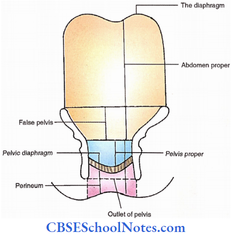
The inlet of the bony true pelvis is the demarcation between the pelvis and abdomen proper. Part of the pelvis between the pelvic diaphragm and skin between the upper parts of the medial thigh is called the perineum.
A bony pelvis above the pelvic inlet about the iliac fossa is called a false pelvis. It forms the posterolateral wall of the lower part of the abdomen proper.
The true pelvis has got inlet, outlet and cavity. The boundaries of each are as follows.
1. Cavity
- Anterior
- Pubic symphysis
- Pubis (Pelvic surfaces of body and rami)
- Posterior – Sacrum
- Pelvic surfaces of ilium and ischium
- Lateral – Pelvic surfaces of ilium and ischium
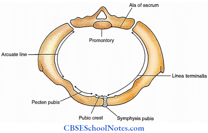
2. Inlet
- Superior margin of pupic symphysis.
- Pelvic brim. It is comprised of,
- Sacral promontory – Anterior margin of superior surface of 1st sacral vertebra.
- All of the sacrum (Lateral part of the base of the sacrum)
- Linea terminalis
- Iliac part – Arcuate line
- Pupic part – Pecten pubis and pubic crest
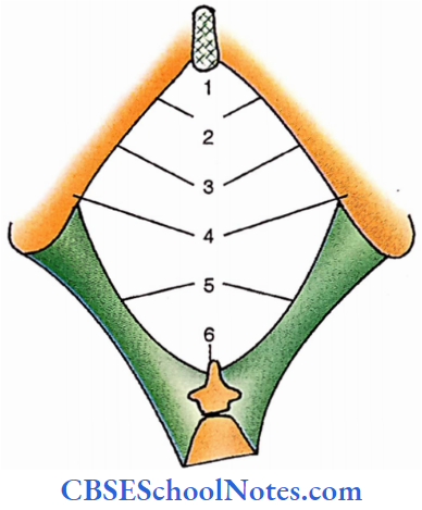
3. Outlet
- Inferior margin of the pubic symphysis
- Subpubic angle
- Ischiopubic rami
- Ischial tuberosity
- Sacrotuberous ligament.
- Tip of coccyx
- Inferior Margin Of Public Symphysis
- Subpublic Angle
- Ischiopublic Rami
- Ischial Tuberosities
- Sacrotuberous Ligaments
- Tip Of Coccyx
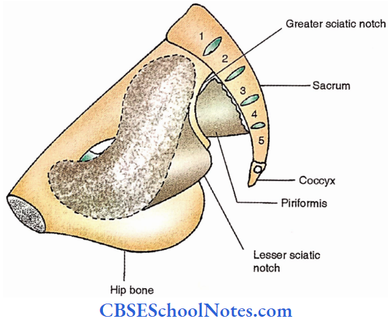
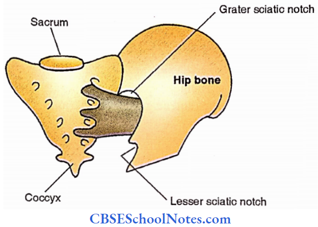
Pelvic Wall
Two paired muscles contribute to the wall of the pelvis i.e., piriformis and obturator items.
Pelvic Wall Piriformis
Origin – Pelvic surfaces of middle three pieces of sacrum
Insertion – Upper border of greater trochanter of femur
Nerve – Branches from sacral plexus (L5, Sl,2)
Pelvic Wall Actions
- Lateral rotation of the extended thigh.
- Abduction of flexed things
Pelvic Wall Obturator internus
Origin – Pelvic surface of obturator membrane and adjacent parts of hip bone below the pelvic brim.
Insertion – Medial surface of the greater trochanter.
Nerve – Nerve to obturator intemus (L5, S1,2)
Obturator internus Action
- Lateral rotation of the extended thigh.
- Abduction of the flexed thigh.
Floor Of Pelvis (Pelvic Diaphragm)
Coccygeus
This is also called iliococcygeus. It is a triangular muscle. Its apex is attached to the ischial spine. Its base extends to the lateral margin of the lower sacrum and adjacent coccyx. Its posterior surface is covered by a sacrospinous ligament.
Levator Ani
It has two components
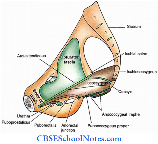
iliococcygeus
It arises from the posterior half of the arcus tendinous, a thickening in the obturator fascia along a line from the back of the body of the pubis to the ischial spine.
Fibres extend backwards and downwards to get attached to the side of the coccyx and interlace with the opposite side from the anococcygeal raphe.
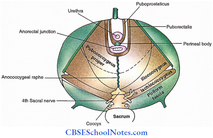
Pubococcygeus
It arises from the posterior surface of the body of the pubis and the anterior half of the acrus tendineus. It is divided into three parts from posterior to anterior.
Pubococcygeus proper- It is the main posterior part of the muscle arising from the anterior half of the arcus tendinous. Fibres pass backwards and medially superior to iliococcygeus interlacing with the apposite half at anococcygeal raphe.
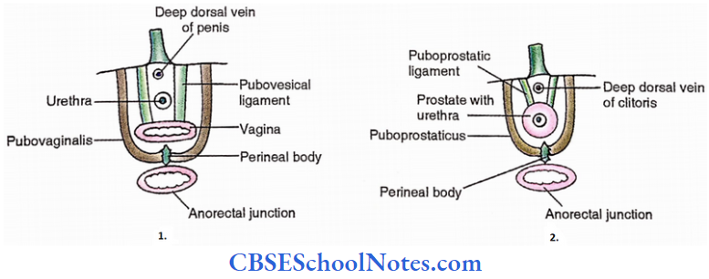
Puborectalis – It arises from the posterior aspect of the body of the pubis and continues with that of the opposite side forming a sling at anorectal junction.
Puboprostaticus or pubovaginalis- It passes backwards from the posterior aspect of the body of the pubis and turns medially to fuse with a perineal body behind the prostate in males (puboprostaticus) and vagina in females (pubovaginalis).
The floor of Pelvis Nerves
From above – Perineal branches of 4th sacral nerve.
From below
- Inferior rectal nerve.
- Muscular branches of the perineal nerve.
The floor of the Pelvis Actions
- To support the pelvic viscera.
- To fix the perineal body.
- Maintenance of continence of bladder and rectum.
- Maintains increased intra-abdominal pressure.
- Acts as sphincter of rectum, vagina and urethra.
- Helps in childbirth during 2nd stage of labour
Pelvic Fascia
According to location, pelvic fascia can be of three types
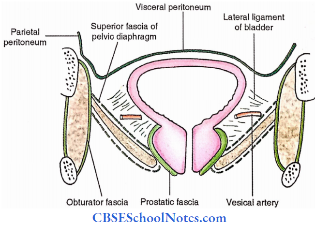
1. Parietal pelvic fascia
This is located over the pelvic wall and floor and named accordingly,
- Over pelvic wall
- Obturator fascia – Covering the obturator intemus.
- Piriform fascia – Covering the piriformis muscle
- Over pelvic floor – Thin epimysium – This is also called superior fascia of the pelvic diaphragm.
2. Visceral pelvic fascia
This covers the viscera of the pelvis and is divided into two types.
- Over distensible viscera – It is thin to allow distension of viscera For Example., over the urinary bladder.
- Over nondistensible viscera – It is well-defined fascia, For Example. over the prostate (Prostatic fascia).
3. Fascia between the pelvic floor and peritoneum
It can be loose or well-defined ligaments.
- Loose areolar tissue – It forms dead space to allow expansion of viscera like the urinary bladder, rectum and uterus (in females).
- Ligaments,
- Condensation around vessels and nerves
- Lateral ligament of uterus (Lateral cervical ligament or Mackenrodt’s ligament) – It extends laterally from the cervical region of the uterus to the lateral pelvic wall. It carries uterine
vessels. - Lateral ligament of the bladder – It extends from the side of the bladder to the region of the arcus tendinous. It carries vesical vessels.
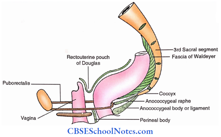
- Fascia of Waldeyer – It is the dense connective tissue between the back of the rectum and the front of the sacrum. It holds superior rectal vessels.
- Lateral ligament of uterus (Lateral cervical ligament or Mackenrodt’s ligament) – It extends laterally from the cervical region of the uterus to the lateral pelvic wall. It carries uterine
- Independent ligaments
- Puboprostatic ligament (in males) or Pubovesical or bulbourethral ligament (in females) Puboprostatic ligament extends from the region of the body of the pubis and the anterior end of the arcus tendinous to the prostatic fascia.
- The prosodic ligament extends from the regions of the female urethra to the back of the body of the pubis.
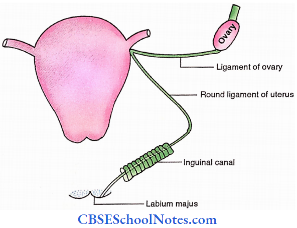
- Round ligament of the uterus – It extends from the lateral angle of the uterus to the labium majus. During its course, it passes through the inguinal canal.
- Uterosacral alignment – It extends from the side of the cervix to the posterior wall of the pelvis. It raises a fold of the peritoneum called the rectouterine fold which forms the lateral wall of the rectouterine pouch.
- Condensation around vessels and nerves
Pelvic Peritoneum
Pelvic Peritoneum Parts
- Visceral layer – It covers the viscera of the pelvis and receives autonomic innervation.
- Parietal layer – It lines the wall of the pelvis and is supplied by the obturator nerve
Pelvic Peritoneum In Male
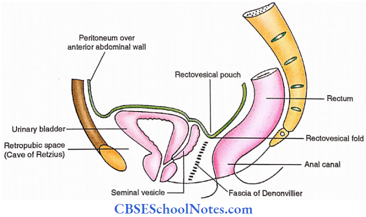
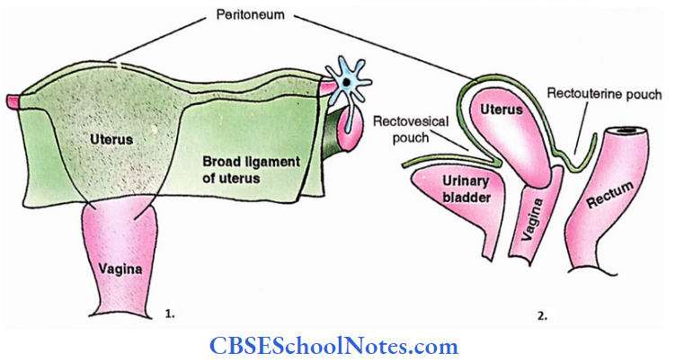
The Peritoneum is reflected from the junction of the upper 2/3rd with the lower l/3rd of the front of the rectum to the upper part of the posterior surface of the urinary bladder. Here it forms a peritoneal pouch called a rectovesical pouch.
After covering the superior surface of the urinary bladder, the peritoneum is reflected over the posterior surface of the anterior wall of the abdomen. This reflection limits the retropubic space of Retzius between the urinary bladder and the pubis.
Pelvic Peritoneum In Female
The peritoneum is reflected from the lower part of the front of the rectum to the posterior fornix of the vagina from where it continues over the posterosuperior and then the anteroinferior surface of the uterus.
Between the rectum and uterus, the reflection forms a rectouterine pouch (of Douglas). From the uterus, it is reflected over the superior surface of the urinary bladder where it forms a ureterovesical pouch between the uterus and the urinary bladder.

Pelvic Vessels
Veins Drain Into Internal Iliac Veins
Veins form plexuses about the viscera. These do not possess valves and intercommunicate with each other and with the internal vertebral venous plexus. These venous plexuses are named after the viscera as follows:
- Rectal venous plexus – about the rectum.
- Uterine venous plexus – to the uterus.
- Prostatic-vesical venous plexus – about both prostate and urinary bladder
Pelvic Vessels Arteries
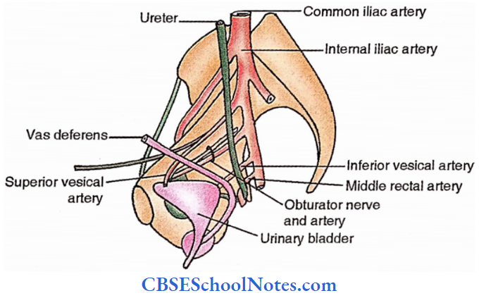
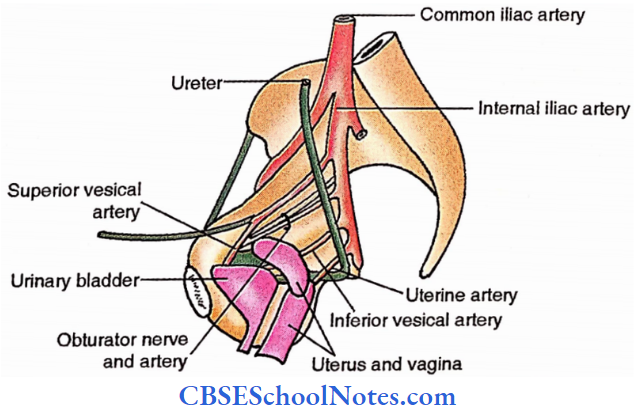
The internal iliac artery splits into anterior and posterior divisions before providing named branches.
1. Anterior division
- Three parietal branches (all pass through foramen)
- Inferior gluteal artery – It passes through the greater sciatic foramen.
- Internal pudendal artery – It passes through the greater sciatic foramen.
- Obturator artery – It passes through the obturator foramen
- Three visceral branches
- Superior vesical artery
- Inferior vesical artery
- Both vesical arteries supply the urinary bladder
- Middle rectal artery. It supplies the musculature of the rectum in males. In females, it is replaced by the uterine artery.
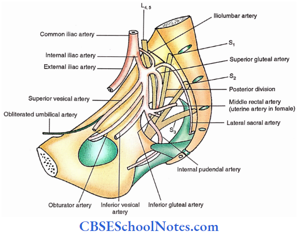
3. Posterior division
Only three parietal branches appear from it.
- Iliolumbar artery – It ascends to supply the lower part of the posterior abdominal wall.
- Superior gluteal artery – It enters the gluteal region by passing between the lumbosacral trunk and 1st sacral nerve (ventral ramus) and then through the greater sciatic foramen.
- Lateral sacral artery – It descends just lateral to pelvic sacral foramina over the roots of the sacral plexus.
Nerves Of The Pelvis
Obturator Nerve
Follows the medial border of the psoas to reach the obturator canal.
Sacral Sympathetic Trunk
Consists of 4 sympathetic ganglia connected by nerve fibres. Descends about the pelvic sacral foramina.
Inferior Hypogastric (Pelvic) Plexus
Lies in front of the sacrum behind the rectum.

Sacral Plexus Formation
Following nerves from its roots,
- Lumbosacral trunk (L4,5) – It is formed by the union of ventral rami of the 4th and 5th lumbar nerves. It descends in front of the ala of the sacrum to join the sacral plexus.
- Ventral rami of upper 4 sacral nerves (S1, S2, S3, S4).
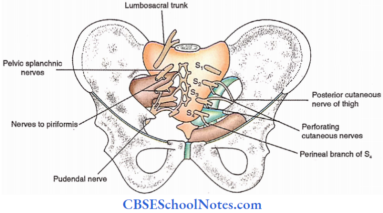
Sacral Plexus Branches
1. Branches from sacral nerves (roots)
- From behind
- Nerves to piriformis (S1,2)- Supply piriformis.
- Perforating cutaneous nerves (S2,3)- Pierce sacrotuberous ligament to appear in the gluteal region.
- The posterior cutaneous nerve of the thigh (S2,3)- Passes through the greater sciatic foramen.
- From front
- Pelvic parasympathetic (splanchnic) nerves (S2,3,4). Also called nervi erigentes. These join the inferior hypogastric plexus.
- Pudendal nerve (S2,3,4) – Passes through the greater sciatic foramen. Crosses the apex of the sacrospinous ligament.
- Perineal branch of S4 – Passes between iliococcygeus and iliococcygeus after supplying both.
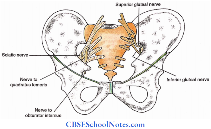
2. Branches from anterior divisions
- Nerve to obturator internus (L5, S1,2)- Passes through the greater sciatic foramen to appear in the gluteal region. Then enters the lesser sciatic foramen to supply the obturator internus and superior gemellus.
- Nerve to quadratus femoris (L4,5, S1)- It passes through the greater sciatic foramen to supply the quadratus femoris and inferior gemellus.
- The tibial component of the sciatic nerve (L4,5, S1,2,3)- It unites with the common peroneal component to form the thickest nerve of the body (Sciatic nerve).
3. Branches from posterior divisions
- Superior gluteal nerve (L4,5, S1) – It passes through the greater sciatic foramen to enter the gluteal region where it supplies the gluteus medius, gluteus minimus and tensor fasciae latae.
- Inferior gluteal nerve (L25, S1,2)- It passes through the greater sciatic foramen to supply the gluteus maximus.
- Common peroneal component of the sciatic nerve (L4,5, S1,2)- It unites with the tibial component to form the sciatic nerve.
Nerves of The Pelvis Coccygeal plexus
- Roots – S4,5, Col
- Supplies
- Coccygeus, Levator ani
- Sacrococcygeal joint
- Skin over the coccyx
Urinary Bladder
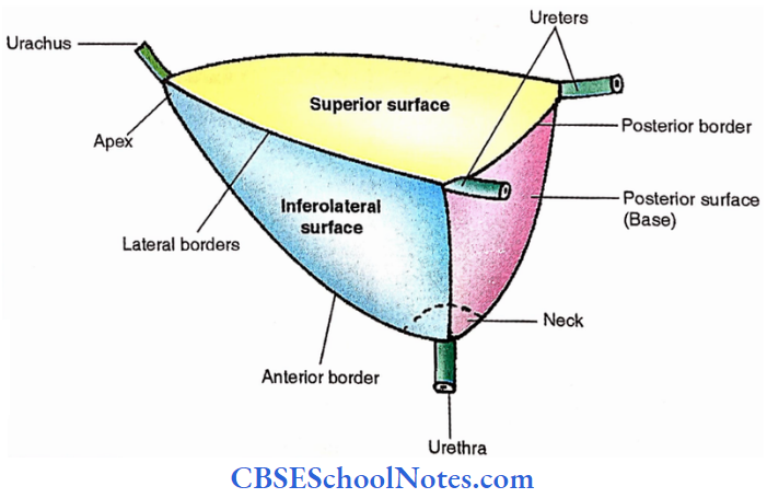
Urinary Bladder Surface Features
It is a muscular vesicle for collection and expulsion of urine.
The shape of an empty bladder is tetrahedral
- 4 Surfaces.
- 1 Porterior (Base or fundus)
- 1 Superior
- 2 Inferolateral
- 4 Borders.
- 2 Lateral
- 1 Anterior
- 1 Posterior
- Neck – It is the lowest conical part.
- Apex – It is the anterior pointed end.
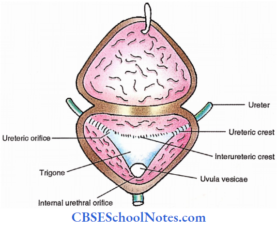
Urinary Bladder Interior
Trigone – A triangular smooth area in the posterior wall.
- 3 Openings – 2 Ureteric orifices at the upper lateral angles of the trigone.
- I internal urethral orifice at the lower angle of the trigone.
- Interureteric crest – It is a transverse mucosal ridge between two ureteric orifices.
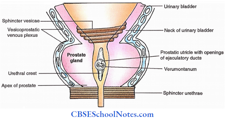
- Uvula vesicae (Male) – Elevation in the posterior wall of the lower angle of trigone produced by a median lobe of the prostate gland.
Urinary Bladder Relations
- Peritoneal relations – Peritoneum covers the superior surface
- Adjacent relations
- Superior -Coils of intestine
- Infero-lateral – Obturator intemus and levator ani.
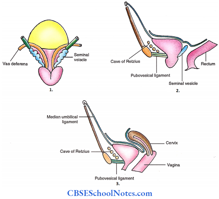
- Anterior-Cave of Retzius, Fat.
- Posterior – Male – Seminal vesicle
- Vas deferens
- Rectum
- Female – Vagina and cervix
Urinary Bladder Capacity
120 – 320 ml (in adults).
280 ml of urine → Refelex micturition
More than 500 ml of urine —> Pain
Urinary Bladder Ligaments
- Median umbilical ligament – Remnant of urachus derived from allantois Lateral ligament – It extends from the side of the urinary bladder to the region of the arcus tendinous. It carries vesical vessels.
- Pubovesical ligament – It is a fibromuscular band on each side extending from the bladder neck to the inferior aspect of the pubic bone.
- It is present in both sexes. It is different from the pubovesical ligament of females which is at a lower level and often named pubourethral ligament.
Urinary Bladder Arteries
Superior and inferior vesical arteries. These appear from the internal iliac artery to reach the bladder through the lateral ligament. Pubic branch of an inferior epigastric artery – Supplies the lower part of the bladder.
Urinary Bladder Veins
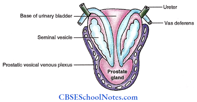
- Male – Prostatic vesical venous plexus – It is located at the junction of the bladder neck and base of the prostate gland. This plexus drains into an internal iliac vein.
- Female – Vesical plexus – It is located at the inferolateral surface of the bladder and drains into an internal iliac vein.
Urinary Bladder Lymphatics
These drain into internal and external iliac lymph nodes.
Urinary Bladder Nerves
- Efferent
- Sympathetic – T12-L2
- Parasympathetic – S2,3,4
- Afferent – Mainly run through parasympathetic fibres
Dissection Steps Of Urinary Bladder And Prostate
Define the peritoneal reflection of the urinary bladder and study the bladder in situ. Identify the rectovesical pouch in males and the rectouterine and uterovesical pouch in females.
- Remove the peritoneum from the superior surface of the bladder. Make a median section through the pubis symphysis in front and pass through the sacrum and coccyx behind.
- This will divide the bladder and rectum in males and the bladder, uterus and rectum in females. Identify and study the posterior relations of the bladder in both sexes.
- In male cadavers, the bladder along with the prostate by blunt dissection, separates it from the surrounding structures and perineum. Examine the gross features of the bladder.
- To study the interior of the bladder open the bladder by incisions passing along the junction of superior and inferolateral surfaces on both sides and identify the trigone of the bladder, openings of the ureters and the internal urethral meatus.
- Identify the prostate at the neck of the bladder. Clean the fascia around it and study its gross features.
- Open the prostate by incising it through the prostatic urethra and identify the lobes of the prostate, uvula vesicae, urethral crest and prostatic sinus. Also identify the prostatic utricle, colliculus seminalis and openings of the ejaculatory ducts.
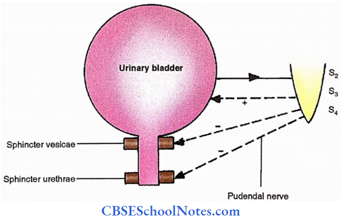
Urinary Bladder Micturition
The reflex phenomenon at the S2,3,4 level
280 ml of urine —> Afferent (nervi erigentes) —> S2,3,4 segments —> Efferent —> (1) Parasympathetic nerve —> Contraction of bladder and relaxation of sphincter vesicae. (2) Pudendal nerve —> Relaxation of sphincter urethrae —> Micturition.
Prostate Gland
Prostate Gland Definition
Fibro – a muscular and glandular organ in males.
Shape: Pyramidal, base-up, apex down.
Prostate Gland Measurements – Anteroposterior – 2 cm, Vertical – 3 cm, Width – 4 cm
Prostate Gland Location: Between the neck of the urinary bladder and the urogenital diaphragm
Prostate Gland Relations
- The base continues with the neck of the bladder
- Inferior – urogenital diaphragm
- Anterior – Cave of Retzius
- Puboprostatic ligament
- Lateral – Levator ani
- Posterior – Fasica of Denonvillier, rectum
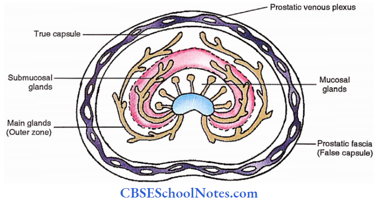
Prostate Gland Capsules
True – covering the surface of the gland
False – (Prostatic fascia) – It is external to the true capsule. In between two capsules is the venous plexus
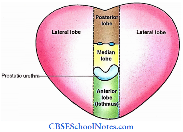
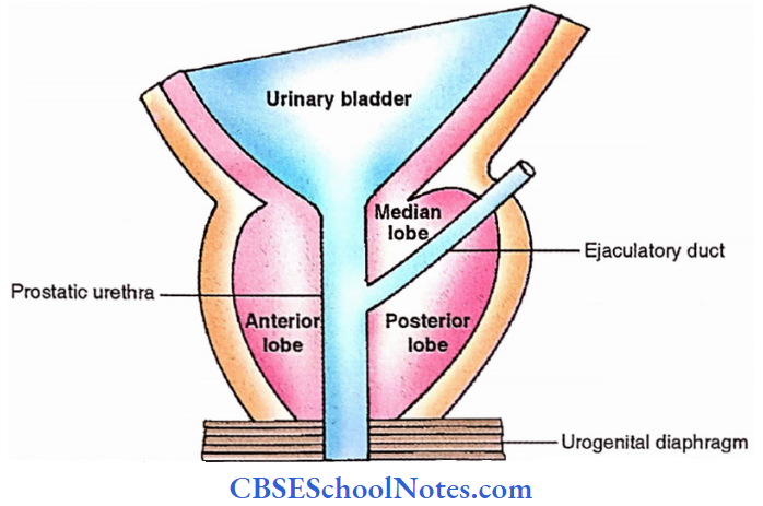
Prostate Gland Lobes
5 in numbers
1. Anterior – Anterior to prostatic urethra
1. Median – Between urethra and ejaculatory duct
1. Porterior- Behind the ejaculatory duct
2. Lateral – Lateral to all the above on each side. The right and left halves fail to fuse completely posteriorly forming a posterior midline groove.
Interior Of The Gland
Interior Of The Gland Zones
- Inner zone
- Zone of mucosal glands- Involved in benign prostatic hyperplasia.
- Zone of submucosal glands
- Outer zone (Zone of main glands)- Involved in malignancy.
Ejaculatory duct. It lies on each side of the midline posteriorly.
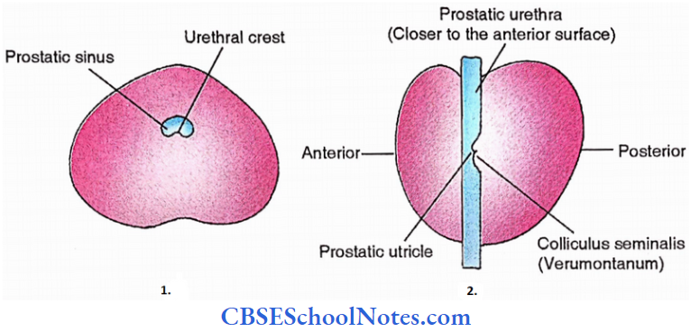
Interior Of The Gland Prostatic urethra
- 3cm in length
- The widest part of the urethra
- Urethral crest- posterior, midline, linear vertical elevation.
- Prostatic sinus – groove on each side of the urethral crest.
- Verumontanum (Colliculus seminalis) – Rounded elevation in the middle of the urethral crest
- Utriculus masculinum (‘Prostatic utricle) – A transverse depression in the verumontanum.
Prostate Gland Arteries
Inferior vesical and middle rectal arteries.
Prostate Gland Veins
Open into prostatic venous plexus which itself drains into internal iliac veins.
Prostate Gland Nerve
Derived from pelvic plexus (inferior hypogastric plexus)
Prostate Gland Lymphatics
Drain into internal iliac lymph nodes.
Prostate Gland Applied
P/R (Per-rectal) examination- The index finger is introduced into the anal canal and the prostate is palpated through the anterior wall of the rectum.
Normally a midline groove is present and the rectum moves over the prostate freely. In cases of cancer of the prostate, no midline groove is felt and the rectal wall is fixed prostate.
Rectum And Anal Canal
Rectum And Anal Canal Situation: Rectum – Posterior pelvic organ
Anal canal – Posterior perineal organ
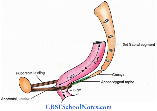
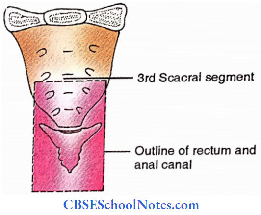
Rectum And Anal Canal Extent: Third sacral segment to the anus
Rectum And Anal Canal Measurements
Total length – 15 cm
Rectum
12 cm – 7 cm – over sacrum and coccyx
5 cm – over anococcygeal raphe
Anal canal – 3cm
Rectum And Anal Canal Curvatures
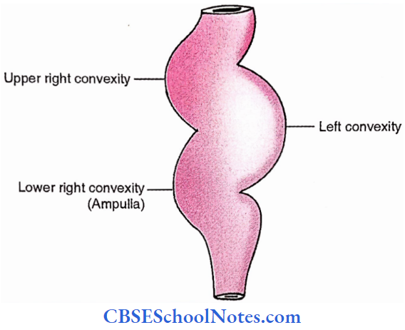
Rectum And Anal Canal Rectum
- Antero-posterior curvature (Sacral flexure). It corresponds with the anterior curvature of the sacrum
- Side-to-side curvature
- Right- Two curvatures (upper and lower), convex to right.
- Left- One curvature (middle), convex to left.
- Junctions of curvatures – These are responsible for folds towards the lumen called transverse folds or rectal valves of Houston or plicae transversals.
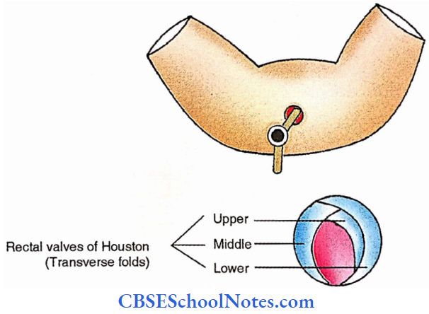
Anorectal junction
90° Backward bend of the anal canal at its junction with the rectum, produced by puborectalis sling.
Subdivisions
- Rectum
- Upper part
- Lower part (Rectal ampulla)
- Anal canal
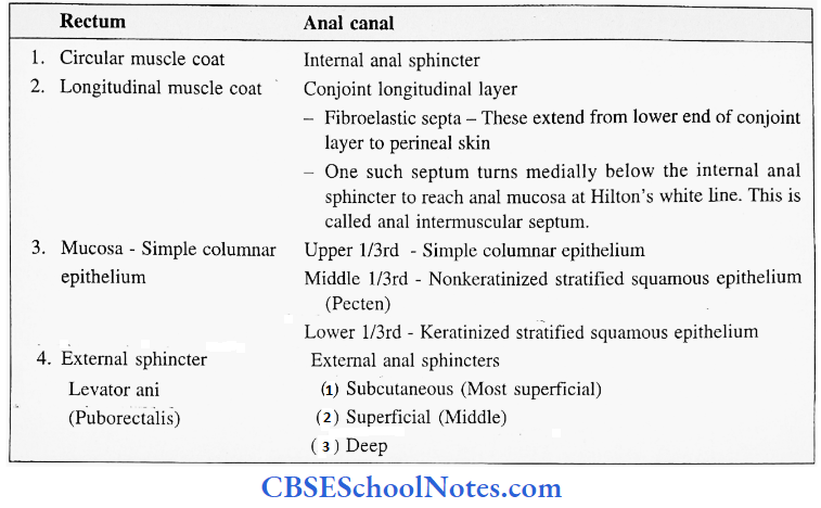
- Upper 2/3 – Derived from the cloaca.
- Lower 1/3 – Derived from the anal pit
Rectum And Anal Canal Relations
Relations of Rectum
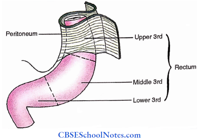
Peritoneal relations
- Upper 1/3rd – Anterior and lateral parts covered with peritoneum.
- Middle 1/3rd – The anterior part is only covered with the peritoneum.
- Lower 1/3rd- No peritoneum related
Relations of Rectum Adjacent relations
Posterior relations
- Midline
- Sacrum and coccyx
- Anococcygeal raphe
- Medium sacral artery and coccygeal body
- Ganglion impar
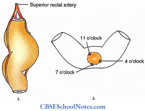
- Branches of superior rectal artery
- Fascia of Waldeyer
- Bilateral
- Muscles – Piriformis, coccygeus
- Sympathetic trunk
- Lateral sacral artery
- Ventral rami of sacral nerves
- Pelvic plexus
- Lymph nodes
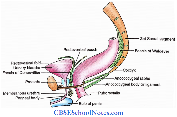
Anterior relations
It is different in male and female
- Male
- Rectovesical pouch with coils of intestine.
- Base of the bladder, seminal vesicle, vas deferens, ureter
- Prostate, fascia of Denonvillier.
- Females
- Rectouterine pouch of Douglas
- Vagina
Relations of Anal Canal
Anterior relations
- Male
- Perineal body
- Membranous urethra
- Bulb of peni
- Female
- Vagina
- Perineal body
- Posterior relations
- Anoccygeal body or ligament
- Tip of coccyx
- Lateral relation
- Ischiorectal fossa

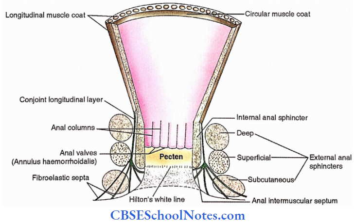
Rectum and Anal Canal Structure
Rectum and Anal Canal Stability
Rectum. The following structures keep the rectum in place:
- Fascia of Waldeyer – Between sacrum and rectum
- Lateral ligament of rectum-It extends from the rectum to the posterolateral pelvic wall. It holds middle rectal vessels
- Pelvic peritoneum – It is reflected from the front of the rectum to the posterior fornix in females and the upper base of the urinary bladder in males and thus forms a rectouterine pouch in females and a rectovesical pouch in males respectively.
- Pelvic diaphragm
Anal canal. It is stabilized by the perineal body and anococcygeal body.

Rectum and Anal Canal Arteries
- Superior rectal artery – It supplies mucosa of the rectum and the upper 2/3rd of the anal canal
- Median sacral artery – It supplies the posterior aspect of the rectum
- Middle rectal artery – It supplies muscles of the rectum.
- Inferior rectal artery – It supplies muscles and lower l/3rd of the mucosa of the anal canal.
Rectum and Anal Canal Veins
- Superior rectal vein – It drains into the inferior mesenteric vein
- Middle rectal vein – It drains into the internal iliac vein
- Inferior rectal vein – It drains into internal pudendal vein
Rectum and Anal Canal Lymphatics
- Rectum and upper 2/3rd of anal canal
- Preaortic lymph nodes
- Internal iliac lymph nodes
- Sacral lymph nodes
Lower l/3rd of the anal canal – Medial horizontal chain of superficial inguinal lymph nodes
Rectum and Anal Canal Nerves
Rectum mucosa and internal sphincter of upper 2/3rd of anal canal receive
Autonomic supply —> Sympathetic – T12 – L2 —> Parasympathetic – S2,3,4
Mucosa ofanal canal (lower 1/3rd) – Inferior rectal nerve
External anal sphincter – Subcutaneous —> Inferior rectal nerve (S3,4 ). It is a branch of the pudendal nerve.
Superficial —> Perineal branch of S4
Deep —> Inferior rectal nerve
Pelvic Part Of The Ureter
Length: 15 cm
Pelvic Part Of Ureter Course
It first runs downwards, backwards and laterally and then medially and forward. During its course, it crosses the following structures from above downwards ;
- Umbilical artery (superior vesical artery)
- Obturator nerve and vessels
- Inferior vesical artery
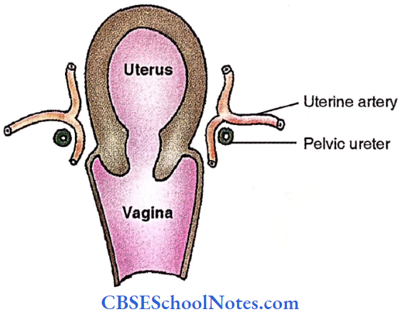
- Middle rectal artery (Uterine artery in females)
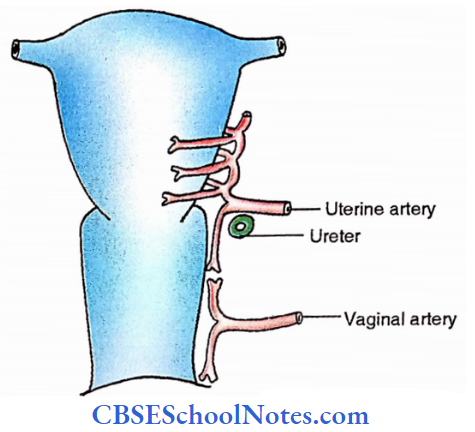
Intra-vesical part of the ureter is located in the wall of the urinary bladder. Its terminal part is crossed by the vas deferens (in males) or uterine artery (in females).
Seminal Vesicle And Vas Deferens
Seminal Vesicle
- Form – Sacculated coiled tube.
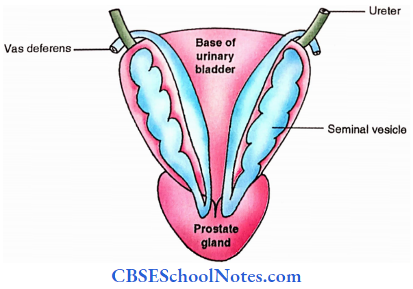
- Location – On the posterior surface of the urinary bladder
- Extent – From terminal ureter to prostate
- Length
- 5 cm, when coiled
- 10-15 cm when uncoiled.
- Diameter – 3-4 mm
Vas Deferens
Vas Deferens Course
Enters the abdominal cavity through a deep inguinal ring. It then descends to enter the pelvis where it crosses the following structures from above downwards:
- Medial umbilical ligament (Obliterated umbilical artery)
- Obturator nerve and vessels
- Inferior vesical artery
- Ureter
Vas Deferens Terminal Part
It is applied to the posterior aspect of the urinary bladder, medial to seminal vesicle. Here it is dilated from the ampulla of the vas.
Formation Of Ejaculatory Duct
Vas deferens pierces the posterior surface of the prostate gland near its upper border and joins the duct of the seminal vesicle to form the ejaculatory duct. It is 2 cm in length. It opens in the posterior wall of the prostatic urethra.
Vascular Supply
Both seminal vesicles and vas deferens are supplied by vesical arteries and drained by vesicoprostatic venous plexus.
Uterus
The uterus is a muscular organ and is part of the female genital tract. It provides a site for the attachment of the fertilized ovum, responsible for its nutrition, growth and protection. At term, its forceful contraction initiates the expulsion of the foetus.

Uterus Weight – 30-40 gm
Uterus Size Height – 7.5 cm (Body- 5 cm, Cervix- 2.5 cm)
Width – 5 cm
Thickness – 2.5 cm
Uterus Parts
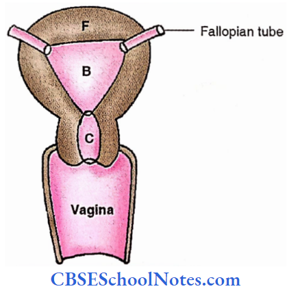
Body – upper 2/3rd expanded part
Cervix – lower l/3rd cylindrical part
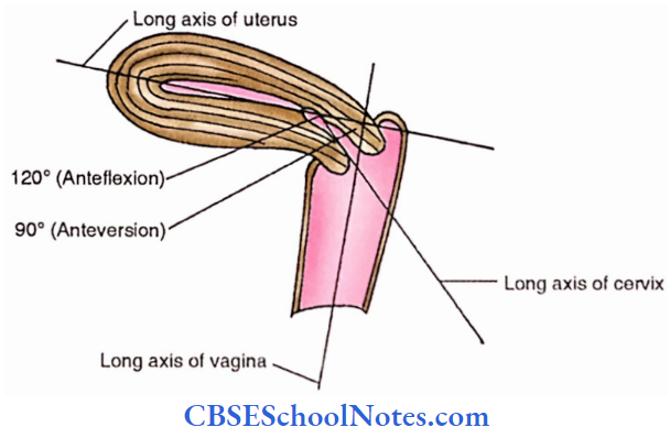
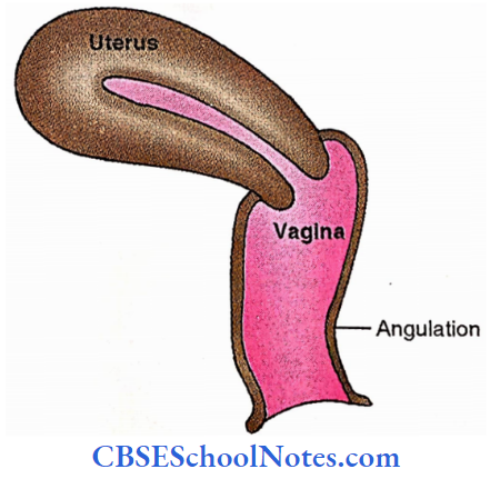
- Anteversion- Forward bending of the uterus concerning the vagina (about 90°).
- Anteflexion- Forward bending of the uterus concerning the cervix (about 120°).
- Long axis – Long axis of the uterus forms an angle of 90 degrees with the long axis of the vagina. This angle opens forward. Roughly, the long axis of the uterus corresponds with the long axis of the pelvic inlet and the long axis of the vagina to the axis of the pelvic cavity and the pelvic outlet.
Fundus: Free upper end of the uterus above the entry points of the uterine tubes. It is dome-shaped, directed anteriorly and covered with peritoneum.
Body Of Uterus
1. Surfaces
- Anterior or vesical surface
- It is flat and related to the urinary bladder. It is covered with peritoneum and forms the posterior wall of the vesicouterine pouch.
- Posterior or intestinal surface
- It is convex and related to the terminal coils of the ileum and sigmoid colon. It forms the anterior wall of the rectouterine pouch and is covered with the peritoneum.
2. Borders
Lateral borders – There are two lateral borders, right and left. Each border is rounded and convex and it provides attachment to the broad ligament of the uterus which connects it to the lateral wall of the pelvis.
The fallopian tube opens in the uterus at its upper end and the uterine artery ascends along it.
3. Cavity
In sagittal and transverse sections it is merely in the form of a slit but in the coronal section, it is triangular with an apex towards the introitus where the uterine cavity becomes continuous with the cervical canal.
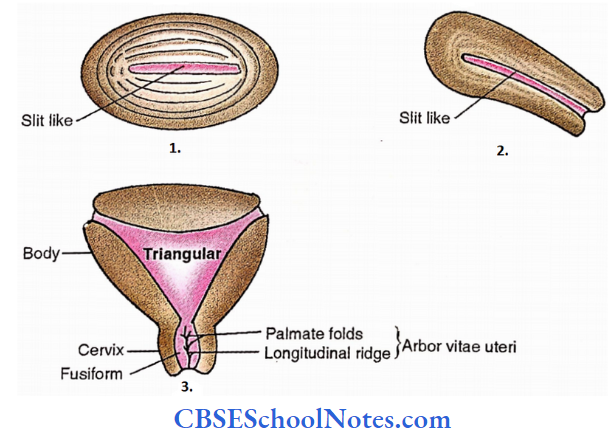
- Transverse Section
- Sagittal Section
- Coronal Section
Cervix Of Uterus
Lower 1/3rd, 2.5 cm, cylindrical, less mobile part of the uterus. In the middle, it is a little wider. The lower part of the cervix projects into the upper part of the anterior wall of the vagina.
Cervix Of Uterus Subdivisions
1. Supravaginal part of the cervix
It is related
- Anteriorly – to the bladder
- Posteriorly – to the rectouterine pouch containing coils of intestine
- Sides – to the ureter and uterine artery embedded in the parametrium of the broad ligament
2. Vaginal part of the cervix
It projects into the anterior wall of the vagina. The spaces between the cervix and vaginal walls are called the vaginal fornices. The cervical canal opens into the vagina through the external os.
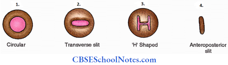
- In the transverse section, it is circular in outline in nulliparous and becomes transverse (having anterior and posterior lips) in multiparous.
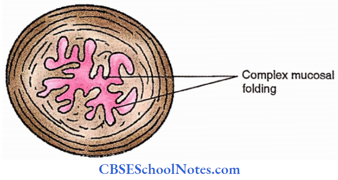
- The cervical canal extends from the external os to the internal os and thus communicates above with the uterine cavity and below with the vaginal cavity. Along its long axis, it is fusiform in shape.
- The anterior and posterior walls of the cervical canal show interlocking mucosal folds which resemble branches of a tree (arbour vitae uteri)
Ligaments Of Uterus
1. Peritoneal ligaments
- Uterovesical fold (anterior)
- Rectovaginal fold (posterior)
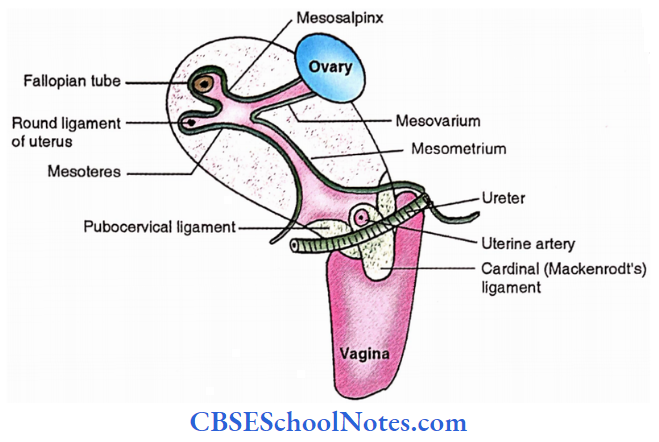
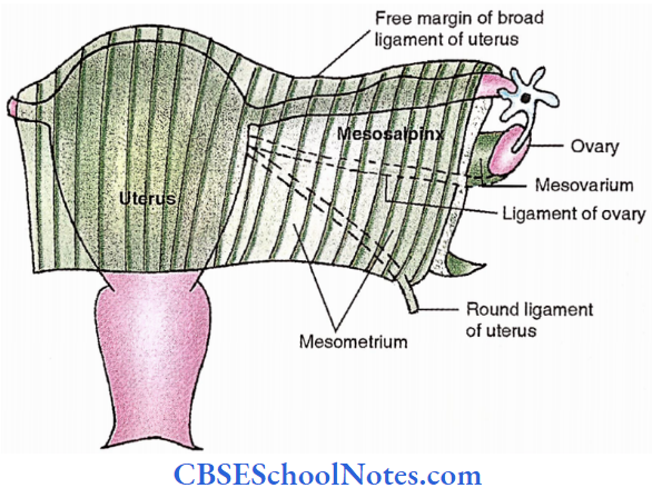
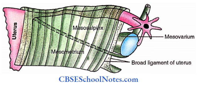
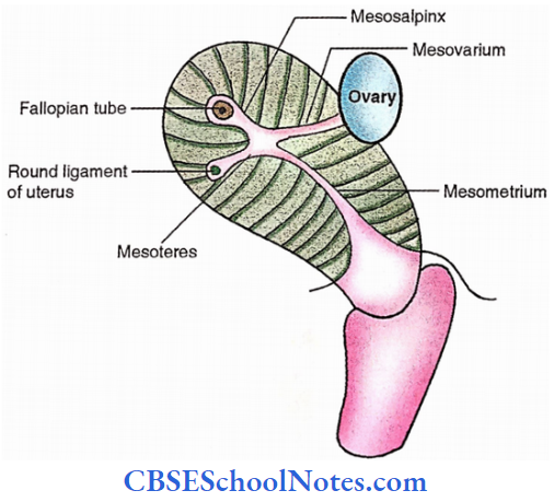
- Broad ligaments (right and left)-Mesosalpinx and mesometrium are the parts of the broad ligament above and below the ligament of the ovary respectively.
Broad ligament contains,
- Uterine tube
- Round ligament of uterus
- Ligament of ovary
- Uterine vessels near the attachment to the uterus
- Ovarian vessels in the infundibulopelvic (suspensory) ligament
- Uterovaginal and ovarian nerve plexuses
- Epoophoron
- Paroophoron
- Lymph nodes and lymph vessels
- Dense connective tissue (parametrium) is present on the sides of the uterus.
Vestigial remnants present in the broad ligament
Epoophoron
It is a remnant of the cranial part of the mesonephric tubule. It is in the form of 10 -15 parallel tubules in the lateral part of the mesosalpinx.
Paroophoron
It is a remnant of the caudal part of mesonephric tubules. It is in the form of a short rudimentary tubule located in the broad ligament between the ovary and the uterus.
Duct of Gartner
When the duct of the epoophoron becomes much larger is called the duct of Gartner. It can be traced along the uterine tube.
Along the lateral margin of the uterus up to the level of internal os then through the cervix to the lateral wall of the vagina to end in the anterior margin of the hymen.
It represents the mesonephric duct. It may present in the form of a cyst in the anterior or lateral wall of the vagina.
Vesicular appendix
These are derived from the cranial end of the paramesonephric duct in the form of a pedunculated cyst attached to the fimbriated end of the uterine tube. These are also called paramesonephric appendices.
2. Fibromuscular ligaments
- Round ligaments of the uterus
- Transfer – 7 cervical ligaments
- Uterosacral ligaments
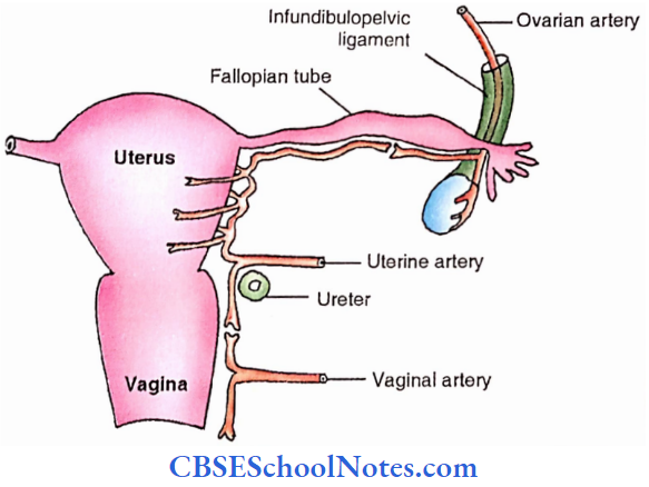
Arterial supply
- Uterine arteries
- Ovarian arteries
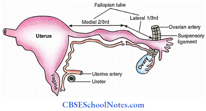
Apart from the uterus the uterine artery also gives branches to
- Vagina
- Medial 2/3rd of uterine tube
- Ovary
- Ureter
- Structures present in the broad ligament
Venous drainage
The veins form the plexus along the lateral border of the uterus and drain into the internal iliac vein through uterine, ovarian and vaginal veins.
Lymphatic drainage
Lymphatics of the uterus begin as three intercommunicating networks- endometrial, myometrial, and subperitoneal. These drain into lymph nodes on the side of the uterus. Lymphatic drainage is described to be in three groups.
- Upper lymphatics (from the fundus and upper part of the body) – pass to aortic nodes. Some of them pass along the round ligament of the uterus to the superficial inguinal lymph nodes.
- Middle lymphatics (from the lower part of the body) – Pass to external iliac nodes.
- Lower lymphatics (from the cervix) – Pass to the external iliac, internal iliac and sacral nodes.
Fibromuscular ligaments Nerve supply
The uterus receives both sympathetic and parasympathetic innervation through inferior hypogastric and ovarian nerve plexuses.
Sympathetic (T12, L1)- Produces uterine contraction and vasoconstriction. Also carries pain sensation from the body of the uterus.
Parasympathetic (S2,3,4)- Produces uterine inhibition and vasodilatation. Also carries pain sensation from the cervix.
However, these actions are modified by hormones which have strong action on the genital tract.
Age and reproductive changes
During fetal life – The cervix is larger than the body and the body projects a little above the pelvic brim.
At puberty– Uterus enlarges and descends in the pelvis. Arbor vitae uteri appear in the cervix.
During menstruation – Little enlarged, more vascular and external os swollen.
During pregnancy– Enlarge (due to both hypertrophy and hyperplasia), with advancing pregnancy it becomes thinner. After parturition it involutes.
In old age -It becomes atrophic and dense in texture. The external os obliterates and the lips disappear.
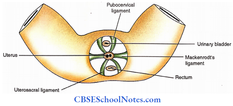
Supports Of Uterus
Primary supports
- Muscular
- Pelvic diaphragm
- Perineal body
- Urogenital diaphragm
- Fibromuscular or mechanical
- Uterine axis
- Pubocervical ligament
- Transverse cervical ligament
- Uterosacral ligament
- Round ligament of uterus
Secondary supports
These are formed by peritoneal ligaments and are of doubtful value.
- Broad ligaments
- Uterovesical fold of peritoneum
- Rectovaginal fold of peritoneum
Role Of Individual Supports
Pelvic diaphragm
It supports the pelvic viscera and also acts against the force due to the rise of intra-abdominal pressure. Some fibres of the pubococcygeus part of the levator ani form a supporting sling and a sphincter for the vagina (indirectly for the uterus and urinary bladder).
During parturition sometimes the pubococcygeus part is tom and it results in loss of support to the vagina which along with the uterus sinks into the vestibule (Prolapse of uterus).
Perineal Body
It is the fibromuscular node to which nine muscles are attached. It acts as an anchor for the pelvic diaphragm and thus maintains the integrity of the pelvic floor.
The efficacy of levator ani as a support is lost when the perineal body becomes unstable due to a tear of the perineal muscle.
Urogenital Diaphragm
The sphincter urethrae form the external sphincter of the urethra. Some of its fibres also get attached to i the wall of the vagina and provide support to it.
Pubocervical Ligaments
These are derived from the pelvic fascia and correspond to the medial and lateral puboprostatic ligaments found in males. Thus they extend from the cervix to the posterior part of the pubis.
Transverse cervical ligaments (of Mackenrodt)
These are fan-shaped condensations of pelvic fascia on each side of the cervix above the levator ani and around the uterine vessels.
They extend from the lateral aspect of the cervix and upper part of the vagina to the lateral pelvic wall. They form a hammock which supports the uterus.
These ligaments are known by many other names – For Example., Lateral cervical ligaments; Paracervical ligaments; Cardinal ligaments; Retinacula uteri; and Sustentaculum of Bonny.
Uterine Axis
The normal position of the uterus is anteverted which is maintained by the uterosacral and round ligaments. This position itself prevents the sagging of the uterus into the vagina.
Increased abdominal pressure further increases its anteverted position by pushing the uterus against the urinary bladder and the pubic symphysis.
Uterosacral ligaments
These are a condensation of pelvic fascia enclosed within the recto-uterine folds (forming the lateral boundaries of the recto-uterine pouch). They connect the cervix to the 2nd and 3rd sacral vertebrae.
These ligaments counter the forward pull of the round ligament on the cervix. Round ligaments and the uterosacral ligaments form a couple that maintains the uterine axis.
Round Ligaments Of Uterus
These are 10-12 cm long, flat fibromuscular bands which lie between two layers of the broad ligament located anterior to the uterine tube.
- Course- Each ligament begins at the lateral angle of the uterus, runs forward and laterally, passes through the deep inguinal ring, traverses the inguinal canal and merges with the areolar tissue of the labium majus after breaking up into fine filaments.
- The round ligament keeps the fundus pulled forward and maintains the angle of anteversion against the backward pull of the uterosacral ligament.
- Canal of Nuck – In the inguinal canal the round ligament is accompanied by a process of peritoneum during foetal life. If it persists after birth, it is known in females as the canal of Nuck.
Uterus Applied Anatomy
- Retroversion of the uterus – When the uterus comes to lie in a straight line with the vagina.
- Prolapse of the uterus – When the uterus sags into the vagina due to weakening of support of the uterus.
- Intrauterine contraceptive device – Insertion of a foreign body into the body of the uterus prevents implantation.
- Caesarian section – When birth through the vagina is difficult then it is performed by cutting the anterior abdominal wall and the uterus (Roman emperor Julius Caesar was delivered by. this method).
- Hysterectomy – Surgical removal of the uterus in various conditions.
- Hysteropexy – Surgical fixation of the unduly mobile uterus.
- Hysterosalpingography – radiological visualization of the uterus and uterine tube after filling them with radiopaque dye.
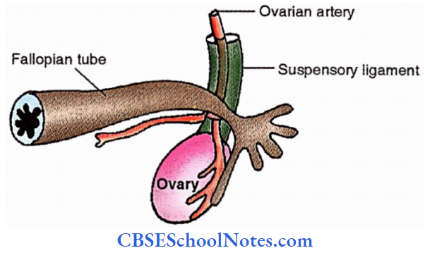
Fallopian Tube
It is also called the uterine tube or oviduct. It allows passage of ova from the ovary to the uterus and spermatozoa from the uterus towards the ovary.
Fertilization usually takes place in the lateral 1/3rd of the tube. It is 10 cm long. Its lateral ostium opens into the abdominal cavity.
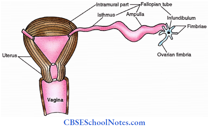
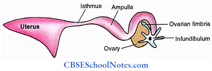
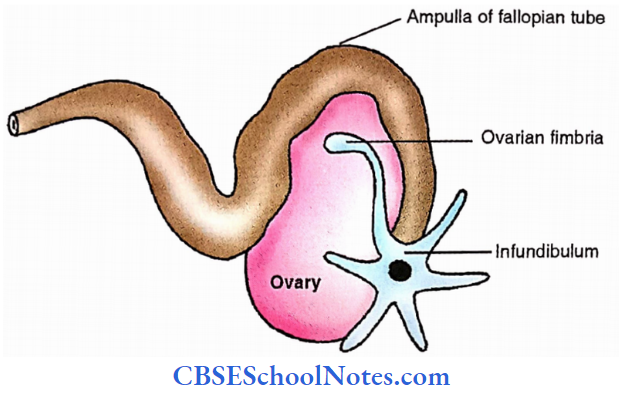
Fallopian Tube Subdivisions
- Infundibulum. The pole of the ovary isItscalledlateraltheendovarianis fimbriated. fimbria. One fimbria being longer and attached to the tubal pole of the ovary is called ovarian fimbria.
- Ampulla – Medial to infundibulum and arches over the upper pole of the ovary. It is thin-walled, tortuous and dilated. It forms the lateral 2/3rd of the uterine tube.
- Isthmus – Medial to ampulla and forms medial l/3rd of the tube. It is narrow, rounded and cord-like.
- Interstitial (uterine or intramural) – It lies within the wall of the uterus and opens at the upper angle of the uterine cavity through a narrow uterine ostium (1mm as against 3 mm abdominal ostium)
Fallopian Tube Course And Relations
- The uterine tube lies in the upper free margin of the broad ligament of the uterus.
- The part of the broad ligament between the attachment of the mesovarium and the uterine tube is called mesosalpinx. It contains the terminations of the uterine and ovarian vessels and the epoophoron.
- Ampulla arches over the ovary and is related to its anterior and posterior borders.
- The infundibulum projects beyond the free margin of the broad ligament.
Fallopian Tube Arteries
- Uterine artery – supplies the medial two-thirds.
- Ovarian artery – supplies the lateral one-third.
Veins run parallel with the arteries and drain into the pampiniform plexus of the ovary and also into the uterine veins.
Fallopian Tube Lymphatic Drainage
Most of the tubal lymphatics join the lymphatics from the ovary and drain with them into the lateral aortic and pre-aortic nodes. Those from the isthmus accompany the round ligament of the uterus and then drain into the superficial inguinal lymph nodes.
Fallopian Tube Nerve supply
It is supplied by both sympathetic and parasympathetic nerves running along the ovarian and uterine arteries.
Sympathetic nerves – are derived from the hypogastric plexuses. Contains both visceral afferent and efferent. Efferents are vasomotor and to some extent stimulate peristalsis.
Parasympathetic nerves – are derived from the vagus (to the lateral half) and splanchnic nerves (to the medial half). Inhibit peristalsis and produce vasodilatation.
Fallopian Tube Applied
- Salpingitis – Inflammation of the fallopian tube.
- Sterility – Inability to produce offspring.
- Tubal insufflation – Transuterine insufflation of carbon dioxide to test the patency of the fallopian tube.
- Hysterosalpingography – X-ray study of the uterus and uterine tube after injecting radiopaque material into those organs.
- Tubal pregnancy – An ectopic pregnancy where implantation has taken place in the uterine tube.
- Tubectomy and tubal ligation – Surgical methods for female contraception.
Steps Of Dissection Of Female Reproductive System
Clean and identify the female genital organs in situ. Trace the peritoneal reflections in the pelvis and identify the rectouterine pouch, ureterovesical pouch, broad ligament and transverse cervical ligament.
- Identify structures about the broad ligamentnamely fallopian tubes, ovaries, ligament of the ovary, round ligament of the uterus and uterine artery.
- Separate the broad ligament from the sides of the pelvis. Pull the uterus towards the pelvic cavity and separate the sides and back of the cervix.
- Separate the vagina from the perineum and remove the uterus along with the fallopian tube and ovaries after cutting the broad ligament. Study the uterus, fallopian tubes and ovary mass.
Ovaries
These are the female gonads and produce female gametes.
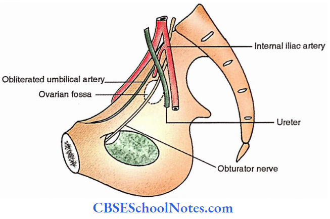
Ovaries Location: Each ovary lies in the ovarian fossa.
Boundaries of the ovarian fossa
- Anterior- obliterated umbilical artery
- Posteriorly – ureter and internal iliac artery
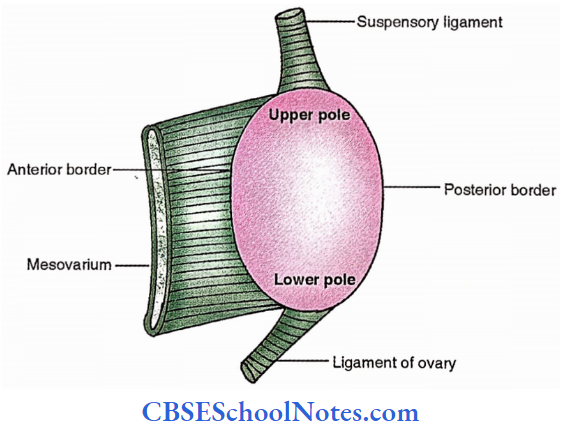
Axis of the ovary is.
Vertical in nulliparous and hence has upper and lower poles.
Horizontal in multiparous and therefore has lateral and medial poles
Size: Length: 3 cm. Width: 1.5 cm, Thickness: l cm
Ovaries Borders
- Anterior – mesovarian border
- Posterior – free border
Ovaries Relations
1. Peritoneum – covers it entirely except at its anterior border where two layers are reflected as mesovarium on the posterior surface of the broad ligament.
- Mesovarium– short fold of peritoneum connecting the anterior border of the ovary with the posterior layer of the broad ligament. It transmits nerves and vessels to and from the ovary.
- Suspensory ligament of ovary (infundibulopelvic ligament) – the lateral part of the broad ligament of the uterus extending from the infundibulum of the fallopian tube and upper pole of the ovary to the external iliac vessels. It contains ovarian nerves and vessels.
2. Visceral relations
- Upper or tubal pole – uterine tube and external iliac vessels; appendix on the right side.
- Lower or uterine pole – pelvic floor, ligament of the ovary.
- Anterior or mesovarian border – uterine tube, obliterated umbilical artery; forms hilus of ovary.
- Posterior or free border – uterine tube and ureter.
- Lateral surface – ovarian fossa lined by parietal peritoneum
- Medial surface – uterine tube.
Ovarian bursa peritoneal recess between mesosalpinx and medial surface of ovary.
Ovaries Arterial supply
- Ovarian artery – A branch of the abdominal aorta, descending over the posterior abdominal wall and entering the suspensory ligament of the ovary. It sends branches to the ovary through the mesovarium.
- Uterine artery – gives some branches which reach the ovary through the mesovarium.
Ovaries Venous drainage
Veins emerge at the hilus and form a pampiniform plexus around the ovarian artery. Near the pelvic inlet, the plexus transforms into a single ovarian vein which ascends on the posterior abdominal wall.
On the right side, it opens into the inferior vena cava and on the left into the left renal vein.
Ovaries Lymphatic drainage
Lymphatics from the ovary freely communicate with those from the fallopian tube and fundus of the uterus. These ascend along the ovarian vessels and terminate in lateral aortic lymph nodes.
Ovaries Nerve supply
It is derived from the ovarian plexus which follows the ovarian vessels. It contains both sympathetic (T10–11) – afferent for pain and vasomotor and parasympathetic (S2,3,4 ) – vasodilator.
Ovaries Applied
- Prolapse – Downward displacement of the uterus. The cervix sometimes protrudes through the vaginal orifice.
- Endometriosis – Ectopic endometrium located in various sites throughout the pelvis or the anterior abdominal wall.
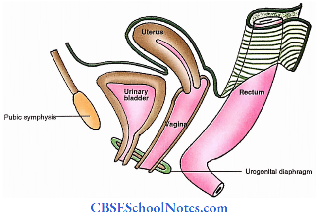
Vagina
It is a fibromuscular canal forming the female copulatory organ extending from the vulva to the uterus. It is located behind the bladder and in front of the rectum and anal canal. In an erect posture, it is directed upwards and backwards.
In the supine position, it makes an angle of 75 degrees with the horizontal plane. Its anterior wall is 8 cm and posterior wall is 10 cm long. The diameter of the upper end (vault) is 5 cm and that of the lower end is 2.5 cm only.
However, it is very distensible and allows passage of the foetal head during parturition. The cervix of the uterus protrudes in its upper end and makes it circular in outline.
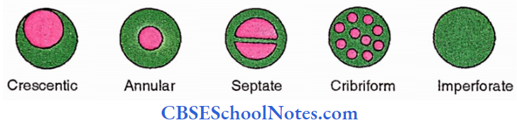
- Below the cervix, the anterior and posterior walls are in contact with each other. In the virgin, the lower end of the vagina is partially closed by an annular fold of mucous membrane called the hymen.
- In married women, the hymen is represented as rounded elevations around the vaginal orifice, the carunculae hymenale.
- Upper 1/3rd
- Middle 1/3rd
- Lower 1/3rd
- Vaginal Orifice
The interior of the upper end of the vagina is in the form of a circular groove that surrounds the protruding cervix and this groove gradually becomes deeper from before backwards.
This groove is divided into 4 parts called the vaginal fornices. The anterior fornix lies in front of the cervix and is the shallowest. The posterior fornix lies behind and is deepest and the lateral fomices lie on either side of the cervix.
Vagina Relations
- Anterior wall – Bladder and urethra
- Posterior wall – Recto-uterine pouch, loose connective tissue and perineal body separate it from the rectum and anal canal.
- Lateral walls – Transverse cervical ligament, pubococcygeus part of the levator ani, urogenital diaphragm and below it to the bulb of vestibule, bulbospongiosus and greater vestibular gland.
Vagina Arterial supply
Being a very vascular organ, it is supplied by
- Vaginal branch of the internal iliac artery
- The cervicovaginal branch of the uterine artery
- Branches from middle rectal and internal pudendal arteries
- Vaginal azygos arteries – (anterior and posterior) arising from anastomoses of the above arteries.
Venous drainage
The rich vaginal venous plexus joins to form a vaginal vein which accompanies the vaginal artery and drains into the internal iliac vein.
Venous Lymphatic drainage
- From upper 1/3rd to external iliac lymph nodes
- From the middle 1/3rd to the internal iliac lymph nodes.
- From lower l/3rd to the medial group of superficial inguinal lymph nodes.
Venous Nerve supply
- Lower 1/3rd is sensitive to pain and is supplied by internal pudendal, inferior rectal and posterior labial branches of the perineal nerve.
- The upper 2/3rd is insensitive to pain and is supplied by sympathetic and parasympathetic nerves derived from inferior hypogastric and uterovaginal plexuses. The fibres which accompany the vaginal artery are known as vaginal nerves.
Venous Applied Anatomy
- Vaginitis – Inflammation of vagina.
- Fistulae (abnormal tubular communication)- due to congenital malformation, injury or abscess. It may be between different organs like,
- vesicovaginal (between vagina and urinary bladder)
- uterovaginal (between uterus and vagina)
- rectovaginal (between rectum and vagina)
- Colpotomy – Incision into the wall of the vagina.
- Colporrhapy – Repair of vaginal wall.