
Patterns Of Inheritance And Disorders Of Genes
It is imperative to be acquainted with the patterns or the genetics of inheritance (dominant, recessive, sex-linked, etc.) before we can actually understand important aspects of a genetic disorder (disorders caused by abnormalities in a gene). In this chapter we shall learn about the patterns in which a defective gene is passed onto new generations and how these methods determine the degree of severity in the outcome of the diseases in the progeny.
This is understanding is helpful for an accurate diagnosis of genetic disorders and evaluating the risk of acquiring the genetic disorders and evaluating the risk of acquiring the genetic diseases in a newborn.
Knowledge of the exact pattern of inheritance aids in suggesting measures that may prevent the inheritance of genetic disorders.
Common traits or disorders follow the following patterns of inheritance:
- Single gene (Mendelian/Monogenic) inheritance.
- Multiple genes (Polygenic/Multifactorial) inheritance.
Single Gene (Mendelian/Monogenic) Inheritance
The traits in this type of inheritance are carried by single genes and the pattern obeys the Mendel’s laws of inheritance. The single gene inheritance is further classified into the autosomal and sex-linked inheritance types.
The patterns of transfer of genes present on autosomes determine Autosomal while sex-linked inheritance is established by the mode of transmission of genes present on sex chromosome (X or Y). Autosomal inheritance is further classified into autosomal dominant and autosomal recessive types. In case of an autosomal dominant inheritance, a dominant gene that is present in the genome of a person (somatic cell) expresses itself even if it is present in a single dose (heterozygous state).
Read and Learn More Genetics in Dentistry Notes
If such a mutation arises in the germ cells (gametes) of a person in the gonads, the trait is carried to the offspring and expresses itself clinically (in phenotype) even when the gene it is present in a single dose (heterozygous state). However, an autosomal recessive trait is manifested clinically only when a gene is present in double dose (homozygous state) on a pair of chromosome.
Genes present on sex chromosome (X or Y) determine the characteristics of sex-linked inheritance whether the trait is transmitted to the next generation by X-linked or Y-linked genes.
Formulation of a Pedigree Chart
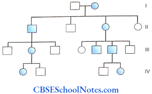
A pedigree chart is a diagram showing the ancestral history of a group of relatives with relation to a particular disease in question in an individual. To investigate a genetic disorder it is important to record the family history of relatives in relation to the occurrence of the particular genetic disorder in them. The information about the health of the whole family is recorded in the form of a pedigree chart. Following are certain conventions used in the preparation of pedigree chart.
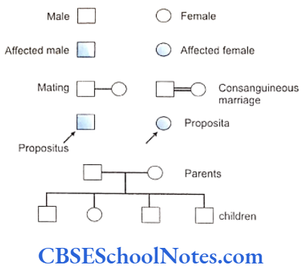
- Squares in the diagram represent males and circles represent females.
- Solid squares or solid circles symbolize affected persons.
- The individual being investigated and for whom the pedigree is prepared is termed as the proband; propositus (male) or proposita (female). The position of proband in the chart is indicated by an arrow.
- A horizontal line which connects a male and a female represents mating.
- The offsprings from a mating are represented in order of their birth from the left to the right.
- Roman numerals in the pedigree represent successive generations, e.g. I, II, III, etc. designated to each of the horizontal rows in the chart.
Autosomal Dominant Inheritance
An autosomal dominant trait or disease can be manifested in an individual if the gene responsible for this kind of trait or disorder is present on an autosome (non-sex chromosome). The expression of the trait or the disorder occurs even if the gene is present in a single dose, i.e. a normal dominant gene or a mutated yet dominantly expressing gene produces its effect even when it is present in only one of the chromosomes of a particular pair (heterozygous state).
On the other hand such genes or traits the express in the heterozygous state are called dominant genes. These genes are transmitted following the laws of autosomal dominant inheritance.
Some common examples of dominant traits and disorders in medicine and dentistry are:
- Achondroplasia
- Dentinogenesis imperfecta type 1
- Amelogenesis imperfecta hypoplastic type 2 (AIH2)
- Amelogenesis imperfecta hypocalcification type
- Hypodontia
- Osteogenesis imperfecta
- Huntington’s disease
- Myotonic dystrophy
- Polycystic kidney disease
- Congenital cataract
- Polydactyly.
Distinctive Features of an Autosomal
Dominant (AD) Trait
- An autosomal dominant trait or disorder is seen in every generation without skipping.
- The disorder may appear for the first time (de-nove) in a individual due to a new or fresh mutation occurring either at the time of gametogenesis in the individual. The new nutation obeys the laws of autosomal dominant inheritance.
- If not arising due to a new mutation, an affected person will always have an affected parent.
- Transmission is observed from male-to-male; from female-to-female; from female-to-male and from male-to-female, i.e. between all the sexes. The traits or disorders equally affect the males and the females. This signifies that the gene responsible for this type of inheritance is located on an autosome.
- Autosomally dominant genes present in a heterozygous state have 50% chance to get transmitted and affect half of offsprings.
- The disorder cannot be transmitted further to next generations by the normal offsprings as they do not have the abnormal gene.
- Theoretically, normal and affected children in a generation should be equal in number.
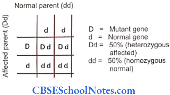
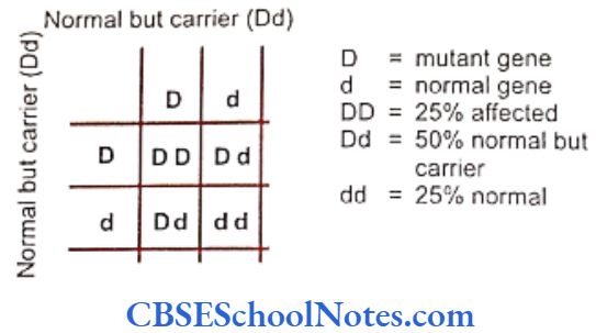
All the observations discussed earlier are explained in the Punnett squares below. We know that two genes present on a pair of homologous chromosomes at the same locus are responsible for determining a particular trait. We also know that the expression of a trait will also depend on the ‘dominance’ or the recessiveness’ of a gene. Certain normal genes as well as the effects of certain mutations in a gene always express in a dominant fashion.
If we represent a normal gene as “d” and a dominant mutant gene as “D” then the genotype of one parent who is homozygous for the normal allele will be “dd” and will be normal. The other person or parent bearing the dominant mutated gene (in heterozygous state) will have a “Dd” genotype. The genetic risk of outcome in such mating can be calculated.
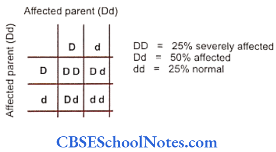
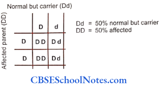
In another situation a mating between both affected parents carrying a normal and a dominantly mutated gene (Dd) produces a different set of affected offsprings.
Offsprings with homozygous (DD) genotype would be severely affected due to presence of the mutant genes in double doses.
Common Autosomal Dominant Disorders
Some of the common medical disorders showing Autosomal dominant inheritance are enumerated below with their brief characteristics. The Autosomal dominant dental disorders are dealt in the appropriate sections of the book later.
Achondroplasia
Achondroplasia is an autosomal dominant trait. It is a class of skeletal growth syndromes characterized by short stature due to slow development of the middle portions of the long bones in the arms and legs. The most common form of achondroplasia is due to a defect of the Fibroblast Growth Factor Receptor (FGFR), and is recognized by exaggerated cranial growth and bossing (depression) at the bridge of the nose.
A second form is pseudoachrondroplasia, which is due to a defect of Cartilage Oligomeric Matrix Protein (COMP) in the joints, and is characterized by more typical development of cranial proportions. The COMP gene is situated on chromosome 19 (19q13.1). Both forms of achondroplasia are described as sporadic, meaning that they occur in different families due to independent mutations.
Thus, most affected children are born to parents of ordinary stature, one of whom has a germline mutation. In the children of two parents with achondroplasia (Dd x dD), most affected offspring are heterozygous (Dd), which suggests that the homozygous dominant genotype (DD) is lethal.
Shown in the accompanying photograph are seven pseudoachondroplasic members of the Ovitz family, a family of Romanian Jews who toured Eastern Europe as a musical troupe before World War II (their taller siblings working backstage), survived imprisonment at Auschwitz, and finally immigrated to Israel. They were photographed on arrival in Haifa in 1949.
Their father was (apparently) of ordinary height and was twice married, both times to women of ordinary height. With his first wife, he had two affected daughters [possibly the two older women in flowered dresses,], and with his second five affected children (three girls and two boys) shown here, as well as three children of ordinary height. This suggests the father had a germ line mutation.
Huntington’s Chorea
Characteristics of the disease: Incidence is approximately 1 in 15,000 people. This is a fatal disorder that usually begins with disorders of movement. Occurs in the middle ages though 10% of disorders are seen before the age of 20 years (juvenile variety). Shows phenomenon of anticipation (explained later).
Characterized by involuntary movements like facial grimacing, limb movements followed by unsteady gait and slurred and unclear speech. Intellectual impairment and dementia precede death.
There is a progressive loss of neurons due to cell death in the CNS. This autosomal dominant disease shows complete penetrance. The mutation is located on the short arm of chromosome number 4 that codes for an abnormal protein called Huntingtin which has been implicated for cell death or apoptosis in the central nervous system.
Autosomal Recessive Inheritance
An autosomal recessive trait is only expressed when the responsible gene or the mutation exists in a homozygous state and as such in double dose. Following is a list of some common autosomal recessive disorders and traits.
- Cystic fibrosis
- Amelogenesis imperfecta (local hypoplastic type)
- Amelogenesis imperfecta (pigmented hypomaturation type)
- Neonatal osseous displasia 1
- Sickle cell anemia
- Phenylketonuria
- Schizophrenia
- Alkaptonuria
- Spinal muscular atrophy
- Albinism.
Distinctive Features of an Autosomal Recessive (AR) Inheritance
Autosomal recessive inheritance is characterized by the expression of gene or traits only when they are present in a homozygous form in the genome.
The presence of an autosomal recessive trait or a mutation in a heterozygous state does not express the disorder and the individual is perfectly healthy. An individual who is heterozygous for an autosomal recessive trait is called carrier.
Autosomal recessive mode of inheritance can be distinguished by the following features:
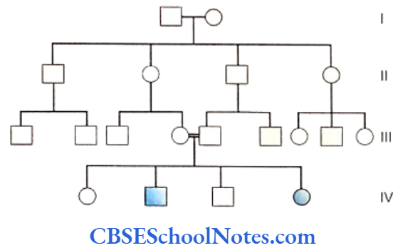
- Brothers and sisters (siblings) in the same generation manifest the trait. The trait is not seen in previous (parents) or in subsequent generations (offsprings). The disorder is seen only in the 4th generation and not in either of the 3rd or 5th generation as depicted.
- Members of both the sexes are equally affected.
- The proband usually have closely related parents. Such an offspring is said to arise from a consanguineous marriage or union. Each of the parents is a carrier (heterozygote) for the particular trait or disease.
- The results of mating between two carriers of an autosomal recessive trait (Dd).
- In case of mating between an affected individual (homozygous, DD) and a carrier (heterozygous, Dd), 50% of the offsprings get affected (homozygous) and 50% obtain a carrier status (heterozygous). This pattern may be easily confused for a dominant inheritance. This kind of inheritance is thus known as pseudo-dominant inheritance.
Some Common Autosomal Recessive Disorders
Many of the disorders in medicine, especially those involving errors of metabolism exhibit autosomal recessive type of inheritance. Dental diseases that inherit in the pattern are discussed later in appropriate sections. Some of the medical disorders are mentioned below.
Spinal Muscular Atrophy
Characteristics of the disease: Affected individuals exhibit progressive weakness of muscles resulting from degeneration of the spinal motor neurons. Type I of the disease is severest characterizing hypotonia and lack of spontaneous movement in infants. Type II and III are milder form of the disease with a later age of onset.
Death usually occurs within first two years of life due to impairment of swallowing and respiratory functions. the milder form of disease results in recurrent respiratory failures but death is often delayed. The gene responsible for SMA disorder is situated on the long arm of chromosome number 5.
Cystic Fibrosis (mucoviscidosis)
Characteristics of the disease: Occurs in 1 out of 2500 people in the population. It is a common and fatal disorder seen in the children of white populations of Europe. Cystic fibrosis (CF) is characterized by the accumulation of thick, sticky, honey like mucous fluid which leads to blockage of airways, intestines and other viscera causing secondary infections.
Death results mostly due to the obstruction of the respiratory tract. The lung tissue gets fibrosed leading to cardiac failure (corpulmonale). Blockage of the pancreatic ducts, malnourishment, cirrhosis of the liver, occasional congential bilateral absence of vas deferens in males (making them sterile) are some of the features associated with the disease.
Patients usually die in childhood or adolescence. But life can be prolonged up to 30 years of age with effective treatment. The gene implicated for CF is present on the long arm of chromosome number 7. The CF gene was cloned in 1989 and was named cystic fibrosis transmembrance conductance regulator (CFTR) gene. The CFTR protein contains 1480 amino acids which act as a chloride channel.
The mutation occurs at the 508th codon resulting in the loss of the amino acid phenylalanine. The defective chloride channel causes increase in the level of extracellular chloride and accumulation of intracellular sodium. The chloride content of secretons make them viscid and sticky. Prenatal diagnosis should be advised to the parents of an affected child for future pregnancies.
Sex-linked Inheritance
Meaning and it variance
Sex-linked inheritances are defined as inheritances linked to inheritance of genes on the sex chromosomes. This pattern of inheritance shows two variances; the Y-linked and the X-linked types of sex-linked inheritances.
Y-linked Inheritance: Only a very few traits transmit as Y-linked inheritances. The H-Y histocompatibility antigen gene, the tests differentiating gene and gene related to spermatogenesis and the hairy ear gene are carried on the Y-chromosome.
Y-linked traits are transmitted from an affected male to all his sons but not to his daughters (female offsprings do not receive any Y-chromosome). Males are the exclusively affected sex by a Y-linked disorder. All sons of affected males inherit the trait and females never transmit or receive the trait.
X-linked Inheritance: The term X-linked inheritance denotes all types of sex chromosome linked inheritance. Given the paucity of genes on the Y chromosomes, inheritances linked to the Y chromosome are only a very few. Thus all sex linked inheritances are grouped in X-linked nomenclature. Recessive or dominant are the two varieties of X-linked inheritances.
X-linked Recessive Inheritance: All genes on the X-chromosomes are not involved in the determination of sex. Some of them are functionally similar to the structural genes present on the autosomes. These genes have functions other than determination of sexual phenotypes, e.g. the gene for color perception or the gene coding for blood clotting factors.
Following are few X-linked recessive disorders:
- Diabetes mellitus
- Hemophilia
- Ectodermal dysplasia type 4
- Amelogenesis imperfecta hypomaturation type (AIH).
- Chondrodysplasia punctata – 1
- Duchenne muscular dystrophy.
Males have only one X-chromosome while females have two of them. Recessive traits manifest only when the responsible genes are present in double doses (homozygous state). Hence it is quite rare to find females with recessive traits linked to the X chromosome. A female who is heterozygous for a particular gene or trait or mutation on the X chromosome does not manifest the trait. The normal allele present on the locus of the other homologous X-chromosome compensates for recessive mutation.
However, a heterozygous female can transmit the gene to the next generation. Hence heterozygous females for recessive trait are called carriers. On the contrary, as the males have only one X chromosome, even a single recessive mutant gene present on the solitary X chromosome would produce the diseased phenotype (compensation is not possible for the mutant gene as they do not have a normal allele). A male with a mutant gene on his X chromosome is called hemizygous for that particular allel. The affected male tansmits the gene to all his daughters who will become carriers and further transmit the disease to 50% of his grandsons through the daughter.
Distinctive Features of X-linked Recessive Inheritance
- Males are affected predominantly. The mutated gene only when present in double doses (homozygous situation) in the female is capable of causing the disorder, in them.
- Traits are transmitted to the sons through unaffected but carrier females.
- The affected males can never transmit the disorder to their sons as the concerned gene is not present on the paternal Y-chromosome transmitted to the son.
- As the mutant gene on X-chromosome is received from a normal but carrier mother, affected males usually have normal parents.
- Females may show minor effects of a sex-linked recessive trait in cells where the normal X-chromosomes get randomly inactivated (Lyon’s hypothesis).
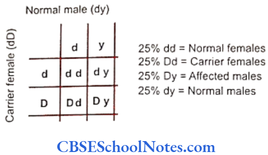
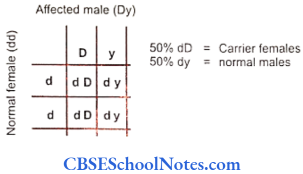
Risks involved in inheritance of X-linked recessive traits are calculated below in the Punnet squares. Symbol “d” represents a normal allele and “D” denotes its mutated form. Therefore the genotype of a carrier female would be “Dd” and the genotype of normal male will thus denoted as “dy” with “d” representing the normal gene present on the single X-chromosome. The small ‘y’ represents the Y-chromosome that lacks in the presence of a homologous gene.
Some common X-linked Recessive Disorders
Some of the common medical conditions transmitted as X-linked recessive disorders are enumerated below.
Duchenne Muscular Dystrophy (DMD)
Characteristics of the disease: The incidence of the disease is rated at 1 in 3500 males. DMD is the commonest and most severe form of muscular dystrophy seen in males. Slow and progressive muscle weakness start presenting soon after the age of 3 years. Proximal muscles of lower limbs are severely affected with weakness and wasting.
Pseudohypertrophy occurs in the calf muscles as they get replaced by fat and connective tissue. Patients cannot walk by the age of 11 years and the boys get wheel chair bound early. Joint contractures and respiratory failure leads to death around 18 years of age.
The gene is located on the short arm of X-chromosome (Xp21). DMD gene encodes a protein known as dystrophin. Dystrophin acts a link protein between muscle fibres and surrounding cell membrane and helps in transmission of power of muscle contraction. Absence of which is responsible for muscle cell degeneration.
This is also associated with an increased permeability of muscle membranes that leads to the escape of muscle enzymes in to the blood. Increased levels of serum creatine kinase (ck) are indicative of the disease along with clinical assessment. Physiotheraphy is helpful.
Hemophilia: This is a disorder of blood coagulation exhibiting an X-linked recessive trait of inheritance. Factors VIII and IX are absolutely necessary for blood coagulation. These factors convert prothrombin to thrombin. Thrombin further converts fibrinogen to fibrin eventually forming the structural framework for bolld to clot. Hemophilia A and B are the two different kinds of the disease encountered commonly.
Hemophilia A (royal hemophilia) is caused by deficiency of factor VIII with an incidence of 1 in 5000 males and several royal families of Europe have suffered this kind of hemophilia.
Hemophilia B, also known as Christmas disease, is caused by the deficiency of IX and has an incidence of 1 in 40000 males.
Characteristics of the disease: Minor trauma or surgery causes prolonged bleeding from the wound. The disease is noted for bouts of spontaneous bleeding that occur into muscles and joint cavities. The genes for hemophilia A and B are located on the long arm of X-chromosome near its distal end. Transfusion of plasma-derived factors VIII or IX often used as replacement therapy. Gene therapy may be available for more effective treatment in future.

X-linked Dominant Inheritance
This type of inheritance is related to the transmission of a dominant or mutant gene located on the X chromosome. As the gene or the mutation is dominant it expresses even when it is present in the heterozygous state. As a result females as well as males manifest the trait when the gene is present in a single X-chromosome.
The X-linked dominant inheritance resembles an autosomal dominant affected male having an X-linked dominant trait will transmit this trait to all his daughters but to none of his sons. Therefore to distinguish this trait from autosomal trait one has to follow the pregeny of affected male.
Disorders that show X-linked dominant characteristics.
- Vitamin D-resistance rickets
- Xg-blood groups
- Amelogenesis imperfecta (Hypoplastic).
Mitochondrial Inheritance
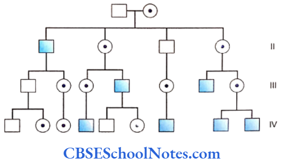
All mitochondria and hence entire complement of mitochondrial DNA (mtDNA) is inherited from the maternal gamete, the oocyte. Mitochondiral inheritance is also known cytoplasmin inheritance as the entire. cytoplasm of the zygote is derived from the maternal gamete (oocyte).
The mitochondrial disorders are only transmitted through the mother to all her sons and daughters. Affected males cannot transmit the disease to their offsprings.
- The mtDNA shows high rates of mutation as compared to the nuclear DNA. Mitochondrial mutations in DNA may cause diseases. Most of these conditions decrease the ATP generating capacity of a cell or tissue. Common tissues to be affected thereby are muscles and the nervous tissue that normally use large amounts of energy for functioning. Hence mutations in mtDNA lead to a wide range of clinical features including hypotonia of skeletal muscles, cardionyopathy, neuropathy, seizures, dementia, encephalopathy, ataxia, stroke, dystonia and acidosis. Some of the mtDNA disorders are named as Leber’s Hereditary Optic Neuropathy; Neurodegeneration Ataxia and Retinitis Pigmentosa (NARP), Mitochondrial Encephalomyopathy Lactic Acidosis and Stroke like Episodes (MELAS), Myoclonic Epilepsy and Rugged Red Fiber Disease (MERRF), etc.
At the onset of a mitochondrial disease only a few mitochondria get mutated while others remain normal. Therefore the expression on the disease varies depending upon the number of mutant or affected mitochondria in the cell. As and when the proportion of mutant mitochondria increases with time, the clinical severity of the disease also increases form “mild” to “severe” degrees. Heteroplasmy is the simultaneous existence of two populations (normal and mutant) of mitochondria within a cell.
Multiple Genes (Polygenic/Multifactorial) Inheritance
Quite a few and fairly common traits and disorders do not follow the pattern of simple Mendelian (single gene) inheritance. Several common traits like intelligence, blood pressure, height, weight, hair color, color of the eye, facial appearance, etc. have a far more complex genetic basis. Only two different types of phenotypic individuals would have resulted in human s(one short and the other tall) if the trait of height were to be determined by just a pair of genes as in case of Mendelian experiments with garden peas.
If we assume ‘T’ to represent the trait of tallness and ‘t’ to denote shortness in a individual, the genotype in the tall would either be ‘TT’ or ‘Tt’. The short would have ‘tt’ as the genetic composition for the trait. However we encounter individuals whose heights show remarkable quantitative variations from one extreme to the other even within a single family. These are Continuous traits or quantitative traits and cannot be clearly classified into distinct groups but are measured quantitatively.
Just as we are unable to predict height or blood pressure, a number of disorders also do not follow Mendelian (monogenic) laws of inheritance and hence cannot be measured. But these diseases have no intermediate forms even though they are governed by multiple factors. These disorders are either present or absent in a person and are called threshold traits, e.g. a person is either a diabetic or a nondiabetic. A few such examples are:
Congenital Malformation Adult Onset Disease
Neural tube defect Diabetes mellitus
Cleft lip Hypertension
Cleft palate Ischemic heart disease
The cumulative or additive effects of several genes which are situated at different loci on chromosomes determine these threshold traits. This kind of inheritance is called polygenic inheritance. Genes in polygenic inheritance do not behave as dominant or recessive but exert a collective effect on the outcome of the trait.
It is a result of interactions between genetic and environmental factors that almost all common physical traits, disorders or congenital malformations are expressed. They don’t depend entirely on a single gene for their existence. Factors like diet, sunlight, diseases, chemical exposure, radiation, etc. may influence activity in genes and its final outcome. Polygenic inheritance is therefore also called Multifactorial inheritance.
Some Important Terms Commonly Used In Relation To Genetic Inheritance
- Pleiotropy
Autosomal dominant gene generally results in a single effect and by and large involves a particular organ or part of the body. However, when a single gene disorder produces multiple phenotypic effects, it is termed as pleiotropy. In cases of the collagen disorder osteogenesis imperfecta, the causative mutation produces defective collagen. However, this diseased collagen leads to many other effects like osteosclerosis, blue sclera and brittle bone etc. - Variable expressivity of gene: Autosomal dominant genes can vary in expression from person-to-person. In clinical trms the spectrum of the expression of a gene may range from its mild, moderte tot he severest of forms of the trait. A common example is that of polydactyly (extra finger). The occurrence of this dominant trait in individuals may vary from having a rudimentary small digit-like structure to a fully developed extra finger. This is explained by the degree of variability in expression of a gene.
- Reduced or incomplete penetrance: It is a variant of variable expressivity. Reduced or incomplete penetrance signifies variable expression of a dominant gene taht is presen tin the heterozygous condition. Indivudual with this condition fail to manifest the disorder clinically. Thus a dominant trait may appear to have skipped a generation in spite of the gene being inherited in the subject. Penetrance of a gene in any generation is expressed in terms of percentage (%) calculated from the number of offspring showing the trait as compared to expected number of affected individuals.
Reduced penetrance or the variation in the expression of gene may be due to influence of the activity of other genes at different loci. It may be also due to difference in environmental factors. - Sex-limited traits: Sometimes the expression of a trait is limited to one sex. The expression of sex-limited traits in humans is represented by the growth of facial hair normally in the males and the development of breast in females.
- Sex-influences traits: The expression of the trait of common baldness hsows sex influence. The trait of baldness behaves as an autosomal dominant trait in males and hence is quire common inmen while it acts as an autosomal recessive trait in females; a bald female is seen very rarely. Thus a trait or a characteristic is said to be sex-influenced when it expresses differently in males and females.
- Codominance: Situations where both the allels representing a trait are expressed fully despite representing a trait are expressed fully despite existing in the heterozygous state they are called codominant. For example, a person with the bolld griup AB possesses both the allelic genes A and B at the loci that are related to blood groups in humans (near the tip of long arm of chromosomal 9). The genes for the antigen A and antigen B are therefore codominant as both get expressed on the red blood cells.
- Intermediate inheritance: A recessive trait or an abnormal recessive (mutant) allele is unable to express itself in a heterozygous state. It only manifests itself in ahomozygous situation. However in certain conditions a recessive gene exhibits intermediate levels of expression even in the heterozygous contidion. The degree of expression remains somwehre between the levels of an abnormal homozygous and that seen in a normal homozygous state for that recessive trait. In sickle cell anemia individuals having the mutation in a heterozygous state possess both abnormal as well as normal hemoglobin in blood.
- Anticipation: If a genetic disease manifests in one generation at an earlier age than the age of occurrence in the previous generation, the diseases is said to exhibit ‘anticipation’. This advancement of the age of, occurrence in seen in every successive. Generation e.g. as seen in Huntington’s disease.
- Genetic imprinting: Sometimes there is a differential expression of a mutated gene in an individual depending on whether the gene in derived from the father or the mother, e.g. Prader willi syndrome (PWS) and Angleman Syndroms (AS). Both these disorders are caused by micro-deletions of chromosome 15. If the mutated 15th chromosome is derived from the father it results in PWS whereas, if the mutated chromosome is maternal in origin it causes AS; both diseases have different clinical manifestations.
- Uniparental disomy: Sometimes both chromosomes of a pair of chromosome may be derived from a single parent instead of the usual one maternal and one paternal copy in a pair. This may give rise to syndrome like the PWS.
- Compound heterozygote: An individual is said to be a compound heterozygote if both the genes located at the respective loci are mutated. Thus the individual is a heterozygote for a particular trait where both the genes are defective or mutated.
Patterns Of Inheritance And Disorders Of Genes Summary
- Common traits or disorders follow either monogenic (single gene) or polygenic (multiple genes) patterns of inheritance.
- Single gene or mutation present on an autosome (autosomal inheritance) or sex-chromosome (sex-linked inheritance) is regulated by monogenic or mendelian inheritance.
- Autosomal dominant inheritance expresses itself even if the dominant gene or mutation is present in a heterozygous state (single dose).
- Autosomal recessive inheritance is manifested only when the responsible gene is present in a homozygous condition (double dose).
- Features of autosomal dominant inheritance.
- As the gene is present on an autosome, both sexes are equally affected.
- Half of offsprings are affected in each generation and the normal offsprings do not transmit the disorder to the next generation.
- Pleiotropy – The condition where a single gene disorder produces multiple phenotypoc effects is called pleiotropy.
- Features of autosomal recessive inheritance.
- The disorder is seen only the siblings in one generation.
- Both sexes are equally affected.
- Parents of affected offspring are closely related (Consanguineous).
- Features of X-linked recessive inheritance.
- Primarily only the males are affected.
- Carrier females are unaffected and transmit the disease to their sons and gets half daughters as carriers.
- Affected males cannot transmit the disease to their sons as the gene is present on the X and not the Y-chromosome.
- Features of X-linked dominant inheritance.
- Males and females are equally affected.
- The trait or the mutation expresses itself even when it is present in a heterozygous condition.
- Affected males transmit the trait to all his daughters but not to any of his sons. Affected females pass the trait to 50% of all her offsprings.
- Resembles autosomal dominant inheritance (pseudo autosomal dominant) except that transmission is skipped in case of male-to-male inheritance.
- Features of mtDNA inheritance.
- Only the mother is responsible for transmitting disease to all her sons as well as daughters.
- Diseased males cannot transmit the disease to their offsprings.
- Polygenic inheritance concerns with the expression of a trait of disorder that is determined by additive effects of interplay between activities of many genes situated at different loci on various chromosomes.
- These traits are also not entirely defined by interaction between genes but also influenced by diverse environmental factors. Multifactorial inheritance is synonymous with polygenic inheritance.