
Genetics Of Dental Caries
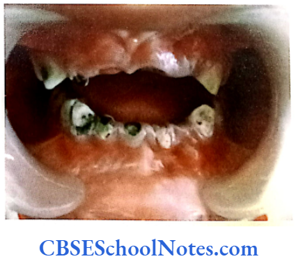
Dental caries is the medical term for tooth decay or cavities. Tooth decay is one of the most common of all disorders of teeth.
Bacteria, that are normally present in the mouth, convert all foods especially sugar and starch into acids. Bacteria, acid, food debris and saliva combine in the mouth to form a sticky substance called plaque that adheres of the teeth. Tooth decay begins if this plaque is not removed throughly and routinely. As stated earlier some of the plaque forming bacteria converts sugar and carbohydrates (starches) in the foods we eat into acids.
These acids dissolve minerals on the surface of the tooth. This erodes the enamel or creates pits on the enamel that are two small to see at first but they get larger over time. Cavities are usually painless until they grow very large and affect nerves or cause a tooth fracture. A tooth abscess can develop. Untreated tooth decay also destroys the internal structure of the tooth (pulp) and ultimately causes loss of the tooth.
The etiology of dental caries has been studied for many years. Multiple factors contribute to a persons risk for caries.
- Environmental factors: Diet, oral hygiene, fluoride exposure and the cariogenic bacteria.
- Host factors: Salivary flow, salivary buffering capacity, position of teeth relative to each other, composition of tooth enamel and host immune response.
In spite of all that is known about this disease there are individuals who still appear to be more susceptible to caries and those who are extremely resistant to dental caries regardless of the environmental risk factors to which they are exposed.’
Similar to periodontics, malocclusion and cleft lip or cleft palate, dental caries is also to be a multifactorial (complex) disease. Though dental caries are seemingly caused out of interaction between environmental and genetic factors, yet the disease is to a great extent influenced by environmental factors. Most scientists agree that the genetic component of dental caries has a minor one in comparison to the overall effect of the environment.
Read and Learn More Genetics in Dentistry Notes
Twin Studies
How We Come to Know that Genetics Plays a Role in the Etiology of Caries
Twins studies were carried out to investigate the role of genetics in the etiology of caries. In case of multi- factorial diseases where genetic and environmental factors play important role in the causation of the disease, twin studies can be used as useful tools to evaluate the roles of the genetic component of the disease. Presence or absence of the trait or disease in a large number of the two types of twins (mono and dizygotic varieties) is calculated in percentage.
Diseases in which the percentage of twin pairs, where both the twins of the pair are affected, is greater in the monozygotic group as compared to the dizygotic group, the diseases can be confirmed to have a definite genetic etiology. The genetic basis of a disease is tested in monozygotic twins who are reared together in the same environment or separated after birth and reared in two different environments. All the similarities in these twins would expectedly be due to com- mon genes and all the dissimilarity will be due to environmental factors.
The early twin studies carried out at the beginning of the 20th century provided some indications that inheritance played a role in caries but the evidence were not conclusive. They only pointed that inheritance was only a contributor to the process (Bachrach et al, 1927; Horowitz et al, 1958; Mansbridge et al, 1958 and Goodman et al, 1959). Twin studies conducted later in the century compared the incidence of caries in monozygotic twins and dizygotic twin groups.
Many studies detected a statistically significant genetic component in the susceptibility to caries and demonstrated that caries experience in monozygotic twins had a greater concordance (Bordoni et al, 1973, Hassell et al, 1995; Townsend et al, 1998). These studies concluded that not only environmental factors clearly have a greater influence but that genetic factors also contribute to the causation of dental caries (Niswander et al, 1975).
A major advance in the understanding of the role of inheritance and the incidence of dental caries was achieved by analyzing twins reared apart. These patients had an average age greater than forty and did not share similar environments from shortly after birth until the time of analysis. The analysis of twins raised apart provides the strongest evidence of a genetic contribution to the incidence of dental caries (Borass et al, 1988 and Conry et al, 1993).
The analyses of dental caries incidence in monozygotic and dizygotic twins also indicate that a large number of different genes contribute to the observed outcomes. Although the twin studies provided some strong evidence of genetic contribution to the risk of dental caries, none provided any evidence of linkage to specific genes (Shuler, 2001).
Risk Factors In Dental Dental Caries
The most important components contributing to the risk of dental caries are as under:
- Microorganisms present in the oral cavity and the host immune response
- Cariogenic diet
- Role of saliva in protection against caries • Morphology of tooth and composition of enamel matrix
- Gene(s) for dental caries.
Many studies have indicated that even if all the environmental factors are identical and monitored to be under controlled and standard conditions, variability in the susceptibility to dental caries still exists and differs within the tested individuals.
This indicates that certain environmental factors are more cariogenic for some individuals as compared to others. This may be explained by the existence of genetic variability among individuals. The genetic influences modify the expression of disease in the individual.
Microorganism Of The Oral Cavity And Host Immune Response
Dental caries will not occur if the oral cavity is free of bacteria. These bacteria are organized into dental plaque. Of the many types of bacteria in the mouth the most caries active appear to be Streptococcus mutans, Lactobacillus spp., Veillonella spp. and Actinomyces spp. These bacteria can be transferred from the mother to child and are present at varying levels in all human mouths. A variety of carbohydrates provide substrates for these organisms to grow on.
Most research on the bacteriology of dental caries has focused on the ubiquitous S. mutans and its ability to ferment sucrose (Loesche, 1986). This organism preferentially ferments sucrose to produce significant amounts of acid and extracellular polysaccharide (plaque). However, most researchers now agree that other organisms present in the mouth are capable of plaque formation and acid production from a variety of fermentable carbohydrate substrates besides sucrose which is present in the normal mixed diet.
The individual’s genotype may influence the likelihood of intraoral colonization of cariogenic bacteria. Scientists have shown that persons may be caries-resistant or caries-susceptible for a particular strain of bacteria. Even if cariogenic bacteria are present in the oral cavity the caries-resistant person usually doesn’t develop caries. This confirms the presence of important genetic elements influencing susceptibility to dental caries.
Streptococcus mutans was first isolated from human carious lesions in 1924 by Clark. This indicated that dental caries had a bacterial etiology and were transmissible infections. S. mutans isolates have been divided into eight serotypes. Human isolates represent serotype c, e and f. The S. mutans serotype c being the most prevalent streptococci isolated from human dental plaque.
Salivary immunoglobulin A (sIgA) is the major antibody present in the saliva. It is the host’s first line of immune defense against Streptococcus mutans. Salivary IgA is synthesized and secreted by plasma cells located in salivary glands. The host’s immune system protects individual from caries by producing various types of antibodies. In humans majority of these antibodies are of the IgA type but types IgG and IgM are also present in the saliva. Salivary IgA acts in the following ways to restrict infection:
- It neutralizes bacterial exotoxins.
- It neutralizes enzymes contributing to the disease processes such as glucosyltransferases. (Caries only form when this enzyme acts in presence of sucrose. Glucosyltransferases breakdown sucrose into its components like simple sugars known as fructose and glucose. Mutans streptococci use these sugars to form plaque. The enzyme glucosyltranferase could be used in making effective vaccines for the reduction in dental caries. Certain components of these enzymes when injected into an individual produce antibodies against the enzymes and inactivate bacterial activity to reduce plaque formation. In the past this enzyme was used as an antigen and was injected in the salivary glands of rats. The immunized rats produced the antibody IgA in their saliva and had fewer dental caries. Thus in future this enzyme may be used as vaccine in humans against caries).
- Inhibits the attachment of bacteria on epithelial or tooth surface.
Because of the above function of the salivary IgA most of the caries vaccines induce salivary IgA antibody response to S. mutans antigens (Han, 2007).
Human studies in the past have measured the levels of antibodies present in blood serum and saliva of caries patients. Some of these studies found a negative association between IgA and caries activities while other reports have shown a positive association. Few reports found no correlation between the two. Thus the association between the levels of IgA and the development of dental caries has been studied with conflicting results.
However, the number of papers is growing that report increases in immunoglobulin IgA contents in the saliva in cases with high caries experience (Weyna et al, 1979 and Dens et al, 1995). One group of research workers studied pair-matched patients between a group of IgA deficient patients and a section of immunocompetent normal subjects by age and plaque index. During a two years period they observed less caries experience in immunodeficient patients than found in normal controls (Robertson et al, 1980). Another study observed that IgA-deficient children showed caries scores lower than those of healthy children (Fernandes et al, 1995).
Association between HLA Antigens and Susceptibility to Dental Caries
Current evidences support the relationship between immune complex genes (HLA) and caries and the association of different levels of cariogenic bacteria and enamel defects. Many studies are now available which show the association between increased risk for caries and immune complex (HLA) genes.
One study reported the strong association between HLA DRw6 loci and DMFS index (Lehner et al, 1981). The same immune complex locus showed a low dose response to Streptococcus mutans antigens. However, few studies could not detect a relationship between HLA DR type and dental caries (De Vries et al, 1985 and Acton et al, 1999).
Celiac disease is an autoimmune disorder of the small intestine that occurs in genetically predisposed people of all ages. Symptoms include chronic diarrhea, weight loss and fatigue. The vast majority of celiac patients have one of the two types of HLA DQ. This gene is a part of the MHC class II antigen-presenting receptor.
The gene is located on the short arm of the sixth chromosome and as a result of this linkage the locus has been labeled CELIAC1. Celiac disease patients exhibit an increased incidence of dental caries. This might be due to the fact that these patients have defective enamel that predisposes the tooth to dental caries (Aine et al, 1990; Aine, 1996 and Aguirre et al, 1997).
However, the cause of this defective enamel in celiac disease patients has not been well-understood. These patients show significant positive correlation between their HLA type and presence of the enamel defect. The HLA-DQ2 and DQ 8 is strongly associated with enamel defects and dental caries in celiac patients.
The association between HLA complex and caries has indicated that a few genes in the HLA complex are responsible for dental caries resulting from altered enamel development and also due to low dose response to cariogenic bacteria (Lehner et al, 1981), i.e. less aggressive immune response to bacterial invasion.
We still do not know whether specific genes dedicated to the development of enamel or responsible immune response to cariogenic bacteria are located close enough (linked) to certain HLA complex. It is expected that the existence of any such association would be determined in the near future.
It is well known that individuals with immune deficiency diseases are susceptible to dental caries and have a greater frequency of harboring S. mutans than do normal persons, e.g. as seen in HIV infection.
Cariogenic Diet
The dietary components that contribute most to the process of caries formation are fermentable carbohydrates. These need to be retained in the mouth long enough to be metabolized by oral bacteria (principally Streptococcus mutans) to produce acid. The acid attacks the tooth enamel and gradually dissolves it (demineralization). A repair process known as remineralization offsets this demineralization process. The balance between remineralization and demineralization deter- mines the occurrence of caries.
The presence of fat in experimental diets has been shown to affect cariogenicity of sugars. The effects have been ascribed to enhance clearance of sugars from the mouth. It is also conceivable that several fatty acids express a potent antibacterial effect. The presence of calcium and phosphorus has been shown to influence the cariogenicity of foods; the effect, however, is restricted to the food containing the minerals. Evidence suggests that pyridoxine (vitamin B6) may exert a cariostatic (stopping caries) effect by enhancing decarboxylation activity in dental plaque.
Studies have indicated that the persons suffering from hereditary fructose intolerance are free from caries (Newburn et al, 1980 and Saxen et al, 1989). They are free from caries because there is absence of sugar in their diet and not because a hereditary fructose intolerance provides some kind of resistance to the production of caries.
Very few studies are available that has investigated the heritability of caries in relation to sucrose. A twin study in 2003 was aimed to determine heritability estimates for dental caries traits and sucrose sweet-ness preference. Results indicated that variations in dental caries traits and sucrose sweetness preferences have a significant genetic contribution mediated independently (Bertz et al, 2003).
Many studies have revealed that higher and more frequent sugar intake may increase the risk of caries formation in children. The high sugar intake reflects a preference for sweet substances. Inherited behavior and taste thresholds may play an important role in the frequency of carbohydrate intake. Genetic sensitivity to taste may be associated with a preference for or rejection of some food by children.
Many studies indicate that children belonging to the group “non- tasters” (with high threshold for taste) were sweet likers and prefer strong tasting food (Verma et al, 2006) while children belonging to group “tasters” are sweet dislikers and preferred weak tastes. The incidence of dental caries was significantly higher in nontasters as compared to tasters.
Role Of Saliva In Protection Against Caries
Saliva is body’s natural protective mechanism against decay. It contains salivary proteins that adsorb strongly onto the teeth, protecting enamel against acid dissolution. This adsorbed protective layer is referred to as the pellicle. Salivary proteins also act as antibacterial agents. Saliva is the primary resource of calcium, phosphate and fluoride; materials used to remineralize the enamel.
Saliva also acts quickly to clear away food debris from the mouth and to buffer the organic acids that are produced by the bacteria. Saliva is therefore a very vital and complex material in the prevention of dental caries. Salivary dysfunction can lead to rapid deterioration of dental enamel. Salivary dysfunction may occur as a result of certain medications or as side effects of medical treatments such as radiotherapy.
There is a strong correlation between the composition of saliva and the production of caries. The formation of dental plaque is the result of interactions between environmental and genetic factors. The caries- susceptible plaque is formed due to presence of certain chemicals in saliva. A group of saliva proteins known as proline-rich proteins (PRPs) are responsible for early plaque and pellicle formation (Mayhall, 1970 and Bennick et al, 1983).
At present eight different kinds of PRPs are known that are thought to be produced by a cluster of genes located on the short arm of chromosome number 12 (Goodman et al, 1985 and Mamula et al, 1985). People show variations in the type of PRPS produced in their saliva due to variations in their genotype at these regions. Some people with certain protein genotypes (especially Pa+ and Pr22) are more susceptible to dental caries (Yu et al, 1986), On other hand individuals with genotypes Pa- and Pr11 are resistant to the dental caries. Similarly low levels of salivary calcium and phosphate have been shown to be associated with the increased risk of caries.
Different individuals respond in a different way to specific biochemical differences in oral environment depending on their genetic constitutions. For example there is significant difference between monozygotic and dizygotic twins in terms of salivary flow, pH, and salivary amylase activity when compared between the two groups.
On the contrary, both the monozygotic twins of a pair will respond similarly (because of same genetic constitution) to these factors whereas such similarity may be lacking between the two individuals of a dizygotic pair (because of difference in their genetic constitutions).
Literature is nearly equally divided both in favor of and against the anticaries role of salivary immuno- globulins, especially sIgA. Many studies have indicated inverse relations between sIgA and caries (Camling et al, 1987 and Rose et al, 1994). Few studies have also reported increased levels of sIgA (Prakash et al, 1994). However, some studies have indicated no correlation at all (Kristila et al, 1994).
Xerostomia or decreased secretion of saliva (due to pathological dysfunction of salivary glands) has been demonstrated to be responsible for increased rate of caries. Studies indicate that a low salivary flow rate (less than 1 ml/min after salivary stimulation) is associated with an increased risk of caries. Sjögren’s syndrome is an autoimmune disorder in which abnormally activated immune cells attack and destroy exocrine glands that produce tears and saliva.
The primary and secondary variants of Sjögren’s syndrome are found to be associated with increased caries risk (Ravald et al, 1998). This is due to the fact that Sjögren’s patients have a decreased flow rate of saliva. Similarly, the condition of scleroderma related xerostomia is also associated with caries tooth (Wood and Lee, 1988).
Several medical conditions including therapeutic radiations administered to the head and neck regions (Nasman et al, 1994) and pharmacological agents with xerostomic side effects (Ryberg et al, 1990) lower salivary flow rate dramatically to pathological levels and elevate the patient’s risk of caries. The evidence therefore indicates that normal salivary flow rate is strongly protective against caries and clinicians should identify individuals with reduced salivary output to modify their treatment.
Dental caries has a higher prevalence rate in females as compared to males. A recent study indicated that biochemical composition of saliva and salivary flow rates are modified in women due to hormonal fluctuations during events such as puberty, menstruation and pregnancy. This makes the oral environment significantly more cariogenic for women when compared to men.
Saliva is a major carrier of topical fluoride. The concentration of fluoride in the ductal saliva, as it is secreted from salivary glands, is low. This concentration of fluoride is not likely to resist cariogenic activity. Drinking fluoridated water, brushing with fluoride toothpaste or using other fluoride dental products, on the other hand, can raise the concentration of fluoride in the saliva present in the mouth to about 100- to 1,000-folds.
Saliva and the extraneous sources thus serve as important sources of fluoride that gets concentration in the plaque and aid tooth remineralization. Fluoride concentrated in plaque and the saliva inhibits demineralization of the sound enamel and enhances the remineralization or recovery of demineralized enamel (Featherstone, 1999). Fluoride also inhibits dental caries by impairing cellular mechanisms of cariogenic bacteria.
High caries prevalence has been reported for individuals suffering from the deletion at 22q11; the High caries prevalence has been reported for 22q11 Deletion Syndrome (22q11 DS). It was observed that patients with 22q11 DS had impaired salivary secretion rates, higher numbers of cariogenic bacteria, increased salivary protein concentrations and reduced output of electrolytes in the saliva compared to the controls. This indicated that the salivary function is affected in 22q11 DS explaining increased caries risk seen in these subjects (Klineberg et al, 2007).
Morphology Of Tooth And Composition Of Enamel Matrix
The morphology of teeth related to their shapes, sizes, pit and fissure morphology, enamel structure and composition, arch forms, dental spacing and order of the teeth are some of the important factors that regulate the “washing” effects of saliva and thereby may profoundly influence the production of caries.
These factors are in fact largely determined by hereditary factors. Both kinds of twin studies (“twins reared together” and “reared apart”) have indicated that the morphologies of teeth, arch forms, dental spacing, malocclusion, etc. have strong genetic contributions.
Teeth are composed of a thin layer (1-2 mm) of dental enamel, which forms the hard protective coating over the tooth. This layer mainly consists of calcium, phosphate and other ions in a structure known as “hydroxyapatite”. Dental enamel is porous and is susceptible to acid dissolution during the process of demineralization. Many genes are known which are active in the formation of enamel (AMELX, ENAM, KLK-4 and MMP20 which encode various proteins like ameloblastin, amelogenin, enamelin, tuftelin- 1, and tuftelin interacting protein 11).
Certain variations in some of these enamel matrix genes may be associated with enhanced caries susceptibility. Some of these genes are linked to specific syndromes where the process of tooth development itself is altered. This is due to an alteration in the proteins responsible for biomineralization of the enamel matrix. Patients with alterations in the morphology of teeth and formation of the enamel are susceptible to caries. Syndromes, which are associated with caries and well-defined altered craniofacial phenotypes, are usually determined by mutation in a single gene.
These syndromes are Turner’s syndrome (Takala et al, 1985), the fragile X syndrome (Shellhart et al, 1986), ectodermal dysplasia, cleft lip and/or cleft palate (Dahllot et al, 1989), diastrophic dysplasia, etc. We do not know much about specific genes responsible for these disorders yet.
Nonsyndromic forms of Amelogenesis Imperfecta (AI) present with abnormal formation of the enamel. The enamel is composed mostly of minerals that are modified and regulated by the activities of the proteins in it. AI is caused due to malfunction of proteins in the enamel: Ameloblastin, enamelin, tuftelin and amelogenin (Refer Chapter 10).
People afflicted with amelogenesis imperfecta have teeth with an abnormal color. The teeth have a higher risk for dental cavities. The AMELX, ENAM, KLK-4 and MMP20 genes provide instructions for making proteins that are essential for normal development of tooth. Mutations in the AMELX, ENAM, MMP20, and KLK-4 genes have been found causative for amelogenesis imperfecta (nonsyndromic form).
Mutations in any of these genes alter the final structure of these proteins or completely prevent synthesis of any protein at all (Refer Chapter 10). As a result tooth enamel is abnormally thin or soft and has high a risk of developing dental caries.
The Search Of Candidate Gene(s) For Dental Caries
Very recently (postgenomic) quite a few studies have tried to locate the candidate gene for caries but with little success. A candidate gene is a gene known to be located in a region of interest in the genome. Product/s of the candidate gene has/have biochemical or other properties the presence or absence of which can be directly related to a disease. Findings form the following significant studies are as under:
- A study on mice indicated that major gene(s) responsible for the regulation of susceptibility to dental caries or resistance are located on chromosomes number 1, 2, 7 and 8 (Nariyama et al, 2004).
- Genome wide genotype data and DMFT scores in a large number of families were evaluated (Vieira et al, 2008). Low caries susceptibility loci were found on chromosomes number 5 (5q13.3), 14 (14q11.2) and X (Xq27.1). The high caries susceptibility genes were identified on chromosome number 13 (13q31.1) and 14 (14q24.3). The presence of genes for caries on the X chromosome may account for the sex differences observed in the incidence of caries. This study was the first of genome wide scans introduced for dental caries.
- In a recently concluded study single nucleotide polymorphism (SNP) assays were performed for 6 candidate genes. The candidate genes selected for this study were the amelogenin (AMELX), ameloblastin (AMBN), tuftelin (TUFT1), enamelin (ENAM), tuftelin-interacting protein (TFIP11) and kallikrein 4 (KLK4) genes. There were no significant associations concluded between single candidate genes and caries susceptibility. A significant interaction between tuftelin and S. mutans was however observed (Slayton et al, 2005).
- The Osteopontin (OPN) gene plays an important role in mineralization. In a recently conducted study OPN was chosen as candidate gene with respect to caries susceptibility as OPN gene was found to be associated with incidences of enamel hypoplasia in primary dentition. Results indicated an association between the OPN gene and caries in the primary dentition (Willing et al, 2006).
- A new study applied scanning of single- nucleotide polymorphism (SNP) markers with relation to selected candidate genes (ameloblastin, amelogenin, enamelin, tuftelin-1 and tuftelin interacting protein 11) that influence enamel formation. One copy of a rare amelogenin allele was found to be associated with caries experience. This result suggested that variations in amelogenin may contribute to caries susceptibility (Deeley et al, 2008).
In conclusion one may say that the search for genetic variables in the etiology of dental caries has just begun.