
Boundaries And Subdivisions Of Perineum
Boundaries And Subdivisions Of Perineum Definition
The region at the lower end of the trunk, in the interval between the two thighs where external genitalia and anus are located.
Boundaries And Subdivisions Of Perineum Boundaries
Superficial – Anterior – The scrotum in males and mons pubis in females.
- Posterior- The buttock.
- Lateral- Upper part of the medial side of the thigh on each side.
Deep – Anterior- Upper part of pubic arch and inferior (arcuate) pubic ligament.
- Posterior- Tip of the coccyx.
- Lateral- Ischiopubic rami, ischial tuberosities, and sacrotuberous ligaments.
Subdivisions of perineum
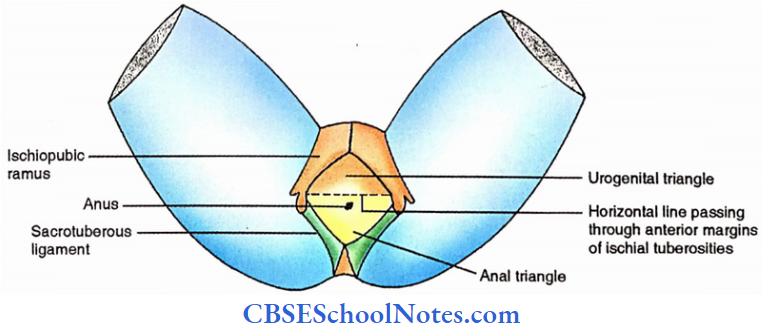
The perineum is divided into two triangular areas (anal and urogenital triangles) by a transverse line joining the anterior parts of the ischial tuberosities and passing immediately anterior to the anus.
1. The anal triangle (region)
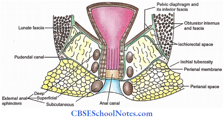
It contains the termination of the anal canal in the midline and ischiorectal fossa on each side. The pudendal canal carrying the neurovascular bundle to the perineum is located in the lateral wall of the ischiorectal fossa.
2. The urogenital triangle (region)
It contains the external urogenital organs. It also contains the superficial and deep perineal spaces.
External Genitalia
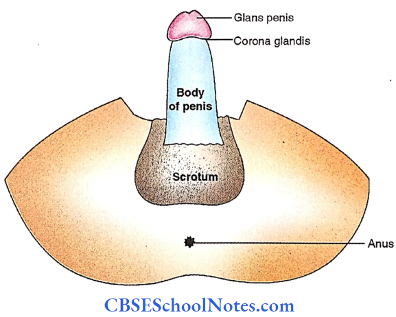
Male External Genital Organs
The male genital organs include the penis, scrotum, testis, epididymis, and spermatic cord.
External Genitalia Penis
It is the male organ of copulation. It consists of (1) the attached part called the root and (2) the free part called the body.
Root Of Penis
It is located in the superficial perineal pouch. It is made up of two crura and one bulb which are masses of erectile tissue. Each crus is attached to the margins of the pubic arch and covered by ischiocavemosus.
The bulb lying between two crura is attached to the perineal membrane and covered by the bulbospongiosus.
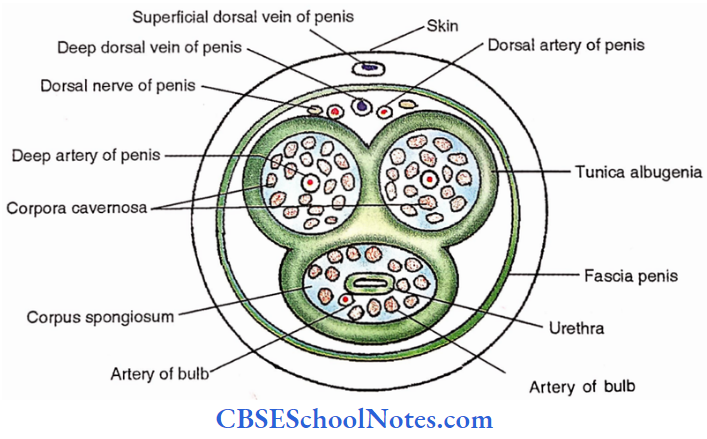
It is pierced by the urethra which shows dilatation in this region called the inrabulbar fossa. The urethra continues anteriorly in the corpus spongiosum located in the body of the penis.
Body Of The Penis
It is continuous with the root of the penis and is completely covered by skin. It has an anterior and posterior surface. It consists of three elongated masses (right and left corpora cavernosa and middle corpus spongiosum) of erectile (cavernous) tissue.
During an erection, the cavernous spaces are excessively filled with blood. Corpora cavernosa are forward continuation of crura and both of them terminate anteriorly in a blunt conical end covered from the front by the glanspenis.
The terminal conical expanded part of the corpus spongiosum. Corpora cavernosa are enveloped by a thick connective tissue covering called the tunica albuginea.
The base of the glans penis is a projected margin (corona glandis) which overhangs an oblique constricted region (neck of penis). Glans is also pierced by the urethra which presents a dilatation in this region called the navicularfossa.
Female External Genital Organs (Pudendum Or Vulva)
These include the following structures;
The mons pubis, the labia majora, the labia minora, the clitoris, the vestibule of vagina, the bulbs of the vestibule and the greater vestibular glands.
Mons Pubis
The rounded eminence present in front of the pubic symphysis formed as a result of the accumulation of subcutaneous fat. It is covered with pubic hairs and in contrast to males, the upper limit of the hair-bearing area is demarcated by a horizontal line.
Labia Majora
These are two thick folds of skin enclosing fat forming the lateral boundaries of the pudendal cleft. Their external surface is covered with hair and the inner surface contains a large number of sebaceous glands.
- The anterior ends of the two are joined together below the mons pubis forming the anterior commissure. The posterior commissure is formed by the union of the relatively less prominent posterior ends in front of the anus.
- The gynecological perineum is the area (about 1 inch) located between the posterior commissure and the anus.
Labia Minora
These are two thin folds of skin within the pudendal cleft. Their anterior ends divide into upper and lower layers.
- The upper layers of two sides join to form the prepuce of the clitoris and the lower layers join to form the frenulum of the clitoris.
- The posterior ends of the two sides join to form the frenulum of the labia minora. A large number of sebaceous glands are found on the inner surface of the labia minora.
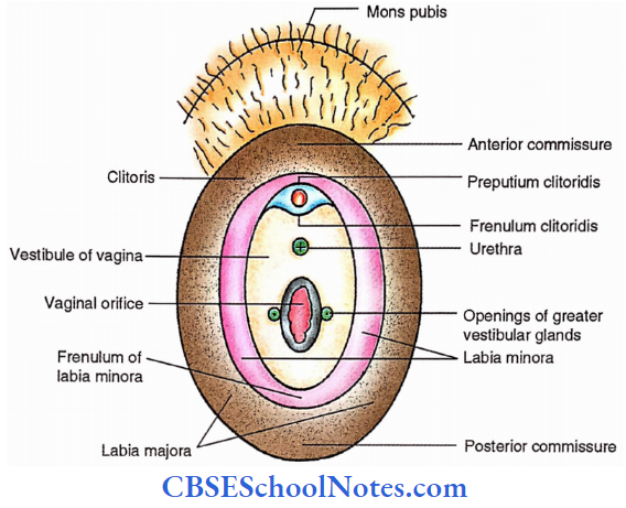
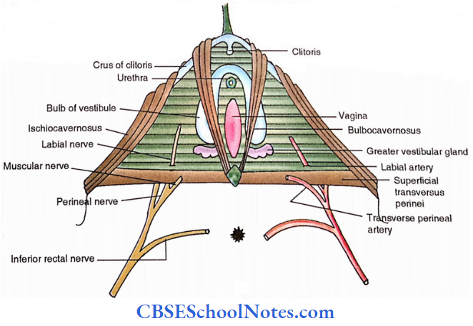
Clitoris
It is located in the anterior part of the pudendal cleft. It is homologous to the male penis and erectile but not pierced by the urethra.
- Like the penis, the body of the clitoris has a pair of corpora cavernosa encloses in a fibrous sheath and separated by a pectiniform septum; however, the corpus spongiosum is absent. Each corpus callosum is attached to the ischiopubic rami.
- The glans clitoridis which caps the s ends of corpora cavernosa is made up of erectile tissue which is continuous posteriorly with the commissural venous plexus uniting right and left bulbs of the vestibule.
- The surface of the glans is very g sensitive and plays an important role in sexual responses.
Vestibule Of The Vagina
The space between two labia minora is called the vestibule of the vagina. It has the following features.
- Urethral orifice. 2.5 cm behind the frenulum of the clitoris
- Vaginal orifice (introitus).
- Orifices of the ducts of the greater vestibular glands.
- Openings of lesser vestibular (mucous) glands.
- Vestibularfossa- depression between vaginal orifice and frenulum of labia minora.
Bulbs Of The Vestibule
These correspond to two halves of the bulb of the penis. They are oval and made of erectile tissue and are located superficial to the perineal membrane on either side of the vaginal and urethral orifices.
The posterior expanded part is partly overlapped by the greater vestibular glands. The tapering anterior ends of the bulbs are united in front of the urethra by a venous plexus called the bulbar commissure.
Greater Vestibular Glands (Of Bartholin)
These are located in the superficial perineal space on either side of the vaginal orifice and are homologous with the bulbourethral glands (of Cowper) in males.
Each gland is partly overlapped by the bulbs of the vestibule and its long duct (2 cm) opens between the hymen and labium minus.
Anal Triangle
Cutaneous Nerves
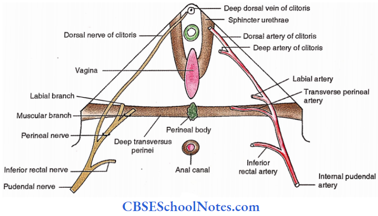
- Inferior rectal nerve (S2, S3, S4): Supplies the skin around the anus and over the ischiorectal fossa.
- Perineal branch of4th sacral nerve: Supplies the skin posterior to the anus.
Superficial Fascia
It contains a large amount of fat which fills the ischiorectal fossa.
Deep Fascia
It forms the inferior fascia of the pelvic diaphragm and the fascia over the obturator intemus below the attachment of the levator ani.
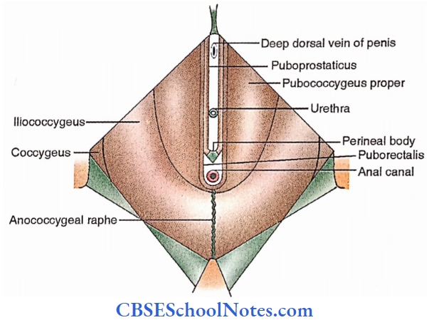
Anococcygeal Ligament Or Body
It consists of fibrofatty tissue intermingled with muscle fibers which are derived from the levator ani and the external anal sphincter. It extends between the tip of the coccyx and the superficial external and sphincter.
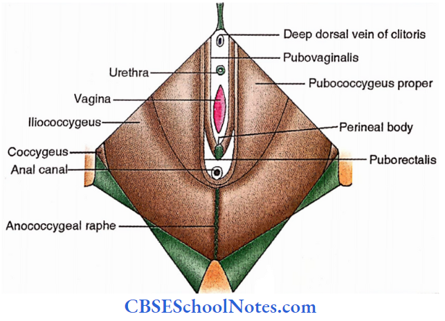
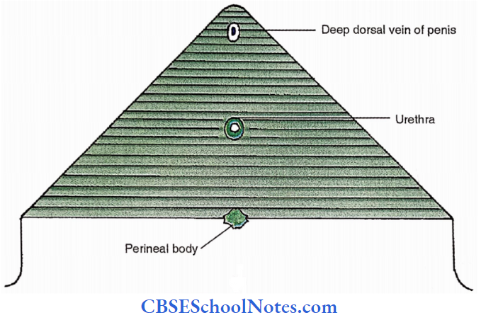
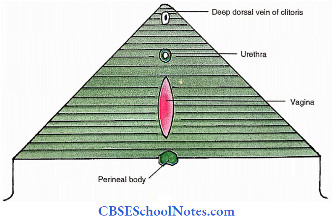
Perineal Body
It consists of a fibromuscular node situated in the median plane about 1.25 cm in front of the anal margin and close to the bulb of the penis. Many muscles of the perineum converge. and interlace in the perineal body.
It provides support to the pelvic organs. It is more important in females and when damaged during childbirth it may lead to prolapse of the urinary bladder, uterus, ovary, or even rectum.
External Anal Sphincter
The external anal sphincters surround the anal canal. These are supplied by the inferior rectal nerve and the perineal branch of the 4th sacral nerve. It is under voluntary control and keeps the anus and anal canal closed.
Ischiorectal Fossa
It is a wedge-shaped space (with the base directed downwards and the edge directed upwards) located on each side of the anal canal and below the pelvic diaphragm.
Ischiorectal Fossa Dimensions
- Length (anteroposterior)- 2 inches.
- Width (side to side)- 1 inch
- Depth (vertical)- 2.5 inches
- Boundaries
- Base – skin
- Edge Apex- line of origin of levator ani from the lateral pelvic wall
- Anterior-posterior border of the perineal membrane
- Posterior- lower border of gluteus maximus and sacrotuberous ligament.
- Lateral-obturator intemus with its fascia and medial surface of the ischial tuberosity.
- Medial- external anal sphincters and levator ani.
Ischiorectal Fossa Recesses
- Anterior recess – extends above the perineal membrane to enter the deep perineal pouch and reach the back of the body of the pubis.
- Posterior recess – extends deep to the sacrotuberous ligament.
- Horseshoe recess – connects the ischiorectal fossae of two sides behind the anal canal.
Spaces And Canals Of The Fossa
Perianal space
It is subcutaneous and contains fat which is enclosed in tight loculi. It is separated from the deep ischiorectal space by a fascial extension called the perianal membrane which passes.
Laterally from the lower end of longitudinal muscle coat (conjoint longitudinal layer). It extends from the white line of Hilton medially to the pudendal canal laterally. The infection of this space is very painful.
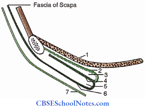
Ischiorectal Space
This a large and deep space filled with loosely packed fat. The infection of this space is the least painful. The lunate fascia arches over the ischiorectal fat. The fascia divides the ischiorectal space into,
- Suprasegmental space – above the fascia.
- Tegmental space – below the fascia.
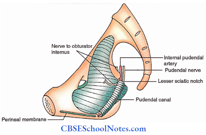
Pudendal Canal
A fascial canal in the lateral wall of the pelvis encloses the pudendal nerve and internal pudendal vessels.
The fascia of the canal fuses with,
- obturatorfascia- laterally
- lunate fascia – above
- perianalfascia- medially
- falciform process of sacrotuberous ligament – below
Contents of the ischiorectal fossa
- Ischiorectal pad of fat.
- Inferior rectal nerve and vessels.
- Posterior scrotal ( or labial, in female) nerves and vessels.
- Perineal branch of 4th sacral nerve
- Perforating cutaneous branches of nerves S2, 3
- Pudendal canal with its contents.
Anal Triangle Applied Anatomy
- The ischiorectal fossae allow distension of the anal canal during defecation.
- Abscess formation- Perianal and ischiorectal spaces are common sites of abscess formation.
- Anorectal fistula (fistula in ano) – Sometimes the abscess may burst medially into the anal canal or rectum or laterally on the surface resulting in anorectal fistula.
- External sinus- If the abscess bursts externally and does not heal, it results in the external sinus.
- Prolapse of the rectum- The ischiorectal pad of fat supports the rectum and anal canal and its loss may result in prolapse of the rectum.
- Ischiorectal hernia- Hmiation of a pelvic organ through the hiatus of Schwalbe (the gap between the tendinous origin of the levator ani and obturator fascia).
Steps Of Dissection Of Ishciorectal Fossa
Place the cadaver in the prone position and expose the lower border of the gluteus maximus. Remove the skin and fascia from the perineum, external anal sphincters, and anococcygeal ligament as well as from the margins of the anus.
- Trace and define the boundaries of the ischiorectal fossa. Expose and clean the posterior margin of the perineal membrane and identify and trace the inferior rectal and posterior scrotal (labial) nerve and vessels in the lateral wall of the fossa.
- Try to identify the gluteal branches of the posterior cutaneous nerve of the thigh and the perineal branch of S4. Remove all fat from the fossa. Clean and define the pudendal canal on the lateral wall of the fossa and trace the pudendal nerve and internal pudendal vessels.
Urogenital Triangle
Cutaneous innervation
- Dorsal nerve of penis (or of the clitoris): It supplies the skin of the whole penis (clitoris) except its root.
- Ilioinguinal nerve and genital branch of genitofemoral nerve: Supply the anterior 1/3rd of the scrotum (or labium majus in females) and the root of the penis.
- Perineal branch of the posterior cutaneous nerve of the thigh: Supplies the lateral part of the
urogenital region and lateral part of the posterior 2/3rd of the scrotum (or labium majus). - Posterior scrotal (or labial) nerves: Supply the medial part of the urogenital region including labium minus in females and the medial part of the posterior 2/3rd of the scrotum (or labium majus).
- Perineal branch of the pudendal nerve: Supply the mucous membrane of the urethra.
- Levator ani
- The superior fascia of the urogenital diaphragm
- Deep Perineal pouch
- Perineal membrane
- Superficial perineal pouch
- Colles fascia
- Skin
Superficial fascia
It consists of two layers:
- Superficial fatty layer– it is continuous with the superficial fascia of the surrounding region.
- Deep membranous layer (Colles fascia) – Attachments
- Posterior- Posterior border of the perineal membrane
- Sides- Pubic arch below the crus of penis (clitoris in female).
- Anterior- Continues with the fascia of the scrotum (dartos), of the penis, and with the fascia of the Scarpa (deep membranous layer of the superficial fascia of the anterior abdominal wall).
Deep Fascia
It consists of two layers lining the urogenital diaphragm from its superior and inferior aspects. The inferior layer is thicker and called the perineal membrane.
Superior Fascia Of Urogenital Diaphragm
- It is thin and less defined than the inferior fascia
- It is coextensive with the perineal membrane and attached anteriorly and posteriorly to it.
- Laterally it is continuous with the obturator fascia.
- It is pierced by the urethra and continuous with the prostatic fascia.
Deep Perineal Space (Pouch)
It is the space between the superior and inferior fascia of the urogenital diaphragm.
Contents of deep perineal pouch
- Urethra (membranous urethra in male)
- Muscles
- Sphincter urethrae
- Deep transverse perineal
- Nerves
- Dorsal nerve of penis (clitoris in female)
- Muscular branch from perineal nerve
- Arteries
- Initial parts of arteries of the penis (clitoris in females) pass to the superficial perineal pouch
- Bulbourethral glands in male


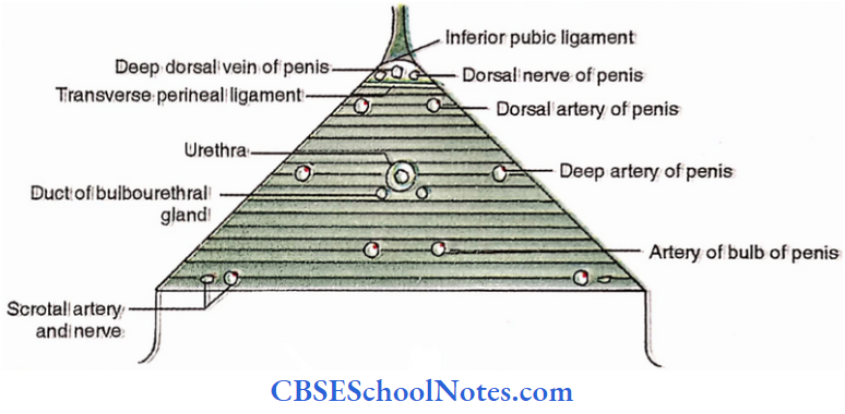
The Perineal Membrane
It is a thick, triangular sheet of fascia lying across the pubic arch.
- Its base (posterior border) is continuous
- Above with the superior fascia of the urogenital diaphragm
- Below with the code fascia.
- In the midline connected to the perineal body.
- On each side- Attached to ischiopubic rami above the attachment of crus of the penis.
- Anterior border (apex) – Continues with the superior fascia of the urogenital diaphragm and is thickened to form a transverse perineal ligament.
- The gap between the arcuate pubic ligament and transverse perineal ligament transmits
- Deep dorsal vein of the penis (clitoris in female)) to the prostatic (vesical in female) venous plexus.
- Dorsal nerve of penis (clitoris in female).
Structures Piercing The Perineal Membrane
In males
- Urethra, 1 inch below – the pubic symphysis.
- Ducts of bulbourethral glands, close to the urethra.
- Artery and nerve to the bulb of the penis.
- Urethral artery.
- Deep artery- of the penis.
- Dorsal artery of the penis
- Posterior scrotal nerves and vessels.
- Branches of perineal nerve supplying superficial perineal muscles
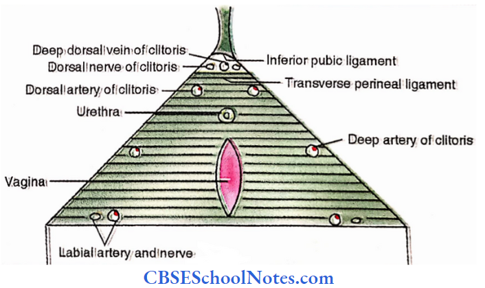
In females
- Urethra
- Vagina
- Artery and nerve to the bulb of the vestibule
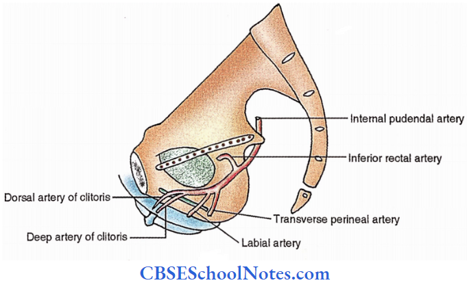
- Deep artery of clitoris
- Dorsal artery of the clitoris
- Posterior labial arteries and nerves.
- Branches of perineal nerve supplying superficial perineal muscles
Urogenital diaphragm
It is formed by
- Deep perineal muscles – sphincter urethrae and deep transverse perineal
- The superior fascia of the urogenital diaphragm
- The inferior fascia of the urogenital diaphragm (perineal membrane)
Superficial perineal pouch
It is a superficial space in the perineal region located superficial to the perineal membrane.
Urogenital Triangle Boundaries
- Superficial – Colles’ fascia
- Deep – Perineal membrane
- On either side- Ischiopubic rami
- Anteriorly- Open and continue with the spaces of the scrotum and anterior abdominal wall.
- Posteriorly- Closed by the union of the perineal membrane with the Colles’ fascia
Urogenital Triangle Contents
- The root of the penis (clitoris in females), is made up of a pair of crura and a bulb (only crura in females)
- Muscles on each side-
- Ischiocavemosus
- Bulbospongiosus
- Superficial transversus perinea
- Nerves
- Branches of the perineal nerve- posterior scrotal (labial in females), nerve to bulb (in males only), and muscular branches
- Long perineal nerve- from the posterior cutaneous nerve of the thigh
- Arteries
- Branches of perineal artery- posterior scrotal (labial in female) and transverse perineal
- Arteries of the penis (clitoris in female) – artery to the bulb (in male only), urethral artery, deep artery of the penis (clitoris in female), and dorsal artery of the penis (clitoris in female).
- Greater vestibular glands in females (or, ducts of bulbo-urethral glands in the male).