
Brainstem Medulla Oblongata
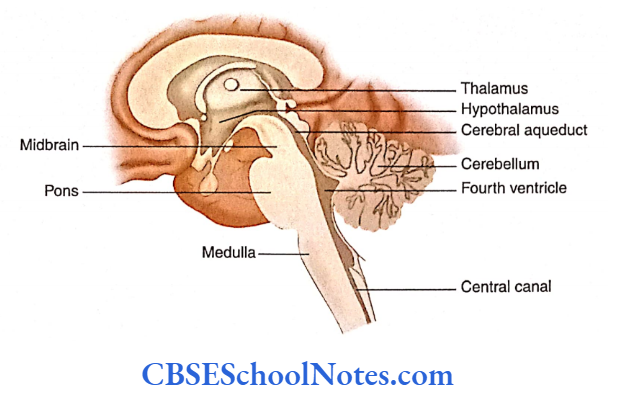
The brainstem consists of the medulla oblongata, pons and midbrain. Superiorly, the brainstem is continuous with the structures forming the forebrain— thalamus, hypothalamus and cerebral hemispheres. Inferiorly, it is continuous with the spinal cord.
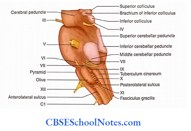
External Features Of Brainstem
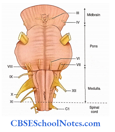
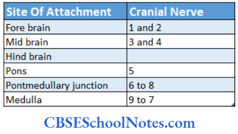
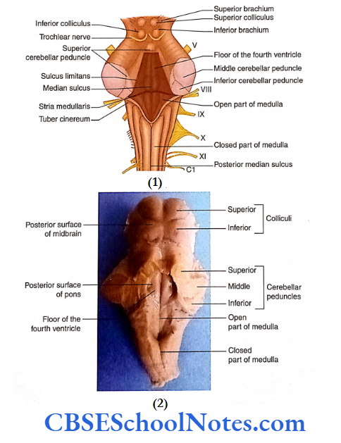
The three parts of the brainstem—midbrain, pons and medulla—are connected to the cerebellum posteriorly with the help of the superior, middle and inferior cerebellar peduncles, respectively.
Posteriorly, the upper parts of the medulla and pons are separated from the cerebellum by the fourth ventricle. The cavity of the fourth ventricle is continuous superiorly with the cerebral aqueduct of the midbrain.
Inferiorly, the cavity of the fourth ventricle is continuous with the central canal of the spinal cord. The brainstem gives attachment to cranial nerves 3 to 7.
Read and Learn More Neuroanatomy
Medulla Oblongata Shape
Cranial nerves 3 and 4 are attached to the midbrain, 5 to the pons, 6 to 8 are attached at the junction of pons and medulla and 9 to 7 are attached to the medulla.
The brainstem gives passage to various ascending and descending tracts connecting the forebrain with the spinal cord.
The brainstem also contains centres for the control of consciousness, respiration and the cardiovascular system (vital centres).
Vital centres control essential autonomic functions such as cardiac activities (cardiac centre), blood pressure (vasomotor centre), respiratory rate (respiratory centre) and vomiting (vomiting centre).
Medulla
The medulla oblongata or simply medulla is about 3 cm long and 2 cm wide. It is conical in shape, broad above and narrow’ below’ It is continuous above with the pons and below with the spinal cord. The medulla consists of two parts:
Lower closed part and Upper open part. The lower closed part contains the central canal, which is in continuation below the central canal of the spinal cord. In the upper open part ofthe medulla, the central canal expands to form the lower part of the cavity of the fourth ventricle.
Medulla External Features
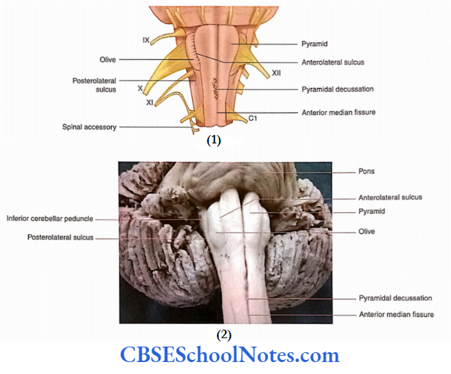
The medulla presents anterior (ventral) and posterior (dorsal) surfaces. On the anterior aspect, there is the presence of the anterior median fissure. This fissure is continuous below the anterior median fissure of the spinal cord.
Medulla Oblongata Anatomy
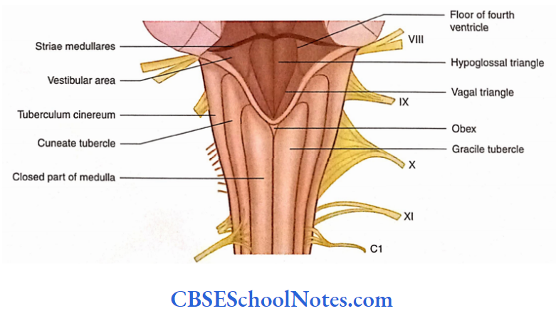
The posterior surface of the closed part of the medulla shows the presence of a midline sulcus known as the posterior median sulcus. This sulcus is present only in the closed part and is an upward continuation of the posterior median sulcus of the spinal cord.
Medulla Anterior Aspect
- On either side of the anterior median fissure, a swelling or bulge is present which is called a pyramid.
- The pyramid consists of corticospinal fibres. In the lower part of the medulla, most of these pyramidal fibres cross to the opposite side and this crossing constitutes pyramidal decussation.
- Lateral to the pyramid, an oval swelling is present which is called the olive. This bulge is due to the presence of the underlying inferior olivary nucleus.
- Between the pyramid and olive, a sulcus is present, known as anterolateral sulcus. This sulcus gives attachment to the rootlets of the hypoglossal nerve.
- Similar to the anterolateral sulcus, another sulcus is present posterolateral to the olive, known as posterolateral sulcus.
- This sulcus is situated between the olive and the inferior cerebellar peduncle. The posterolateral sulcus gives attachment to 9, 10 and 11 cranial nerves.
Medulla Oblongata Anatomy
Medulla Posterior Aspect
- The posterior surface of the lower part (closed part) of the medulla lies between the posteromedian and posterolateral sulci.
- This surface is in upward continuation of the fasciculi gracilis and cuneatus of the spinal cord and presents elevations, the gracile and cuneate tubercles on either side of the posteromedian sulcus.
- Beneath the gracile and cuneate tubercles, the corresponding gracile and cuneate nuclei are present.
- Between the fasciculus cuneatus and rootlets of the accessory nerve, a swelling may be seen which is referred to as the tuberculum cinereum.
- The posterior surface of the upper part of the medulla contributes to the lower part of the floor of the fourth ventricle.
Medulla Internal Structure
To study the internal structure of the medulla, transverse sections (TS) are studied at the following levels:
- TS of the medulla at the level of the pyramidal decussation
- TS of the medulla at the level of sensory decussation.
- TS of the medulla at the level of olives.
Transverse Section of Medulla at the Level of Pyramidal Decussation
This level of pyramidal decussation is the transitional zone between the spinal cord and the medulla. An extensive rearrangement of grey and white matter is observed at this level
Medulla Oblongata Diagram
White Matter at the Level of Pyramidal Decussation
- At this level, a majority (about 75-95%) of fibres constituting the pyramid cross the median plane in front of the central canal.
- After crossing the median plane, the pyramidal fibres pass backwards and laterally, to reach the lateral white column (funiculus) of the spinal cord and form the lateral corticospinal tract.
- The remaining 25% of the pyramidal fibres remain uncrossed and descend below in the anterior white funiculus as the anterior corticospinal tract
- The gracile and cuneate fasciculi start terminating in the caudal end of the gracile and cuneate nuclei, respectively.
- The posterior and anterior spinocerebellar tracts are similar to those of the spinal cord.
Nuclei (Grey Matter] at the Level of Pyramidal Decussation
Medulla Oblongata Diagram
- Because of the pyramidal decussation, the central canal and central grey matter are displaced more posteriorly.
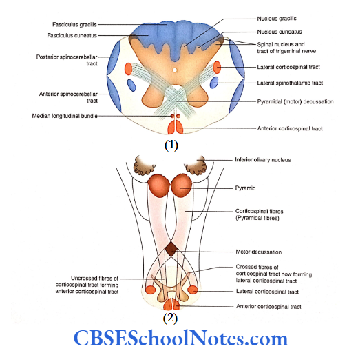
- Due to the passage of decussating pyramidal fibres, the anterior horn gets detached from the central grey matter. This detached part forms the spinal nucleus of the accessory nerve.
- The caudal end of the nucleus of the spinal tract of the trigeminal nerve and its tract are seen at this level. Below this, the nucleus becomes continuous with the substantia gelatinosa of the spinal cord (dorsal grey horn).
- Nuclei gracilis and cuneatus start appearing at this level and lie within the substance of fasciculi gracilis and cuneatus, respectively. These nuclei appear as the posterior extension of the central grey matter.
Transverse Section of Medulla at the Level of Sensory Decussation
This level of medulla is situated just above the level of pyramidal decussation in the rostral portion of the closed part. Extensive changes are seen in the arrangement of grey and white matter at this level.
Medulla Oblongata Diagram
Arrangement of Grey Matter
- The readers are requested to read the following discussion.
- Nuclei gracilis and cuneatus: These nuclei, which had started appearing at an earlier level, are now large.
- They lie deep to fasciculi gracilis and cuneatus, respectively. At this level, nuclei gracilis and cuneatus are seen to have detached from the central grey matter.
- Accessory cuneate: This nucleus is situated just dorsolateral to the nucleus cuneatus. The nucleus of the spinal tract of the trigeminal nerve: It is situated ventrolateral to the nucleus cuneatus.
- Nucleus ambiguus: It is situated in the area of reticular formation medial to the nucleus of the spinal tract of the trigeminal nerve. The lowest part of the inferior olivary nucleus appears at this level posterior to the pyramid.
- The small mass of grey matter lies on the anterior aspect of the pyramid. This is the arcuate nucleus.
- Inside the central grey matter (surrounding the central canal), three nuclei are present:
- Hypoglossal nucleus,
- The dorsal nucleus of the vagus and
- The nucleus of the solitary tract
- Arrangement of White Matter
Functions Of Medulla Oblongata
The white matter is organised in the form of the following fascicles or bundles:
Fasciculi gracilis and cuneatus: These fasciculi are situated posterior to nuclei gracilis and cuneatus.
The second-order neuron fibres arise from gracile and cuneate nuclei and cross to the opposite side in front of the central grey matter. These fibres are known as internal arcuate fibres.
Sensory decussation: The internal arcuate fibres of two sides decussate in the median plane. This is called sensory decussation.
After decussation, these fibres run upwards close to the median plane as the medial lemniscus. The fibres constituting the medial lemniscus terminate in the thalamus.
Medial longitudinal bundle: This bundle lies posterior to the medial lemniscus and anterior to the hypoglossal nucleus. It consists of ascending and descending fibres connecting various cranial nerve nuclei (3, 4, 6 and 8).
Spinal tract of the trigeminal nerve: This tract lies superficial to the nucleus of the spinal tract of the trigeminal nerve on the surface of the medulla.
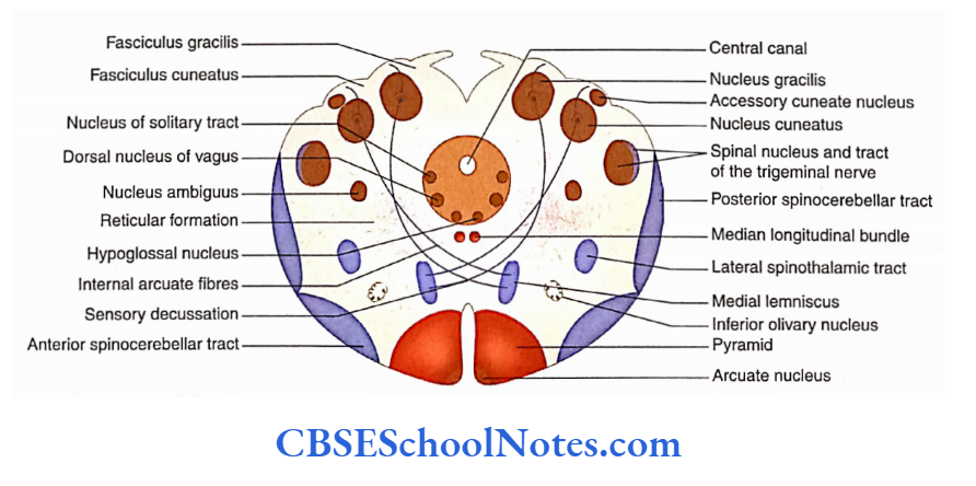
Anterior spinocerebellar and posterior spinocerebellar tracts: These tracts are situated in the ventrolateral area of the medulla, superficially.
Reticular formation: This consists of a network of scattered nerve cells and nerve fibres. The reticular formation is present throughout the brainstem.
Functions Of Medulla Oblongata
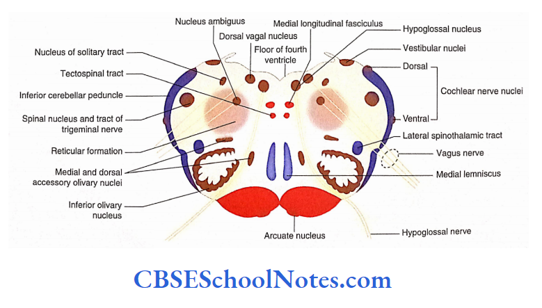
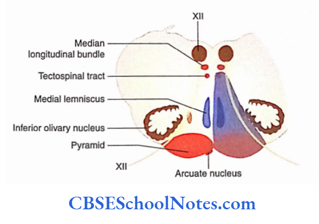
Transverse Section of Medulla at the Level of Olive
- This level of the transverse section of the medulla passes ventrally through olives and dorsally through the lower part of the fourth ventricle.
- The arrangement of grey and white matter shows many changes. The most striking change is that the central canal disappeared while the fourth ventricle is seen.
- The central grey matter now spreads over the ventricular floor.
Arrangement of Grey Matter
The total amount of grey matter has increased at this level because of the appearance of many new nuclei.
Hypoglossal nucleus, dorsal vagal nucleus and nucleus solitaries have changed their position due to the appearance of the fourth ventricle.
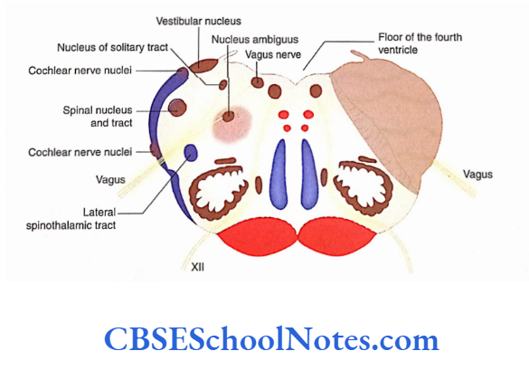
Vestibular nuclei: Lateral to these nuclei, the cauda ends of vestibular nuclei are seen.
Cochlear nuclei: The ventral and dorsal cochlear nuclei are situated on the ventral and dorsal aspects of the inferior cerebellar peduncle, respectively ends vestibular nuclei are seen.
Cochlear nuclei: The ventral and dorsal cochlear nuclei are situated on the ventral and dorsal aspects of the inferior cerebellar peduncle, and respectively nucleus of the trigeminal nerve and its tract are situated ventromedial to the inferior cerebellar peduncle.
Nucleus ambiguus: The nucleus ambiguus is situated in the area of reticular formation.
Olivary nuclei: The ventrolateral area of the section is occupied by the olivary nuclei.
Arcuate nucleus: This is seen on the anteromedial aspect of the pyramid.
Functions Of Medulla Oblongata
Arrangement of White Matter
Inferior cerebellar peduncle: This peduncle is situated in the posterolateral corner of the section. It is mainly formed by the fibres of the dorsal spinocerebellar tract and olivocerebellar tract.
Ventral spinocerebellar tract: The ventral spinocerebellar tract is situated near the surface, ventral to the inferior cerebellar peduncle.
Ascending tracts: Ascending tracts such as anterior spinothalamic, lateral spinothalamic and spinotectal tracts are deeply placed.
In the same area, descending tracts lie, for example tectospinal, rubrospinal, reticulospinal and vestibulospinal.
Pyramids: They occupy the same anterior part of the medulla on either side of the anterior median fissure.
Medial lemniscus, medial longitudinal fasciculus and tectospinal tract: Posterior to the pyramid and in the para-median position, the medial lemniscus, tectospinal tract and medial longitudinal fasciculus are placed in the anteroposterior direction.
Reticular formation: It is present in the ventrolateral part of the transverse section (same place as seen at the level of sensory decussation).
Lesions of the medulla may result due to a variety of causes, i.e. injury, congenital anomalies, raised pressure in the posterior cranial fossa usually due to tumour, demyelinating diseases and vascular lesions. The vascular lesions are the most common.
Injury to Medulla
- The medulla may be injured due to a hard blow on the back of the head or upper neck.
- This kind of injury is usually fatal because the medulla contains vital centres such as respiratory and cardiovascular. Damage to the respiratory centres can rapidly lead to death.
- If the injury is nonfatal, it may affect the cranial nerves that take origin from the medulla. This may affect the function of the cranial nerves on the same side of the injury.
- As the tracts are closely packed in the medulla, a nonfatal injury may produce the lesion of these tracts.
- This may result in paralysis of the muscles on the opposite side (due to damage to the corticospinal tract) and loss of sensation on the opposite side (due to damage to ascending sensory tracts).
Vascular Lesions of Medulla
- Vascular lesions are the most frequently seen along with the lesions of the medulla. The vascular lesion may occur due to thrombosis or haemorrhage.
- Haemorrhage in the medulla is serious because escaping blood destroys the vital centres in the reticular formation, i.e. centres controlling functions of respiration, circulation and consciousness.
- However, thrombosis causes a smaller (localised) lesion in the medulla, and the signs and symptoms of these lesions help to locate the site of destruction.
- Two well-known syndromes, resulting from the occlusion of medullary vessels, are medial medullary syndrome and lateral medullary syndrome.
Brainstem Medulla Oblongata Summary
- The brainstem consists of the medulla, pons and midbrain. It is continuous above the forebrain and below the spinal cord.
- The brainstem is connected posteriorly to the cerebellum with the help of superior, middle and inferior cerebellar peduncles.
- The medulla, pons and cerebellum are collectively known as hindbrain.
- The brainstem gives attachment to cranial nerves 3 to 8.
- The medulla is conical in shape, and related anteriorly to the basilar part of the occipital bone is the posterior cranial fossa.
- As indicated the medulla is divided into an upper open part and a lower closed part.
- The ventral surface shows the presence of anterior median fissure, pyramid, anterolateral sulcus, olive and posterolateral sulcus.
- The anterolateral and posterolateral sulci give attachments to cranial nerves 9 to 12.
- The dorsal surface of the lower (closed) part of the medulla shows the presence of posteromedian sulcus, gracile tubercle, cuneate tubercle and posterolateral sulcus.
- The transverse section at the level of pyramidal decussation resembles the spinal cord. The most striking feature is
pyramidal decussation and appearance of the lateral corticospinal tract. - The dorsal grey horn of the spinal cord is replaced by the nucleus of the spinal tract of the trigeminal nerve.
- The caudal ends of gracile and cuneatus nuclei start appearing in the posterior grey column.
- The transverse section at the level of sensory decussation shows three nuclei in the central grey matter: hypoglossal, dorsal vagal and nucleus of the solitary tract.
- Nuclei gracilis and cuneatus are now a separate mass of grey matter on the posterior aspect of the section.
- The nucleus of the spinal tract of the trigeminal is situated in the lateral part of the sections. The nucleus ambiguus is situated in the area of the reticular formation.
- The fibres arising from gracile and cuneate nuclei are known as internal arcuate fibres.
- The internal arcuate fibres of two sides decussate in the median plane, which is known as sensory decussation. After decussation, these fibres form the medial lemniscus.
- The medial longitudinal bundle is located anterior to the hypoglossal nucleus. In the anterior area of the section are pyramids on either side of the anterior median fissure containing corticospinal fibres.
- The anterior and posterior spinocerebellar tracts are situated in the ventrolateral area of the medulla near the surface.
- The section passing through the open part of the medulla (lower part of the fourth ventricle) shows the most striking change; that is, instead of the central canal, the fourth ventricle is seen.
- Due to the opening of the central canal, the position of the hypoglossal nucleus, vagal nucleus and nucleus solitarius has changed.
- Note the location of vestibular and cochlear nuclei in the lateral part of the section. The nucleus ambiguus and spinal nucleus of the trigeminal have maintained the same position as in the lower section.
- The olivary nuclear complex (inferior, medial and dorsal olivary nuclei) is located in the ventrolateral area of the
section. - The arrangement of white matter is almost the same as at a lower level. The appearance of the inferior cerebellar peduncle is a new feature at this level.
- Lesions of the medulla may result due to a variety of causes: injury, congenital anomaly and vascular lesions.