
Bone Tissue
Bone like cartilage, is also a specialized type of connective tissue. Similar to all other connective tissues, it also consists of ground substances, fibres and cells.
- However, the bone is classified as specialized connective tissue because of the presence of minerals (calcium salts) in its intercellular matrix.
- The presence of calcium salts makes bone tissue hard, which is suited for providing support and protection to the vital organs(lungs, heart and brain).
- The hardness of bone is responsible for locomotion as it forms a skeletal framework and also provides attachment to muscles on its surface.
- Although bone is the hardest tissue of the body, it constantly changes shape about stress is applied.
- If pressure is applied to the bone, it leads to resorption, whereas tension applied to it results in the formation of new bone. These facts are used by orthodontists to treat malocclusion of teeth.
Before we study the histological structure of the bone, it would be useful to understand the gross structure and composition of bone.
Bone Tissue Remember
Bone is a specialized connective tissue. It is special because its intercellular matrix is mineralized making it hard. The hardness of the bone enables it to perform various functions associated with it.
The Macroscopic(Gross) Structure Of A Long Bone
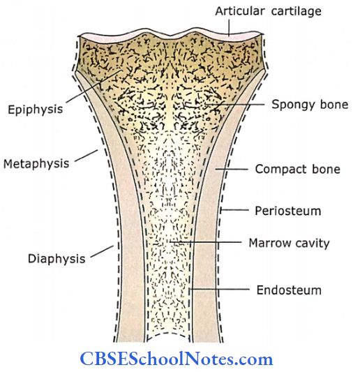
A longitudinal section of a long bone consists of two knobby ends(epiphyses), joined by a long shaft(diaphyses). The expanded portion of the bone between epiphysis and diaphysis is called metaphysis.
- Diaphysis consists of a thick wall of dense bone in which no spaces are visible to the naked eye exi amination; hence, called compact bone. Diaphysis encloses a central cavity known as the marrow cavity.
- Both the ends of a long bone(epiphyses) are covered by a thin layer of compact bone and filled internally by the meshwork of thin and small rods and curved plates.
- This meshwork looks like a sponge; therefore, this kind of bone is called spongy or trabecular bone. The articular areas of epiphyses(which are in contact with another bone to form a joint) are covered by hyaline cartilage(articular cartilage) in living bone.
- Articular cartilage provides a smooth area to facilitate the movements between two bones forming a joint.
The entire outer surface of bone, except the area covered by articular cartilage, is covered by a connective tissue membrane called periosteum.
Similarly, the marrow cavity and spaces of spongy bone are also lined with a membranous layer called an endosteum. The marrow cavity and spaces of spongy bone are filled by bone marrow, which is highly vascular tissue.
- In adults, bone marrow tissues are of two different kinds, i.e., yellow marrow and red marrow. The red marrow is present at the ends of the bone and is involved in the formation of blood cells, while yellow marrow is in the shaft of long bone and is predominantly made UP of adipose tissue.
- From the above description, it is evident that bone tissue can be macroscopically classified into two distinct types, i.e., compact and spongy.
- Students should note that the above description is of a living bone (bone present in a living person). The bones, which are handled in the classroom, are dry and devoid of many structural components of a living bone.
- For example, a dry bone is not covered by hyaline cartilage at its epiphyseal ends. Similarly, it is also devoid of periosteum, endosteum, bone marrow, blood vessels and nerves.
Bone is not only a living tissue but it is also a dynamic tissue. It is continuously engaged in building new bone and breaking down old bone.
- Each living bone is not just a bone tissue, but somewhat similar to an organ. It is evident by the fact that a bone consists of not only bone.
- Tissue proper also consists of many other tissues like fibrous membranes(periosteum and endosteum), cartilage (articular cartilage), bone marrow (adi pose and haemopoietic tissues), nerves and blood vessels.
- Similar to any other organ of the body, bone is also involved in various functional activities(locomotion, support and protection of delicate organs, formation of blood and storage of calcium).
Composition Of Bone Tissue
Like any other connective tissue, bone tissue is also comprised of the following three basic components: cells, fibres and ground substance. In addition to this, the intercellular matrix(fibres and ground substance) of bone tissue is mineralized.
1. Cells
There are four types of cells in bone tissue, i.e., osteogenic cells, osteoblasts, osteocytes and osteoclasts.
Osteogenic Cells
These cells are present in the cellular layer of the periosteum, endosteum and Haversian canals(see below). These are stem cells which, after cell division, give origin to osteoblasts. These cells are derived from embryonic mesenchymal cells.
Osteoblasts
These are bone-forming cells. They synthesize and secrete matrix(collagen fibres and ground substance). They are also responsible for the calcification of the matrix.
- Thus, an osteoblast has a well-developed rough endoplasmic reticulum, Golgi complex and mitochondria that are needed for synthesis and secretion of matrix.
- These cells also possess receptors for parathyroid hormones. During active bone formation, osteoblasts secrete a high level of alkaline phosphatase, thus its level increases in blood. By measuring blood alkaline phosphate levels, one can monitor bone formation.
Osteocytes
These are the main cells of bone tissue. They are formed from osteoblasts that have become entrapped in matrix secretion at the time of formation of new bone.
- They occupy lacunae within the matrix and send cytoplasmic processes into the canaliculi, where they form gap junctions with the processes of adjacent osteocytes.
- The cytoplasm and nucleus of an osteocyte show the features of a resting cell. Osteocytes not only play a role in the maintenance of the surrounding matrix.
- But they also respond to various pressures and tensions applied to bone. They release osteocalcin and insulin-like growth factors, which help in the remodelling of bones.
Osteoclasts
These cells are involved in bone resorption. They are huge cells containing up to 50 nuclei and measuring about 50-150 pm in diameter.
- The cytoplasm shows many lysosomes containing acid phosphatase. The osteoclast’s plasma membrane shows deep foldings(ruffled border) towards the site that comes in contact with bone.
- Here, the cell releases powerful lysosomal enzymes and acids that help in the destruction of the mineralized matrix. As a result, a shallow depression (Howship’s lacuna) can be observed in the bone immediately below the osteoclast.
- It is considered that osteoclasts arise from the fusion of many monocyte cells in the blood. In both origin and function, osteoclasts are closely related to macrophages.
- However, according to new evidence, osteoclasts have bone marrow precursors in common with monocytes termed mononuclear phagocyte system.
Cells Remember
- Bone Cells: Bone tissue consists of four different types of cells, i.e., osteogenic cells, osteoblasts, osteocytes and osteoclasts.
- Osteocytes are mature bone cells derived from osteoblasts that have become entrapped in matrix secretion at the time of formation of new bone.
- Osteoclasts are multinucleated large phagocytic cells that play a role in bone resorption.
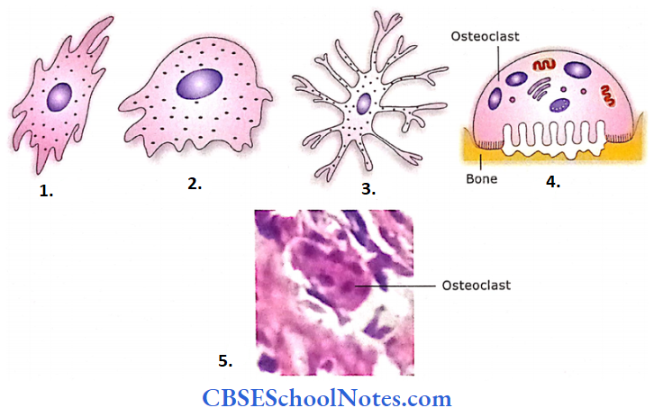
- Osteogenic cell
- Osteoblast
- Osteocyte
- Osteoclast
- Photograph of a multinucleated osteoclast
2. Fibers
Bone consists of type 1 collagen fibres that are synthesized by osteoblasts. These fibres are embedded in the ground substance. The collagen fibres are responsible for providing tensile strength to the bone.
- The collagen fibres within a lamella (see below) are oriented parallel to each other, but the fibres in one lamella are at an angle to those in an adjacent lamella.
- Although the major structural component of bone matrix is type 1 collagen, to a lesser extent type 5 collagen is also found.
3. Ground Substance
It consists of a small amount of amorphous ground substance rich in proteoglycans. The proteoglycans of bone compared with the cartilage-have shorter core protein and fewer side chains.
- The glycosaminoglycans are of three different types, i.e., hyaluronic acid, keratan sulphate and chondroitin sulphate.
- Several multiadhesive glycoproteins are also present in the bone matrix. these are osteocalcin ( this captures calcium from circulation and stimulates osteoclasts in bone remodelling).
- Osteonectin osteopontin and sialoprotein 1 and 2, are responsible for the attachment of collagen fibres to mineralized ground substances.
The ground substance and fibres form the organic part of the matrix, 90% of which is fibres and 10% ground substance. The inorganic component in the bone matrix consists of mineral salts.
Minerals
The principal constituent of the inorganic matrix is crystals of calcium phosphates. However, it also contains calcium carbonate, calcium fluoride, citrate, magnesium and sodium.
Most of the bone minerals are in the form of rod-like crystals that are arranged along the length of collagen fibres. A layer of water and ions surrounds each crystal.
Further Details
In bone, the matrix consists of about 25% water, 25% collagen fibres and 50% mineral salts. The hardness of bone is due to crystallized inorganic mineral salts which are responsible for the compressive strength of bone.
- The flexibility of bone depends upon the presence of collagen fibres and is also responsible for its tensile strength.
- If a long and thin bone-like fibula is treated with a weak acid the inorganic mineral salts will be removed from the bone but collagen fibres will remain.
- The bone thus will lose its rigidity and will become so flexible that it can be tied into a knot.
- On the other hand, burning fibula will destroy the organic matrix (collagen fibres) but mineral salts will remain in the bone and thus the shape of the bone will be maintained.
- This bone will become as brittle as chalk. Thus bony hardness is due to inorganic salts while strength and flexibility are due to collagen fibers.
Ground Substance Remember
The predominant organic component of bone matrix is type 1 collagen, while inorganic components are crystals of calcium hydroxyapatite.
Microscopic Structure Of Bone
Bone tissue is made up of lamellae. It would be useful to understand the lamellar organization of bone before we study the histology of compact and spongy bone.
Lamellar Organization of Bone
As stated earlier, on naked eye examination, two different types of bones are observed, i.e., spongy and compact. Both these types of bones are made up of bone tissue layers or lamellae.
- A lamella(layer) is the basic unit of adult bone. It would be useful to understand the lamellar organization of bone before we study the histology of compact and spongy bone.
- A lamella is a thin plate of bone and is made up of collagen fibres and mineral salts embedded in a gelatinous ground substance. These lamellae are arranged one upon another.
- The orientation of collagen fibres in each lamella is parallel to each other, but the collagen fibres in adjacent lamellae are oriented almost at a right angle to one another.
Small spaces are seen between adjacent lamellae that are called lacunae. Each lacuna is occupied by an osteocyte.
- The adjacent lacunae are interconnected with one another and with a central canal with the help of numerous canaliculi that radiate from each lacuna.
- Each lacuna is present between two adjacent lamellae, but the canaliculi travel through lamellae. Canaliculi arc minute canals that contain cytoplasmic processes of osteocytes and are filled with extracellular fluid.
- This lamellar arrangement Of bone is found in both spongy and compact bones. Even a small spicule or trabeculae of spongy bone consists of several lamellae placed over one another.
Microscopic Structure Of Bone Remember
Bone tissue (both compact and spongy) is made up of lamellae. A lamella is a thin plate (layer) of bone made up of collagen fibres and mineral salts embedded in a gelatinous ground substance.
These lamellae are arranged one upon the other. A lamella (layer) is the basic unit of adult bone.
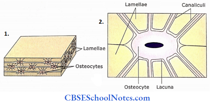
- The bone lamellae are arranged one upon another. Osteocytes are present in lacunae and send their processes to canaliculi.
- Enlarged view of lacuna, osteocytes and canaliculi. The lacuna is present between two adjacent lamellae.
Structure of Spongy Bone
Spongy bone tissue is present at the epiphyses of long bones. A11 short flat and irregular bones also consist of spongy bones. Similarly, a thin rim around the marrow cavity of the diaphyses of long bone consists of spongy bone.
- On naked eye observation, a spongy or cancellous bone is made of a three-dimensional meshwork of trabeculae or spicules. These trabeculae are made up of inter-anastomosing.
- Thin, curved plates and rods. Between bone tissues (plates and rods) numerous interconnecting spaces are filled with bone marrow.
- If we examine the cross-section of a trabecular rod or P’ate under a microscope, it appears to be made of several lamellae placed over one another.
Between these lamellae are small spaces(lacunae) containing osteocytes. Radiating from these lacunae are canaliculi, which are occupied by the processes of osteocytes.
- The osteocytes situated in lacunae derive their nutrition through the canalicular system from blood vessels present in the bone marrow.
- As trabecular rods and plates are not more than 0.4 mm in thickness, there is no need to have blood vessels within bone tissue. (However, in the case of compact bone, blood vessels are present within the bone tissue, i.e., in the Haversian canal.)
Structure of Spongy Bone Remember
Although the spongy bone also consists of lamellae, these lamellae are not organized in the form of Haversian canal systems (osteons).
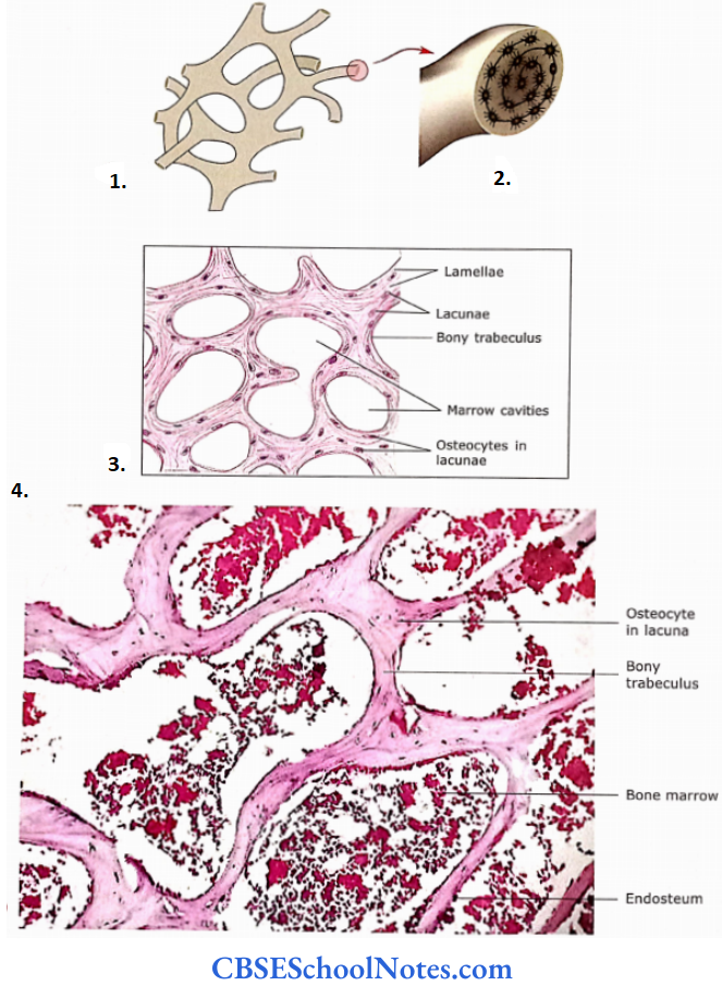
- Spongy bone trabeculae.
- Section of a trabecula.
- A section of spongy bone.
- Section of spongy (cancellous) bone showing network of bony trabeculae.
Structure of Compact Bone
Compact bone forms the bulk of the diaphysis of long bones. It also forms a thin layer on the external surface of all other bones in which the core is made up of spongy bone.
- For Example., short, flat and irregular bones. This bone is called as compact because no space is visible on naked eye examination.
- On its outer and inner surface, it is covered by periosteum and endosteum, respectively.
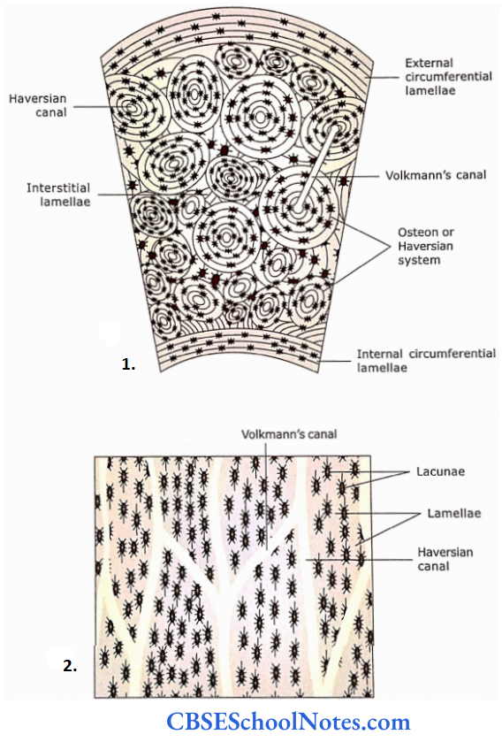
The compact bone is also made up of lamellar bones. However, here lamellae are present in three different patterns:
- Haversian system of lamellae
- Interstitial lamellae
- Circumferential lamellae
Haversian System of Lamellae
- It is also known as osteon.
- In this system, 4-15 concentric lamellae are arranged around a central canal that is called as Haversian canal. This canal contains a small amount of loose connective tissue, capillaries, nerves and lymphatics.
- Between the concentric lamellae are lacunae that contain osteocytes.
- The radiating canaliculi from lacunae connect the Haversian canal with all the lacunae present in an osteon.
- This canalicular route facilitates the passage of nutrients and oxygen to reach the osteocytes from the blood capillaries in the Haversian canal.
- The blood vessels and nerves from the periosteum go inside the compact bone through Volkmann’s canal and communicate with Haversian canals.
- Volkmann’s canals not only communicate periosteal vessels with vessels in Haversian canals but also with vessels in adjacent Haversian canals and with vessels in the marrow cavities.
- Volkmann’s canals are usually identified based on two features: firstly, they are horizontally or obliquely placed concerning the long axis of bone and secondly, are not surrounded by concentric lamellae.
- The Haversian system is cylindrical with its long axis parallel to the long axis of bone. This is because osteons in a long bone are oriented in the direction of the line of stress.
- Surrounding each osteon there is a thin layer of mineralized bone matrix called a cement line.
- It is distinctly visible compared with surrounding lamellae due to the absence of collagen fibres in it.
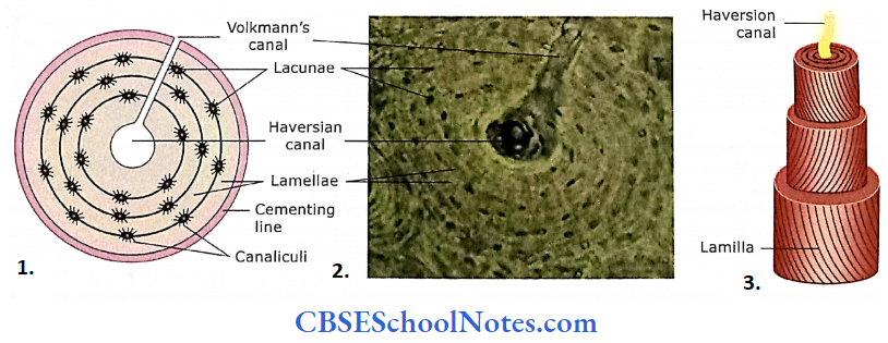
- Schematic diagram.
- As seen in the ground section of a dried compact bone.
- The Haversian canal is surrounded by concentric lamellae.
Interstitial Lamellae
As stated earlier, older bone tissue is constantly being replaced by new bone tissue. Because of this, the fragments of older osteons are seen in areas between osteons.
- These areas also show the lamellar arrangement of bone, i.e., between lamellae are lacunae, which are occupied by osteocytes.
- Radiating from lamellae are canaliculi. These kinds of lamel are called interstitial lamellae.
- Ground section of compact bone (transverse section)
- Ground section of compact bone (longitudinal section).
Circumferential Lamellae
The circumferential lamellae are of two different kinds. The outer circumferential lamellae are present on the outer surface of bone just beneath the periosteum.
They completely encircle the bone. Similarly, the inner circumferential lamellae encircle the marrow cavity.
Structure of Compact Bone Remember
The compact bone consists of four different types of lamellar systems, i.e., outer and inner circumferential, Hav Persian system (osteon) and interstitial lamellae.
Periosteum
Like perichondrium covering the cartilage, the bone on its external surface is also covered with a membrane called periosteum.
- It consists of two layers: an outer fibrous layer made up of collagen fibres and an inner cellular layer made up of cells. These cells in a young developing bone are osteogenic.
- In an adult(developed) bone, the cellular layer is not well developed and may have a few cells called periosteal cells. These cells may convert to osteoblasts when the need arises, i.e., in case of repair of bone after a fracture.
- The blood vessels of the periosteum pass into compact bone, through Volkmann’s canals, to supply the nutrients to the outer layers of bone.
- When a tendon is attached to bone, the collagenous fibres of the tendon after passing through the periosteum continue into the outer layers of bone. These fibres are called as fibres of Sharpey.
Endosteum
The endosteum is the thin lining of the bone that faces the marrow cavity and the spaces of spongy bone. This layer is mostly one cell thick and consists of cells that are concerned with bone formation.
Bone resorption or resting cells. The resting cells are flat or squamous in shape and may change to osteoblasts when the need arises.
Endosteum Remember
The periosteum is made up of two layers, i.e., outer fibrous and inner cellular. The inner cellular layer consists of osteogenic and osteoblast cells.
Endosteum Clinical Applications
Some Important Bone Diseases
Scurvy
As seen certain amino acids and vitamin C are necessary for collagen synthesis. Scurvy is caused by a dietary deficiency of vitamin C.
- This leads to the synthesis of inadequate amounts of normal collagen and organic matrix.
- In a patient suffering from scurvy, spongy bone consists of a reduced number of trabeculae. In the case of compact bone, the cortex of long bone is thinner than normal.
Rickets
This disease is due to inadequate mineralization of bone matrix in young individuals. This is due to an inadequate dietary supply of calcium and phosphorus.
- Vitamin D is needed for the absorption of calcium. Thus, if there is a deficiency of vitamin D, calcium absorption would be affected.
- Most cases of rickets are due to inadequate intake of vitamin D in infancy or childhood. Patients with rickets suffer from the bowing of long bones.
- This is because of loss of rigidity in weight-bearing long bones, as there is inadequate mineralization of bone matrix.
Osteomalacia
This disease is seen in adults and is due to an inadequate supply of minerals or vitamin D. The deformities due to osteomalacia are the same as seen in rickets.
Osteoporosis
Normally a balance is maintained between bone formation and bone resorption. When bone resorption becomes higher than bone formation, the bone becomes thin and unable to resist stress.
- This leads to frequent fractures. This disease is due to poor calcium or phosphorus ratios, which is seen in persons above 50 years of age.
- Osteoporosis is more common in women after menopause. After menopause, there is a reduction in estrogen levels which is considered the cause of osteoporosis.
Osteogenesis Imperfecta
This is a genetic disease. Here, there is a mutation of genes responsible for the synthesis of collagen. Due to inadequate and abnormal collagen, bones become weak and brittle.
- Persons suffering from this disease are prone to frequent fractures. The formation of collagen begins as procollagen molecules.
- Each rope-like procollagen molecule is made up of three chains, two alpha-1 chains and one alpha-2 chain.
- Alpha 1 chain is produced by the gene COL1A1 and alpha 2 chains by COL1A2 gene. The COL1A1 gene is located on the long arm of chromosome 17 and the COL1A2 gene is located on chromosome number 7.
- Mutation of these gene(s) leads to osteogenesis imperfecta. No genetic cure is available at present. Putting metal rods in long bones can prevent fractures of long bones.
Comparison Between Bone And Cartilage

What is the ground section of a bone?
Ground sections of a bone are prepared from dry bones. Thin slices of dried bone are cut with the help of a saw and then further reduced in thickness on a grinding stone.
- A ground specimen must be so thin that light can easily pass through it. These sections are then mounted on glass slides usually unstained.
- As the ground sections of a bone are unstained, it is difficult to observe the details of a section under a microscope. These sections are best observed under minimal light.
- For this, bring the condenser completely down and regulate the light passing through the diaphragm(from completely open to closed position) till you start seeing lacunae, canaliculi and lamellae.
- You should also recollect that a dried bone is devoid of endosteum, periosteum, osteocytes in lacunae and blood vessels and nerves in Haversian canals. Hence, all the above structures will not be seen in a ground section.
All the spaces in a ground section(Haversian canals, lacunae and canaliculi) are filled with dust particles, during the preparation of the section, and therefore appear black.
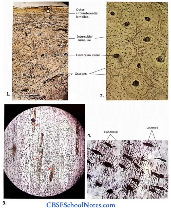
- Photomicrograph of a transverse unstained ground section of the compact bone.
- TS ground section of compact bone(magnified view).
- Longitudinal section of compact bone at low magnification.
- Canaliculi originating from lacunae.