
Genetics Of Malocclusion
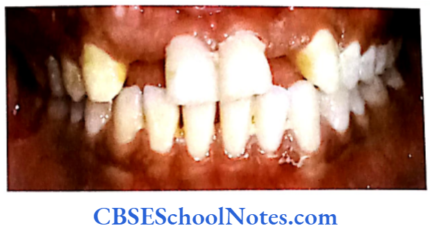
Occlusion means alignment of the upper and lower teeth together. Normally all upper teeth fit slightly over the lower teeth. The upper teeth keep the cheeks and lips from being bitten and the lower teeth protect the tongue. Malocclusion of teeth denotes improper or misalignment of teeth. The maxillary and mandibular teeth are incorrectly positioned with relation to each other. Malocclusion also occurs because of altered relation between the upper and lower jaws.
Classification of Malocclusion
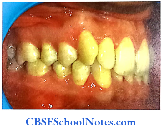
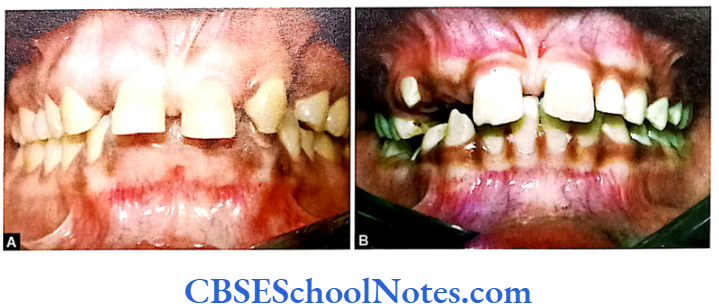
- Class 1 malocclusion is the most common variety of malocclusion. The bite is normal as per the permanent Ist molar relationship but the teeth are crowded or not positioned correctly. The upper teeth slightly overlap the lower teeth.
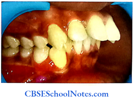
- Class 2 malocclusion is also called as retrognathism. It occurs when the upper jaw/ upper teeth are forwardly placed (lower teeth/ lower jaw are placed distally)
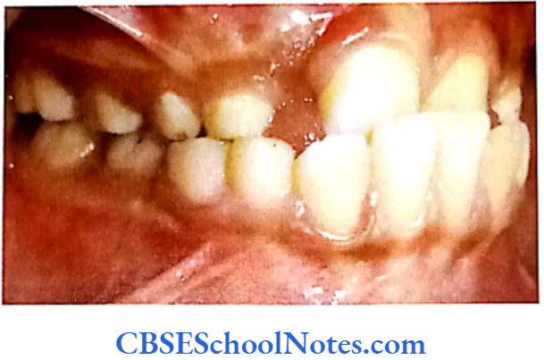
- Class 3 malocclusion is also called prognathism. It occurs when the lower jaw protrudes or just forward causing the lower jaw and teeth to overlap the upper jaw and teeth from beneath the upper jaw.
Malocclusion Causes
Malocclusion is caused due to:
Malocclusion Acquired Factors
- Alteration in the shape or size of jaws; if teeth in a small jaw jostle for space and grow crowded in small area pushing each other.
- Alteration in the shape or size of teeth.
- Tooth loss.
- Thumb or finger sucking, use of pacifier and mouth breathing (due to enlargement of tonsils) and tongue thrusting.
Read and Learn More Genetics in Dentistry Notes
Malocclusion Genetic Factors
- Inherited conditions include:
- Inheritance of too many or too few teeth.
- Inheritance of too much or too little space between the size and shape of upper and lower jaws. These teeth.
- Inheritance of irregular mouth and jaw size and shape.
- Abnormal formations of the jaws and face, e.g. cleft palate.
The etiology of malocclusion is a complex subject and not fully understood. The above brief description of etiology indicates that the bony factors (size and shape of maxillary and mandibular arches) and dental factors (size and shape of teeth, failure of eruption, supernumerary teeth and early loss of teeth) can be determined by both environmental and genetic factors.
For example, the failure of eruption of upper lateral incisor may be due to acquired as well as genetic causes. The presence of supernumerary teeth may lead to the failure of eruption of incisor. Supernumerary tooth again may feature due to an inheritance (as supernumerary tooth may also be present in a parent of the patient).
On the other hand failure of eruption of central incisor may be also due to early loss of many deciduous teeth (due to caries) that leads to forward drift of first permanent molar teeth resulting in the crowding of teeth. Thus it sometimes becomes difficult to assess the clear-cut role of acquired and genetic factors in the causation of malocclusion as there is a complicated interplay between various factors.
Malocclusion may result due to inheritance of disproportionate size of the teeth and jaw, resulting in crowding or spacing of teeth with a small jaw leading to crowding and large jaw giving abnormal spacing between teeth. The other important factor leading to malocclusion is the disproportion between skeletal variables are primarily genetic in nature.
It should be noted that genetic influence on the shape and size of jaws is not due to a single gene defect but is mostly determined by the additive effects of many genes (i.e. polygenic in nature). The environmental factors play an influential role on shaping of the genetic factors. Thus, the etiology of malocclusion is mostly multifactorial in nature (due to interaction between genetic and environmental factors).
Though it is simple to analyze the genetics of a single gene (Mendelian trait), analysis of multifactorial traits (cleft palate, cleft lip, caries, periodontitis and malocclusion) are difficult to analyze. For the analysis of the multifactorial inheritance one has to evaluate the role of genetic as well as environmental factors and the interaction between the two. The time-tested methods to analyze the role of genetic and environmental factors for a particular multifactorial disease in humans are familial and twin studies.
Family And Twin Studies
In familial studies one has to observe the similarities as well as differences between the mother and the child, father and the child and between sibling pairs. Correlation coefficients of the trait are obtained between parents and offspring or between sibling pairs and half sibs. For most measurements of facial skeletal dimensions correlation coefficients for parent-child pairs are about 0.5 which is the upper limit of correlation between first-degree relatives.
The correlations for parent-child in relation the dental char- acteristics range from 0.5 to 0.15. This is also reflected in the Harris and Johnson (1991) study which revealed high heritability of craniofacial (skeletal) characteris- tics and low heritability of dental characteristics. This indicated that malocclusion resulted mainly from facial skeleton deformities that could be inherited while pure dental variations were due to environmen- tal factors. Many other family studies have indicated the role of heredity in determination of the craniofa- cial and dental morphology (Korkhaus, 1930; Rubbrecht, 1930; Trauner, 1968 and Peck et al, 1998).
As stated earlier twin studies are useful in identifying the genetic and environmental factors determining multifactorial traits. Differences in features between monozygotic twin pairs implicate environmental factors while similarities points towards genetic influences as the primary causal factors of the disease. The similarities in disease features between dizygotic twin pairs are thought to result from environmental influences and genetic factors.
A comparison of the differences observed within twin pairs in the two categories should provide a measure of the degree to which monozygotic twins are more alike to each other than dizygotic twins between themselves in a pair. Studies with twins reared apart are more useful as compared to twins reared together in a common environment as these studies overcome the problems of twins displaying similarities because of being reared in a common environment.
Following is a brief review of family and twin studies in relation to the heritability of craniofacial and dental characteristics. Most of the familial and twin studies indicated that heredity plays a significant role in the etiology of malocclusion.
Lundstrom (1948) reported that characteristics like width and length of dental arch, height of palate, spacing and crowding of teeth, tooth size and degree of overbite are genetically determined. A study on triplets by Kraus et al (1959) investigated the cephalometric parameters and concluded that the morphology of craniofacial bones are under strict genetic control.
Nevertheless, environment plays a major role in determining how these bony elements combine with each other to produce normal occlusion or malocclusion. This observation explains why sometimes differences are observed between a pair of monozygotic twins in spite of having the same genetic constitution.
Markovic (1992) conducted a cephalo- metric twin study and concluded that 100% of monozygotic twin pairs demonstrated concordance for malocclusion while 90% of dizygotic twin pairs were discordant. This is strong evidence in support of genetic etiology of malocclusion.
Schulze and Weise (1965) reported the polygenic nature of mandibular prognathism (class 3 malocclusion). They found that the concordance in monozgotic twin pairs was very high as compared to dizygotic twins. Litton et al (1970) also investigated the poly- genic nature of class 3 malocclusion (mandibular prognathism). The Harris (1975) study indicated that craniofacial skeletal patterns of children with class 2 malocclusion were heritable. In a large familial group Nakasima et al (1982) studied the inheritance of class II and class 3 malocclusion.
High correlation coefficient values were seen between parents and their off- spring in the class 2 and class 3 groups. Thus, there appears to be a strong familial tendency in the development of class 2 and class 3 malocclusions. They concluded that the hereditary pattern must be taken into consideration in the diagnosis and treatment of patients with these classes of malocclusion. Environmental factors have also been suggested as contributory to the development of mandibular prognathism.
Among these are enlarged tonsils (Angle, 1907), nasal blockage (Davidov et al, 1961), congenital anatomic defects (Monteleone and Davigneaud, 1963), hormonal disturbances (Pascoe et al, 1960), endocrine imbalances (Downs, 1928), posture (Gold, 1949) and trauma/disease including premature loss of the first permanent molars (Gold, 1949).
Development of both the maxillary and mandibular arch shapes in an Australian twin study was found to be under genetic influence. However, authors (Richards et al, 1990) also have reported the existence of some independent variables that determine the shape of mandibular and maxillary archs. Hughes et al (2001) quantified the extent of variations in different occlusal features in Australian children of European descent with complete primary dentitions but no permanent teeth present in the mouth.
Occlusal traits including interdental spacing, incisoral overbite and overjet, arch breadth and arch depth were studied. Estimates for overbite and overjet were 0.53 and 0.28 respectively and estimates for arch dimensions ranged from 0.69 to 0.89. These results indicated a moderate to relatively high genetic contribution to the observed variations.
The aim of a study by Eguchi et al (2004) was to quantify the relative contributions of genetic and environmental factors to variations in dental arch breadth, length and palatal height in a sample of Australian twins. The study brought forth information that the heritability load for dental arch breadth ranged from 0.49 to 0.92, those for the arch length from 0.86 to 0.94 and those for palatal height were 0.80 and 0.81 respectively. These results indicate a high genetic contribution to the variation in dental arch dimensions in mainly teenage twins.
A twin study by Corruccini et al (1980) indicated the importance of environmental factors in the etiology of malocclusion. The researchers showed that heritability of overjet is almost zero. The study on Australian twins conducted by Townsend et al (1988) for occlusal variations has shown the importance of environmental influences in shaping overjet.
The cross bite was found to be determined by environmental factors. They also indicated that genetics may perhaps play an important role for overjet, a lesser role in overbite and least involved in determining molar relationship.
The masseter muscle electrical activity and its morphology were analyzed in twin studies and it was found that both its activity and morphology were genetically influenced. These twin studies revealed for the first time that soft tissue function and morphology could also be inherited (Lauweryns et al, 1992 and 1995).
A study by Dempsey et al (1996) on tooth size in Australian twins indicated a relatively strong genetic and environmental influence on the development of the canines and first premolars. The findings of the canine and first premolar mesiodistal dimensions support the evolutionary theory that indicates the presence of dominance variation in morphological features that have been subjected to strong selective pressure in the past.
Twin studies have shown that tooth crown dimensions are strongly determined by heredity (Osborne et al, 1958). The molecular genetics of tooth morphogenesis with the homeostatic MSX1 and MSX2 genes have been linked to stability in dental patterning. It has been reported that inheritance of tooth size fits the polygenic multifactorial threshold model.
Markovic (1982) found a high rate of concordance for hypodontia in monozygous twin pairs while he observed discordance patterns in dizygous twin pairs. This and other previous studies concluded that a single autosomal dominant gene with incomplete penetrance could explain such a mode of transmission.
Niswander and Sugaku (1963) analyzed data from family studies and have suggested that the development of supernumerary teeth, most frequently seen in the premaxillary region, appears to be genetically determined. The etiology of ectopic canines is controversial with divergent opinions regarding its genetic or environmental mechanism of occurrence.
Mossey et al, 1994 indicated existence of an association in inheritance between ectopic maxillary canines and class 2 malocclusion demonstrating the involvement of genetic factors in the inheritance of the trait. The study of Camilleri et al (2008) addressed the hypothesis that genetic factors play a role in the etiology of ectopic maxillary canines. Sixty-three probands were identified and information on the dental status of 395 of their relatives was determined.
Only two of seven pairs of monozygotic twins were concordant for ectopic canines. This is consistent with environmental or epigenetic variables affecting the phenotype. The low concordance rate is consistent with the low penetrance determined by segregation analysis studies further supporting the existence of environmental etiological factors.
Environmental influences during the growth and development of face, jaws and teeth are mainly exerted through pressures and forces acting on these structures. Mossey (1999) states, “it is difficult to determine the precise contribution from hereditary and environmental factors in a particular case.
For example, the simultaneous appearance of proclined maxillary incisors and digit sucking may lead to the assumption that the digit was the sole causative factor, but the effect of the digit may very well be either potentiated or mitigated by other morphological or behavioral features in that particular individual.
A similar argument may apply in cases of mouth breathing where the influence of the habit and associated posture is very much dependent on the genetically determined craniofacial morphology on which it is superimposed, and the reason for the habit developing may well be dependent on the morphology in the first place. These scenarios are classical examples of the interaction of genotype and environment, and ultimately success of treatment will depend on the ability to ascertain the relative contribution of each.”
Linkage Studies
Linkage studies identifying chromosomes or gene(s) responsible for craniofacial and dental variables of malocclusion in humans are very limited.
- The role of X and Y-chromosomes on craniofacial morphology has been investigated by Gorlin et al (1965). X and Y-chromosomes exert growth- promoting effects on human tooth crown size. The X-chromosome appears to regulate mainly enamel thickness while Y-chromosome affects both enamel and the dentine. Amelogenin is a matrix protein secreted by ameloblasts and it is thought to direct the growth of hydroxyapatite crystals. The gene for amelogenin is located on X and Y-chromosomes (Lau et al, 1989). The amelogenin gene is located on the distal portion of short arm of X-chromosome and to the pericentromeric region of Y-chromosome. The gene on the X-chromosome is the predominantly functional one. Its mutation leads to amelogenesis imperfecta. This dental deformity (amelogenesis imperfecta) is associated with malocclusion (vide infra).
- Mutation in the novel enamelin gene (ENAM) is responsible for autosomal recessive amelogenesis imperfecta and localized enamel defects. It was discovered by Hart et al (2003). In this study 20 consanguineous families with amelogenesis imperfecta were identified with probands suggesting an autosomal recessive transmission. Linkage studies indicated the presence of amelogenesis imperfecta gene on chromosome 4q region. The mutation of this gene resulted in generalized hypoplastic amelogenesis imperfecta phenotype and a class 2 open bite malocclusion. A strong association was thus established between amelogenesis imperfecta and malocclusion due to homozygous mutation in ENAM gene.
- Ravassipour et al (2005) conducted an investigation to evaluate the association of the AI enamel defect with craniofacial features characteristic of an open bite malocclusion. Open bite malocclu- sion was found to have occurred in individuals with AI caused by mutations in the AMELX and ENAM genes even though these genes are con- sidered to be predominantly or exclusively expressed in the teeth. The purpose of this investigation was to evaluate the association of the AI enamel defect with craniofacial features characteristic of an open bite malocclusion. Affected AI individuals with cephalometric values meeting criteria of skeletal open bite malocclusion were observed in all major AI types. The pathophysi- ological relationship between AI associated enamel defects and open bite malocclusion re- mains unknown.
- The study by Frazier-Bowers et al (2007) investigated whether the class 3 trait was present in several of the pedigrees affected with AI. Their results suggested that the class III trait factor co-segregated with AI in the experiment population.
- The Klinefelter males (47, XXY) show pronounced mandibular prognathism and reduction of cranial base angle (Brown et al, 1993). Similarly, 45,X females show imbalance of growth in the craniofacial skeleton (Peltomaki et al, 1989). They show retrognathic face with short mandible and flattened cranial base angle. There is a tendency to have a large maxillary overjet and crossbite. The X-chromosome seems to be responsible for altering the growth of cranial base by acting on cartilaginous joint at the base of the skull. This has a direct effect on the shape of the mandible.
- A linkage study for the growth of maxilla in mouse was conducted by Oh et al (2007). They found that the gene(s) regulating the shape of maxillary complex was situated on the mouse chromosome 12 at 44cM.
- A recent study by Fraziers-Bowers et al (2007) identified the chromosomal locus responsible for the class 3 trait. DNA samples were processed and subjected to a genome-wide scan and linkage analysis. The linkage analysis of these families revealed that a region on chromosome 1 was suggestive of linkage with the class 3 trait.
- The growth hormone receptor (GHR) gene was found to be associated with mandibular height in a Chinese population. Zhou et al (2005) evaluated the relationship between craniofacial morphology and single-nucleotide polymorphisms (SNPs) in GHR in a healthy Chinese population. Their results indicate that the GHR gene polymorphism 1526L is associated with mandibular height in the Chinese population.
As far as genetics of malocclusion is concerned, much progress is still awaited. We do not know the exact mechanisms of genetic or environmental inter- action that combine to produce malocclusion. Similarly, interactions between genetic and environmental factors are least understood. A clear understanding of mechanisms involving environmental factors would help in designing therapies along with the manipulation of environmental factors for orthodontic treatment. Multifactorial traits determined by the additive effects of many genes need to be investigated better to find out the exact number and specific locations of genes involved in their etiology.
More precise research tools and methods should be applied to understand aspects of genetics associated with malocclusion. It is hoped that sequencing of human genome and the use of single nucleotide polymorphisms (SNPs) will help to attain the desired levels of understanding.