
Genetics Of Cancer
The term “cancer” is derived from the Greek word for crab. Hippocrates likened the spreading of cancerous tumor to the shape of a crab’s claws. Cancer is a disease characterized by uncontrollable and unwanted growth of body cells due to the loss of their normal regulatory controls.
Majority of cancers manifest in the form of solid tumors. A cancerous tumor is often a collection of many abnormal cells, most of which divide wildly. Cancerous tumors infiltrate neighboring tissues by forcing their way between normal cells and may spread to distant places in the body through blood or lymph vessels.
Characteristics Of Cancer Cells
Cancer cells are different from the normal cells. Cancer is a disorder involving dynamic changes in the structure as well as the function of the cellular genome in the cancer cells. Following changes are observed in the cancer cells:
- Unrestricted cellular proliferation-Cells affected with cancerous changes lose their usual control over growth and division. The unrestrained growth and division of the cancer cells hamper the normal functioning of the body as a whole by disrupting metabolic activity of the organism and also by causing local effects by the growing mass.
- Transformation-Cancerous cells are transformed cells. These abnormal cells are transformed and become independent of factors usually needed for normal cell growth and proliferation.
- Ability to invade-One of the potent properties of cancer cells is their ability to invade from their site of origin into the surrounding healthy tissue.
- Metastasis-Cancer cells characteristically scatter away from their origin and disseminate to distant parts of the body where they seed and proliferate.
- Suppression of apoptosis-The program for normal cell death (apoptosis), that usually operates in a healthy cell is altered and suppressed in cancer cells.
- Angiogenesis-Cancer cells have the ability to induce new vessel formation or neo- vascularization in the tumor mass to facilitate the availability of oxygen and nutrients.
Oncogenes And Tumor Suppressor Genes
Factors causing or aiding cancer can be grouped as environmental and genetic. Certain environmental components account for the occurrence of approximately 80% of all human cancers and are hence preventable.
Read and Learn More Genetics in Dentistry Notes
Factors Responsible for the Causation of Cancer
Cancer may develop either due to environmental as well as genetic factors.
Environmental Factors of Cancer
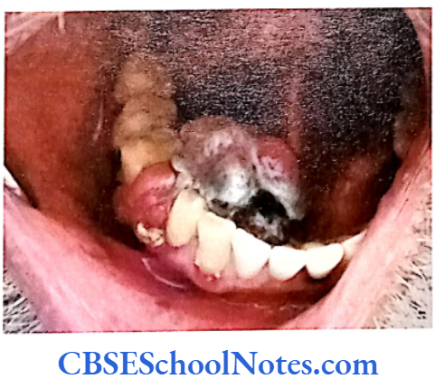
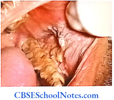
Chemicals: Many chemicals like polycyclic aromatic hydrocarbons (3, 4 benzpyrene), aromatic amines (B-naphthylamine), vinyl chloride and arsenical compounds are known carcinogens and may cause cancers of lung, skin, bladder and liver. Substances like tobacco and alcohol are associated with cancers (squamous cell carcinoma) of the oral cavity.
Radiations: Similarly ultraviolet light (exposure to sunlight) is proven to be carcinogenic for skin (malignant melanoma and basal cell carcinoma) in fair skin people. High dose of ionizing radiation is well known carcinogen especially in people working with radioactive materials. Ionizing radiation is responsible for leukemia and cancers of skin, thyroid, bone and breast. Melanomas are common forms of cancer that involve oral mucous membrane in addition to their usual site of occurrence, the skin.
Viral infection: Many viruses are considered strong carcinogenic agents. About 15% of all human cancers are due to viruses. Several human tumors have known viral etiology, e.g. infection with the human papilloma virus (HPV) causes carcinoma of cervix. HPV infection is incriminated in development of squamous cell carcinoma that constitutes about 95% of all oral cancers. Epstein-Barr virus is responsible for formation of Burkitt’s lymphoma and nasopharyngeal carcinoma. This virus is associated with oral lesions called hairy leukoplakia, especially in patients suffering from AIDS. Certain leukoplakias of the oral cavity may be precancerous conditions. Hepatitis C and B virus produce liver cancer and RNA retrovirus leads to T-cell leukemia and lymphoma.
Bacteria and other microorganisms: Cancer may also result from bacterial infection (H. pylori can produce lymphoma and gastric carcinoma), toxins of fungi (aflatoxins can cause cancer of liver) and parasites like schistosoma can cause bladder cancer.
Production of Cancer by Carcinogens
The Production of Cancer by a Carcinogen Involves a Multistep Process
In the first step the presence of a carcinogen causes a lesion in the cell’s genome (in the DNA of the target cell) that leads to the transformation in the cell. In the second step this transformed cell divides repeatedly (clonal proliferation). This uncontrolled cellular proliferation is the main event leading to formation of carcinoma.
Role Of Genes In Cancer
In the third step the clonal proliferation of tumor cells acquire autonomous growth, i.e. they no longer require the stimulation by carcinogen or other intrinsic factors and rapidly proliferate themselves. In still later stages tumor cells acquire the ability to invade the surrounding tissue, metastasize to distance places in the body and induce vascularization of the tumor.
What is Cell Proliferation and How is it Controlled?
Unrestricted cell proliferation is the main characteristic of cancer. Carcinomatous changes in a cell are brought about by disruption in the normal mechanisms that control cellular proliferation and differentiation. Thus in order to understand the dynamics of cancer we need to understand what is cell proliferation and how it is regulated. Normal proliferation, differentiation and growth in cells are sequentially controlled by the following events:
- Binding of growth factors to specific receptors on the cell membrane.
- Activation of growth factor receptors that further activates signal transducing proteins on the inner surface of the plasma membrane.
- An appropriate signal is then transmitted to the nucleus through certain messenger proteins across the cytoplasm.
- DNA transcription is initiated by the activation of transcription factors that bind at specific regions on the genome to activate transcription.
- Cell enters into mitosis after passing through the checkpoints and eventually undergoes nuclear and cytoplasmic division.
The events mentioned above operate under strict genetic control. Abnormal proliferation of ceils may result from mutations that alter the functions of genes governing cellular proliferation. Uncontrolled cellular proliferation can be studied vis-à-vis the mechanisms of normal cellular life cycle.
Role Of Genes In Cancer
Signal Transduction In Cell Proliferation
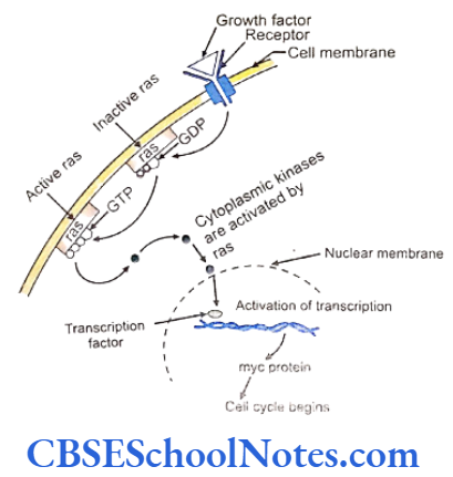
Several of growth factors (GFs) and growth factor receptors (GFRs) exhibit significant role in normal cellular growth and differentiation. Various types of these factors initiate specific course of action. It has been observed that factors like the epidermal growth factor (EGF) stimulates epidermal cells, fibroblast growth factor (FGF) stimulates fibroblasts, platelet derived growth factors (PDGF) stimulates proliferation of connective tissue, etc.
These factors bind to the receptors on the cell membrane in order to exert their action. The sequential activation of successive events through cascading pathways resulting in cellular activity, growth, differentiation or proliferation is termed as signal transduction. Thus extracellular growth factors trigger cellular events through complex pathways. Each of the steps in the pathway is controlled through specific genes and their activity.
Binding of a growth factor to its specific receptor leads to the activation of the receptor.
- A series of cytoplasmic proteins get activated by the receptor in a cascade of reaction. These proteins are called signal transducing proteins. Many of such proteins are present on the inner surface of plasma membranes.
- Two important signaling proteins are produced by the ras and abl genes.
- During the resting state of the cell the ras families of proteins bind to GDP (guanosine diphosphate) constitutively.
- On stimulation by growth factor receptors, inactive ras becomes active by releasing the attached GDP and binding to a new GTP (guanosine triphosphate) molecule.
- The activated ras further turns on cytoplasmic kinases that pass signals to the nucleus for cellular proliferation.
[The life of activated ras proteins is very short. The enzyme guanosine triphosphatase (GTPase) hydrolyzes GTP to GDP and inactivates ras proteins thereby downregulating cytoplasmic kinases. As a result the cell no longer responds to a signal till further activation. The abl gene induces different signal transducer proteins. This gene is located on chromosome 9. - Cytoplasmic kinases enter the nucleus and activate a large number of genes immediately and very early (myc, myb, jun, fos and rel gene). The activity in these genes further regulate transcription of specific DNA segments.
- The myc protein is the one to get frequently bound to specific DNA sites after a cell receives a signal for proliferation.
- The activity of myc proteins induces transcriptional activation of several growth related genes including cyclin D (see later in cell cycle). The quantity of myc protein reduces back to basal levels once the cell enters the cell cycle.
Signal Transduction (Genes And Cancer)
Defective signaling in growth regulating pathways can lead to abnormal growth. Overexpression of growth factors can result in nonneoplastic disorders like psoriasis. Abnormalities at the level of growth factor receptors can lead to conditions like insulin-resistant diabetes (insulin receptor) and dwarfism (fibroblast growth factor receptor). Development of carcinomas, however, involves multiple steps and show other features such as invasion and metastasis.
These multiple steps include unregulated expression of growth factors, receptors or components of other signaling pathways. As discussed in earlier chapters, abnormal expression of components regulating signaling pathways is caused by mutations in the responsible genes. These mutant genes are called oncogenes.
Growth Factors and Cancer
Genes coding for growth factors may acquire oncogenic properties after mutation. For example, the gene coding for PDGF after mutation over expresses the growth factor which give rise to cancers like osteosarcoma and astrocytona.
Growth Factor Receptors and Cancer
Genes coding for growth factor receptors have been found to be mutated in several carcinomatic conditions. Such mutations are believed to induce continuous signals for cell growth and proliferation, even in the absence of growth factors.
Role Of Genes In Cancer
Signal-transducing Proteins and Cancer
The ras genes that produce signal transducing protein are susceptible to mutations. Such mutations are responsible for almost 30% of all human tumors. As a consequence of mutation the enzyme GTPase is unable to hydrolyze the active GTP back to inactive GDP. Thus the ras protein remains constantly active and the cell continues to proliferate. Similarly a mutation in the GTPase protein itself leads to a defective enzymatic action that fails to restrain the activated ras protein. This eventually results in cancer.
Transcription Factor and Cancer
Several early and immediate gene products link the activities of growth factors to other factors that results in transcription of the DNA. The myc gene binds to DNA and activates many transcription factor elaborating genes involved in growth. Mutations in transcription factors with overexpression contribute to sustained proliferation.
Cell Cycle Control
The Cell Cycle
The cell cycle includes all events of cell growth, cell activity, replication of the DNA content and cell division that gives forth the daughter cells. This process is divided into four sequential phases.
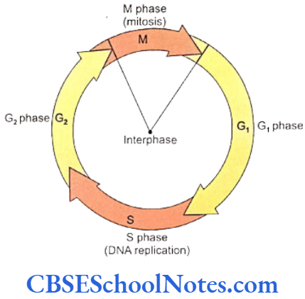
- G1 phase (Gap phase or presynthetic phase) – This phase starts immediately after completion of cell division. The chromosomes gradually become thinned and extended. Cells are responsive to growth signals. They may or may not enter the next cell division depending whether signals are positive or negative with respect to cell division. Cells like neurons that are highly differentiated and lose their capability to divide further are shifted to GO phase. Cells in the GO phase usually subserve their functions and perish after their life is over. These cells may reenter into mainstream G1 phase for replication under special circumstances.
- S phase (Synthetic phase) – It is called S phase as DNA replication or synthesis occurs in this phase.
- G2 phase (premitotic phase) – G2 phase is short where chromosome begins to get condensed in preparation for the cell division phase that comes next. All the above three phases described above constitute the interphase of the cell cycle. Cells usually spend the bulk of their functional lives in the interphase.
- M phase (Mitotic phase) – The M phase results in complete nuclear and cytoplasmic division of a cell into the daughter cells.
Cell Cycle Checkpoints
As cells transit from one phase to the next in the cell cycle, all the events during such transitions are scrutinized and regulated at a number of specific and regular points within the cell cycle. These locations are known as checkpoints. These check- points verify the structural integrity of the genome, ensure that the DNA is free of any breaks and monitor the cellular environment as a prerequisite for a given phase in the cell cycle. The check points of the cell cycle are shown below.
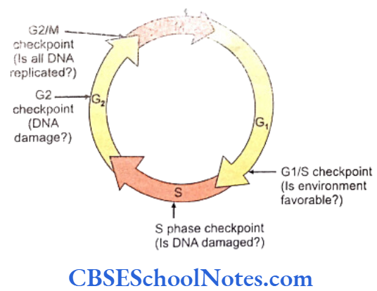
- Restriction point-This restriction point (R) occurs late in the G1 phase between the middle and the termination of the G1 phase. It is the time when a cell verifies whether it has appropriately been instructed by the growth signals to proceed to the S phase for DNA replication. Growth signals sufficient to trigger cells to go into S phase would induce replication of DNA or else the cells would shift to the GO phase.
- G1/S DNA damage checkpoint – The G1/S phase transition forms a major checkpoint for detection of any damage in the DNA molecule entering the synthetic phase.
- S phase DNA damage checkpoint – This checkpoint is strategically located at the later part of the S phase. Defective synthesis of DNA is detected at this stage and appropriately dealt with.
- G2/M checkpoint – Acts as a DNA damage check point.
- Centrosome duplication checkpoint- A defect in duplication of the centrosome or chromatid segregation arrests cells at the G2/M transition.
- Mitotic checkpoint – The M phase checkpoints observes the formation of normal mitotic spindles. The detection of any chromosome that is not attached at a spindle blocks the onset of anaphase.
How is Cell Division (Cycle) Controlled?
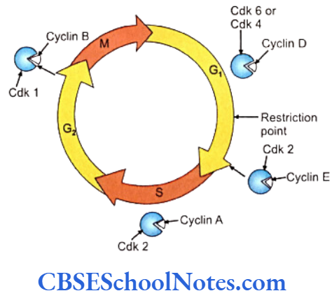
The steps in cell division are usually controlled by proteins called cdk-cyclin complexes. The cdks (cyclin dependent kinases) belong to a family of kinases. The kinases act as catalytic subunits and are named cyclin dependent kinases (cdks) as their activity is dependent on certain cyclins. Cyclins are types of regulatory subunits. The catalytic and regulatory subunits always occur as associated pairs.
Thus a specific cdk is fully activated only when its cyclin partner is expressed in association. As exemplified at specific cell cycle stages, the G1 phase cdk4 and cdk6 act in association with cyclin subunits D1, D2 and D3, while the cdc2/cyclin B complex (cdc = cell division cycle) is expressed in G2/M phase of cell cycle.
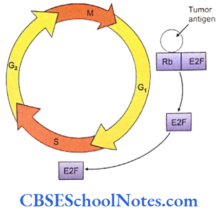
Multiple proteins actively regulate different stages of cell division. The cdks control the phosphorylation of regulatory proteins at different stages in the cell cycle progression, e.g. the retinoblastoma (RB) tumor suppressor gene product (pRb) is a key regulatory protein of the G1 phase that is phosphorylated by a cdk/cyclin complex.
Role Of Genes In Cancer
- In the first part of the G1 phase (very early interphase) pRb is bound to E2F. The transcription factor E2F needs to be in an unbound form for the cells to transit form the G1 to the S phases.
- The pRb with E2F forms a complex that inhibits expression of other transcription factors needed for the initiation of S phase.
- Cells remain in the G1 phase or G0 phase in now unable to phosphorylate the pRb and the cells presence of the complex.
- The synthesis of D cyclin is activated subsequent to the action of growth factors. This event stimulates cells to reenter the cycle from GO or G1 phases.
- Cdks and cyclins couple to form active complexes regulating further steps in the cell cycle. The cdk 4/cyclin D and cdk6/cyclin D become active in the early phase of G1 and cdk2/cyclin E complex function in late phase of G1.
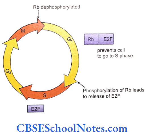
- As the cdk/cyclin complexes phosphorylate the protein pRb in the pRb and E2F complex, the trapped E2F is released.
- The free E2F subsequently activates transcription of genes that are essential for initiating the S phase.
- The inactivation of certain other genes caused by PRb-E2F complex is also disinhibited.
- Thus with the inactivation of pRb the cell now enters in S phase.
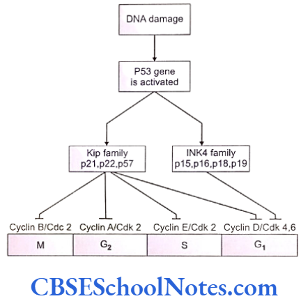
Alternatively the cdk-cyclin kinases also can be inactivated to retain a cell in the G0 or G1 phases. Such inactivation of cdk/cyclin complex can be achieved with the binding of certain inhibitory proteins to the cdk/cyclin complexes. The inactivated complexes are now unable to phosphorylate the pRb and the cells fail to transit into the S phase. There are two families of the cdk/cyclin inhibitor proteins.
- INK4 family (Inhibitors of cdk4 family) – INK4 family of proteins specifically bind and inactivate cdk4 and cdk6. The p16, p15, p18 and p19 are the four members of the family.
- Cip or Kip family (cdk interacting protein or kinase inhibitory protein). The p21, p27 and p57 are the three members of the family. p21 binds to all complexes of cdk2, cdk4 and cdk6 and forms the universal cdk inhibitor that can block all stages of G1 and S phase.
Cell Cyclic Control Genes And Cancer
The cyclin D-dependent kinases integrate the extracellular signals towards progression of the cell cycle. Alterations in pRb and cyclin D-dependent kinases may lead to inappropriate and unbalanced phosphorylation of pRb. This may result in uncontrolled signaling and proliferation of cells.
Cancer Cell Proliferation
Deletion or mutation of the suppressor gene CDKN2 has been implicated in multiple cancer states. The RB (retinoblastoma) gene that acts as a tumor suppressor gene has been found to be associated with cancer. Retinoblastoma is a tumor of the retina seen in children. The retinoblastoma (RB) gene is located on the q arm of chromosome 13.
Retinoblastoma arises when both the copies of the RB genes are deleted or inactivated. Usually the child inherits one defective allele and acquires a fresh mutation in the normal allele in childhood. Thus these tumors are generally sporadic in occurrence resulting from new mutation; homozygous mutations can also be inherited giving rise to the condition.
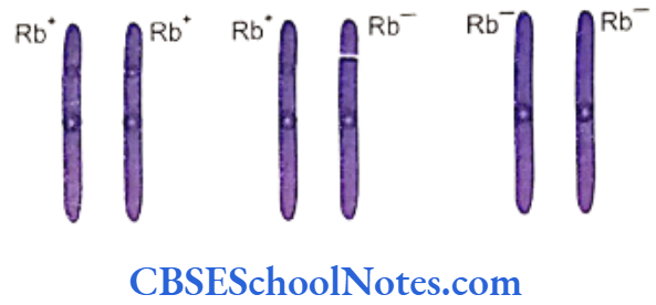
A Condition like retinoblastoma arises due to the loss of protein pRb (product of gene RB) that leads to unrestrained cellular proliferation. Absence of pRb has been linked to osteosarcoma and lung cancer.
RB gene yields a nuclear phosphoprotein (pRb) that influences crucial activities in the cell cycle. The protein pRb is usually kept bound to the E2F group forming an inactive pRb/E2F complex in cells that don’t proceed towards cell division. Once the cdk/cyclin complex phosphorylates the pRb faction of the pRb/ E2F complex, it sets the E2F component free to bind and activate the next set of transcription factors.
Cancer Cell Proliferation
Protein pRb can also bind certain viral tumor antigens like SV40T and E1A. In this situation pRb doesn’t bind E2F and remains as a pRb-tumor antigen complex. The free E2F helps the cell to pass from G1 to S phase as the viral antigen induces unrestrained growth by blocking normal activity of pRb.
Cell proliferation can be arrested if-
- pRb is not phosphorylated (remains coupled to E2F).
- D-cyclin is absent (disabled cdk complex).
- p16, p21 and p27 inactivates cdk-cyclin complex.
On the contrary the loss of the above functions may lead to unrestrained growth or tumor formation.
G1/S Checkpoint
Damage caused by double strand breaks (DSBS) in the DNA activates specific events at this check-point. Ionizing radiation or genotoxic chemicals usually cause such damage in the DNA strands. Escape of undetected damaged DNA on to daughter cells in somatic and germ cells may have devastating consequences.
Cancer Cell Proliferation
Operation of the G1/S cell cycle checkpoint is governed by the tumor suppressor gene TP53. This gene gives rise to the protein p53 that acts as receiver of stress signals including DNA damage. Any damage to DNA leads to the activation of p53 which then acts as a transcription factor inducing cell destruction.
The levels of p53 are generally low in normal cells. To function as transcription factor p53 protein must be activated by phosphorylation and acetylation. The factor Mdm2 prevents phosphorylation and acetylation of p53 and removes p53 from the nucleus. This leads to degradation of p53 by proteosomes in the cytoplasm.
The level of p53 thus is kept low by continuous export of p53 by Mdm2 from nucleus to cytoplasm followed by degradation. On the other hand DNA damage itself results in phosphorylation and acetylation of p53. Once p53 is phosphorylated, Mdm2 cannot bind to the modified (activated) p53. The activated p53 remains in the nucleus.
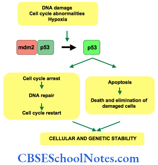
Transcription of a number of genes is brought about by the p53 protein to trigger cell cycle arrest Cyclin B/Cdc 2 Cyclin A/Cdk 2 Cyclin E/Cdk 2 Cyclin D/Cdk 4,6 and apoptosis p53 induced gene p21C1P1 binds to cdk2/cyclin E resulting in the arrest of cell cycle at G1/S checkpoint.
Activities of p53 have earned it the name “Guardian of Genome”. Mutations in TP53 gene are responsible for about 50% cancers in humans. p53 is located on chromosome 17p13.1. Virtually all types of cancers are associated with defects in the p53 gene. Such defects or mutations generally exist in a homozygous pattern.
Cancer Cell Proliferation
Mutant p53 alleles are inheritable. Individuals having such mutations are susceptible to develop malignant tumors. Carriers of heterozygous defects are said to have the Li-Fraumeni syndrome and may develop a varied type of tumors (carcinomas, lymphomas, brain tumors, sarcomas, etc).
p53 protein has a vital function of arresting the cell cycle on detection of breaks and damages in the DNA content of a cell. The protein gets accumulated in the nuclei and inhibits the cell from crossing over to the S phase.
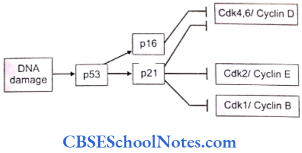
As mentioned above, p53 induces the action of p21 to complete the arrest. p53 also helps in the repair of broken DNA molecules by activating certain other transcription factors and DNA repair enzymes. In the event of a complete repair, the cell is allowed to advance to the next step in the cycle.
In case of an incomplete or failed repair, p53 stops the cell division and induces apoptotic mechanisms in the cell.
S Phase
The S phase checkpoints are also invoked by structural changes in the DNA. Cell cycle is stopped with the action of the dephosphorylated pRb. Arrest of progression of cell cycle may also be brought about by p21 that blocks cdk.
G2/M Checkpoint
This checkpoint is located at the junction between the G2 and the M phase and it is triggered by DNA breaks in the genome. Cell cycle is inhibited at this stage by inactivation of the Cdc2/cyclin B complex. The inhibition of the complex stops the transit of a cell from G2 to mitosis. The action of the checkpoint is to maintain the Cdc2/cyclin B1 complex in an inactive state with the help of p53 protein.
The induction of p21 and its binding to Cdc2/cyclin B in the nucleus causes cell cycle arrest at G2/M checkpoint.
Viruses And Cancer Genes
The study of viral carcinogenesis has shed light on the genetic mechanisms involved in cancer formation. Retroviruses (RNA viruses) are the ones that cause most of cancers in animals and a very few in humans. Some of the DNA viruses also cause a few cancers in man. The understanding of replication in the retrovirus helps us analyze events occurring in cancer.
Retroviruses
The retroviral genome consists of a diploid, double stranded RNA molecule. These viruses are incapable of replication till they infect a cell and use the cellular machinery of the host to replicate its genome and assemble other constituents of its structure. The presence of the key reverse transcriptase enzyme forms a double stranded DNA copy from the viral RNA.
Tumor Suppressor Genes And Cancer
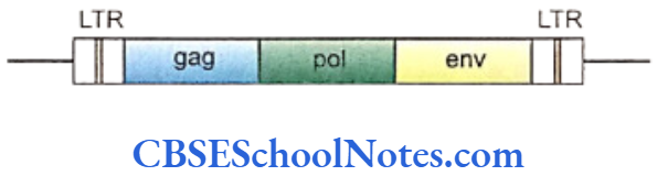
The transcribed DNA strands easily incorporate into the host DNA and are called “provirus”. A provirus constitutes three genes that are adequate for complete viral replication.
- gag – the gene codes for structural protein of the virus.
- pol – this segment codes for the enzyme reverse transcriptase. Genetics of Cancer 165
- env – the sequence codes for protein of the outer envelope.
The promoter and enhancer elements are integrated at each end of the genome. These elements are The promoter and enhancer elements are integrated constitutively associated with the provirus and are termed long terminal repeats (LTRS).
The normal replication of the host DNA also results in the replication of the viral genome by default. Transcription of the integrated viral genome, on the other hand, gives rise to different cellular components of the virus. All viral components assemble in the cell and come out as viral progeny in multiplied numbers after lysing or destroying the host cell.
Once a provirus is integrated in the host genome, it stays in the cell for its life; even passing to the daughter cells of the infected host cell. Proviruses have also been found integrated into DNA of gametes (eggs and sperms) where they reach after infecting the germ cells in an individual.
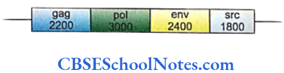
In addition to the gag, pol and env genes found in common retroviruses a potent fourth gene exists in certain viruses (as identified during the study of Rous sarcoma virus). This gene is capable of carcinomatous transformation in a host cell. It is called the src gene with its action of coding for a protein kinase and inducing cancerous transformation of host cell well-documented. A viral gene that can transform the infected host cell is termed oncogene.
Oncogenes carried on viruses are called viral oncogenes or V-onc whereas oncogenes located in host cell genome are called cellular oncogenes or C-onc genes.
Some DNA sequences in the host cell are homologous or identical to the viral oncogenes and thus are called proto-oncogenes. As discussed earlier proto-oncogenes regulate normal cell growth and do not cause cancer in normal circumstances. The cellular proto-oncogenes are potentially carcinogenic and can be transformed to act as oncogenes by point mutation, amplification and chromosomal translocation.
Tumor Suppressor Genes And Cancer
How Viral Oncogenes are Formed
It is interesting to note that retroviral oncogenes originates from cellular genes in the host. Any error in the replication of retroviral genome, after their integration in host genome, gives rise to retroviral oncogene. This viral oncogene is structurally similar to its cellular counterpart (the viral oncogene sis is almost similar to the gene for platelet-dependent growth factor [PDGF]) but different in its function.
Conversion of Proto-oncogenes to Cellular Oncogenes (c-onc)
Proto-oncogenes can be converted into cellular oncogenes in the following ways:
- By increase in the amount of proto-oncogene product: This can be achieved in two ways.
- Integration of the viral oncogene to the host DNA close to a proto-oncogene may induce uncontrolled expression of proto-oncogene through the action of the LTRS of the v-onc.The LTR of the Epstein-Barr viruses have been observed to overexpress myc gene in infected human cells leading to Burkitt’s lymphoma.
- Multiple copies of the proto-oncogenes can be formed in a cell through gene ampli- fication. Activity of several copies of the gene yields a large amount of the transcriptional product that leads to transformation of proto-oncogenes to cellular oncogenes.
The N-myc gene is amplified manifolds in neurofibromatosis. C-myc, N-myc and L-myc amplifications are associated with lung carcinomas and c-neu or erb-B2 genes with types of breast carcinomas.
- Mutation in coding sequence:
Oncogenes are also formed by mutations in the proto-oncogenes. As discussed, these proto- oncogenes are potential oncogenes and are activated through mutations to produce cancer.

- Mutations in the ras gene account for approximately one-third of all human cancers.
- Chromosomal translocation:
A good percentage of human cancers are caused due to translocation of chromosomes with rearrangement of the genome. Chronic myeloid leukemia and Burkitt’s lymphoma are two common examples of the group. The Philadelphia chromosome seen in the white blood cells of the CML patients is formed by rearrangement of chromosomal segments as a result of translocation A reciprocal translocation between the long-arm of chromosome number 22 and the 9th chromosomes transfers cellular abl oncogene from chromosome 9 that fuses with bcr (break-point cluster region) gene of chromosome 22. This fused gene (chimeric gene) manufactures protein that contains about 900 amino acids of ber region and 1100 amino acids of c-abl region. The resultant condition gives rise to abnormal proliferation of the cells.
Similar translocation between the long arm of chromosome 8 containing the c-myc gene and the chromosome 14 at the locus carrying the gene for immunoglobulin heavy chains is implicated in the formation of Burkitt’s lymphoma. This rearrangement brings the translocated c-myc gene under the regulatory influence of the immunoglobulin gene resulting in about 20 fold increase in the levels of c-myc transcription.
Apoptosis
Apoptosis or cell death is a programmed event to maintain a balance between the generation of new cells and the death of senescent or defective cells.
Apoptosis occurs due to several events that causes damage to the growth regulating genes, loss of check- point genes and also when the telomerase enzyme is unable to protect the integrity of the terminal part of the chromosomes after each cell division. (With each cell division, the tails of each chromosome gets shortened due to progressive loss of nucleotides to reach a certain limit where the cells automatically undergo self-destruction. This threshold is referred as the Hayflick’s limit. This phenomenon occurs due to gradual dwindling in the levels as well as activity of the telomerase enzyme that is needed to repair damaged ends of the chromosome).
Cells usually die when they grow old and are unable to furnish proteins like the telomerase enzyme that are needed to cross the checkpoints in the cell cycle. Old cells also suffer breaks in the DNA and are directed towards self-destruction. Cells also die if they are subject to sustained injuries such as heat, oxidative stress, UV irradiation damage or get killed when they become vulnerable and infected with a virus or other intracellular pathogen that destroys the cell.
Thus apoptosis is a form of programmed cell death initiated by extracellular or intracellular signals in which enzymes are activated that break down the cytoplasmic and nuclear skeleton, degrade the chromosomes, disintegrate the DNA and shrink the cells. Initiation of the process of apoptosis begins either with extracellular or intracellular signals.
Execution of cell death is effected with the release of caspases (cysteine containing aspartase specific protease). Caspases are the ultimate destroyers of cell. These are a family of proenzymes that are activated in a cascade. The targets of these proteases are the DNA, several cytoskeletal proteins, DNA repair enzymes, etc. The caspase family includes at least 13 proteins and is divided into 3 groups.
Morphological changes occur in the dying cells. Surrounding cells like macrophages remove the dead cells.
Tumor Suppressor Genes And Cancer
Genetics Of Cancer Summary
- Cancer cells are characterized by:
- Uncontrolled proliferation.
- Transformation to abnormal cells.
- Capacity to invade surrounding tissues.
- Property to metastasize to distant places.
- uppression of apoptosis.
- Induction of angiogenesis.
- Cancer is due to the loss of the normal mechanisms which control cellular proliferation and differentiation.
- Cellular proliferation is under the control of genes.
- Genes responsible for causing cancer are known as oncogenes.
- Tumor suppressor genes (TSG) apply brakes to unrestrained cell growth as they induce tumor suppressor activity. Thus cancer may develop due to loss of function (mutation) of TSGS.
- About 100 oncogenes and about 30 TSGs are now known.
- Signal transduction is a process by which extracellular growth factor regulate cell growth and differentiation by a complex pathway. [Growth factor activation of receptor cytoplasmic proteins (ras and abl) are activated – ras binds to GTP cytoplasmic kinases are activated Kinases enter the nucleus – activate myc which regulate transcription of DNA – cell cycle begins].
- Genes that code for growth factors and growth factor receptors, after mutation may acquire oncogenic properties.
- The mutation of ras gene (which codes for signal transducing protein) and myc gene coding for transcription factor may cause cancer.
- The cell division cycle is divided into four
- The transition from one phase to the next is sequential phases, i.e. G1, S, G2 and M phase. regulated by checkpoints.
- Cell cycle is controlled by cdk/cyclin complexes.
- Various proteins act as key regulatory proteins during cell cycle. For example the product of retinoblastoma (RB) tumor suppressor gene (pRb) is a regulator protein of G1 phase that is phosphorylated by cdk/cyclin complex.
- Cell cycle checkpoints are under genetic control and surviallence. In case DNA (gene) is damaged, cell cycle progression is checked till the damaged DNA (gene) is repaired.
- Any alteration (mutation) in these genes will lead to the formation of tumor due to uncontrolled cell proliferation.
- The maintenance of the G1/S cell cycle checkpoint is dependent on the tumor suppressor gene (TP53). The mutation of TP53 gene is responsible for about 50% cancers in human.
- The gene p21 when induced by p53 binds to cdk2/ cyclin E which leads to cell cycle arrest at G1/S checkpoint. Thus mutation of p53 and p21 may lead to nonfunctioning of G1/S, S and G2/M checkpoints leading to formation of tumor.
- Retroviruses (RNA viruses) and DNA viruses are responsible for causing cancers in man.
- The oncogenes present in viruses are called viral oncogenes (V-onc) those in host cells are called cellular oncogenes (C-onc). In host cells there are DNA sequences homologous to the viral oncogenes and are called proto-oncogenes. Proto- oncogenes are responsible for promotion of normal cell growth.
- Apoptosis is triggered by DNA damage (programmed cell death). A cascade of proteolysis is initiated with intracellular or extracellular activation of apoptotic pathway. The proteases involved are called caspases. Cell death is brought about by the action of caspases.