
Urinary System
The urinary system consists of two kidneys, two ureters, one urinary bladder and one urethra. Two kidneys produce urine, while ureters conduct urine from the kidneys to the urinary bladder, which stores the urine. The urethra drains the urine from the urinary bladder to the exterior.
Kidney Functions:
- Excretion: The major function of the kidney is excretory, i.e., it eliminates the waste material (urea) and excess metabolites (electrolytes, water, glucose and amino acids). However, when these metabolites are not in excess it retains them by reabsorption.
- Endocrine function: Kidney also has an endocrine function, i.e., it secretes renin (involved in the regulation of blood pressure and retention of sodium) and erythropoietin (regulates RBC production) in embryonic life.
- Conversion of vitamin I) into calcitriol: Proximal convoluted tubules of the kidney are involved in the conversion of vitamin D into active hormone [calcitriol, l,25-(OH)2
vitamin DJ that regulates plasma calcium level.
To have a comprehensive understanding of the histological structure of the kidney, readers are advised to learn the gross structure of the kidney structure of nephron and the blood supply of the kidney.
Gross Structure Of Kidney
The naked-eye view of a hemisected kidney shows the outer cortex and inner medulla.
- The cortex lies just beneath the connective tissue capsule. The medulla is made up of 8-12 conical structures called pyramids.
- The cut face of the pyramid displays a striated appearance it consists of numerous parallel tubules and blood vessels.
- The broad base of each pyramid is directed toward the cortex and apex (renal papilla) facing into a minor calyx.
- There are about 8-12 minor calyces, which join to form two or three large extensions called major calyces.
- Major calyces unite to form a funnel-shaped renal pelvis, which is present at the hilus of the kidney. The renal pelvis becomes narrow and forms the ureter.
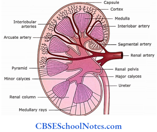
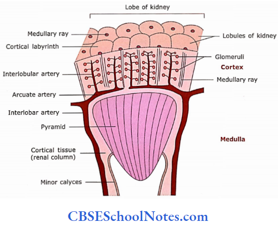
The cortex at the margin of each pyramid extends inward between the pyramids as renal columns. Some of the striated patterns from the base of the pyramid may extend into the cortex, which are called medullary rays. Each medullary ray is the collection of straight tubules and col¬lecting ducts extending in the cortex. A lobe of the kidney is defined as a renal pyramid with its overlying cortex and part of its laterally associated renal columns
A kidney lobule is defined as a medullary ray with its laterally associated cortical tissue. Thus, a lobule consists of a col¬lecting duct and all the nephrons draining to it. As there are about 20,000 medullary rays in the cortex, approximately the same number of lobules are estimated to be present. The cortex of the kidney consists of about 2-3 million tubular structures called nephrons.
Kidney Remember:
The lobe of the kidney includes the renal pyramid with its overly¬ing cortex and parts of its later ally-associated renal columns, While on the other hand, the kidney lobule consists of the centrally placed medullary ray with its laterally placed cortical areas. Thus, the number of kidney lobes is equal to number of pyramids present in the kidney (i.e., 8-12). While, the number of kidney lobules is equal to the number of medullary rays present in the cortex (i.e., about 20,000).
General Structure Of Nephron
The functional unit of the kidney is (lie nephron. Bach nephron has several parts, i.e.,
- Renal corpuscle
- Proximal convoluted tubules
- The loop of Henle and
- Distal convoluted tubule.
1. Renal Corpuscle:
- The blind end of each nephron is expanded in the cortical region into a double-walled cup called Bowman’s capsule made up of an outer parietal epithelium and an inner visceral epithelium. The cup encloses a tuft of capillaries called a glomerulus.
- Bowman’s capsule and glomerulus together constitute the renal corpuscle. The renal corpuscle has two poles, i.e., the vascular pole (where arterioles arc present) and the urinary pole, from where proximal convoluted tubules take origin.
- An ultrafiltrate of the glomerular blood enters the space between two layers of Bowman’s capsule. The filtrate then passes to the proximal convoluted tubules.
2. Proximal Convoluted Tubule:
The proximal convoluted tubule is quite tortuous and begins at the Bowman’s capsule. It makes many convolutions in the cortex near the Bowman’s capsule from which it arises, it then enters the medullary ray and continues as the descend¬ing thick segment of the loop of Henle.
3. Loop of Henle:
The loop of Henle consists of a descending limb (both thick and thin segments), a hairpin turn and an ascending limb (thin and thick segments).
- The upper part of the descending limb is thick and is in continuation of the proximal convoluted tubule.
- The lower part of the descending limb is a thin segment, which is continuous with the hairpin turn.
- The ascending limb has a lower-thin and upper-thick segment. The thick segment ascends back into the cortex.
4. Distal Convoluted Tubule:
As the ascending thick segment of the loop of Henle comes close to the vascular pole of its originating renal corpuscle, it continues as distal convoluted tubules. After this, it makes many shorter loops in the cortex before it opens in the collecting tubule.
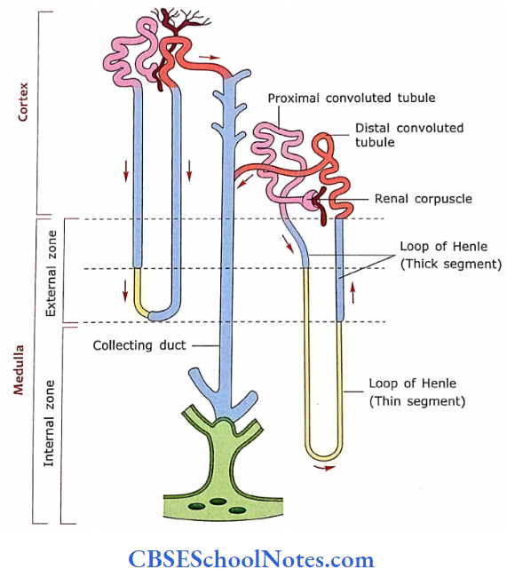
Two different types of nephrons are located in the cortex.
A nephron whose glomerulus is located adjacent to the base of the pyramid is called a juxtamedullary nephron. All other nephrons are called cortical nephrons.
- The collecting tubules are present in cortical tissue and drain into increasingly larger tubules called collecting ducts.
- A collecting duct travels first in the medullary ray and then in the pyramid to its apex.
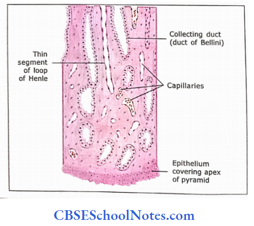
- At the apex of the pyramid, several large collecting ducts open, which are called as ducts of Bellini.
- A nephron, its collecting tubule and its collecting duct together form a unit called a uriniferous tubule.
- The nephrons are embryologically derived from metanephros while collecting tubules and ducts from the ureteric bud.
Nephron Remember:
The nephron is the functional and structural unit of the kidney. It consists of renal corpuscle (glomerulus and Bowman’s capsule), proximal convoluted tubule, loop of Henle and distal convoluted tubule that opens in collecting tubule. Each kidney contains about 2 million nephrons.
Relationship between Parts of Nephron and Zones of Kidney:
- Renal corpuscles are located at varying levels in the cortex and only in cortical tissue.
- Renal corpuscles close to the capsule (cortical nephrons) send their tubules down to the outer zone of the medulla.
- Renal corpuscles in the juxtamedullary area send their tubules deep into the inner zone of the medulla.
- Descending and ascending thick limbs occupy the outer zone, while the thin limbs occupy the internal zone almost to the apex of the pyramid.
- Thus, cortical tissue contains renal corpuscles, proximal and distal convoluted tubules and initial parts of collecting tubules.
- The medullary ray contains thick segments of the loop of Henle and collecting ducts. The medulla contains a thick and thin segment of the loop of Henle, vasa recta and collecting ducts.
The cortex of the kidney Remember:
The cortex of the kidney contains renal corpuscles, proxi¬mal and distal convoluted tubules and initial parts of the collecting tubules, while the medulla contains thick and thin segments of the loop of Henle, vasa recta and collecting ducts.
Renal Blood Supply of Kidney
The renal artery after entering the hilus of the kidney divides into a few segmental arteries, which in turn form interlobar arteries. Each interlobar artery runs between two pyra¬mids through the renal column.
- At the cortico-medullary junction (at the base of the pyramid), the interlobar arteries turn to arch over the base of the pyramid. These arteries are known as arcuate arteries.
- The arcuate arteries give off branches, which ascend into the cortex between lobules as interlobular arteries. These arteries are located between medullary rays.
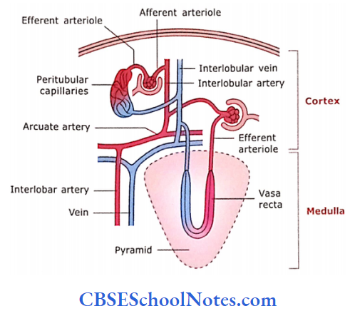
- Many intralobular arteries arise from the interlobular artery and are called as afferent arterioles of glomeruli. They form the capillary network of glomeruli.
- Blood from glomeruli is drained by efferent arterioles. The efferent arterioles give rise to a second network of capillaries, which are called as peritubular capillaries
- The peritubular capillaries of cortical glomeruli surround the local uriniferous tubule and do not go into the medulla.
- However, the peritubular capillaries of juxtamedullary glomeruli descend into the medulla along with loops of Henle. In the medulla, they form capillary loops called recta before returning to the cortex. The ascending limb of the vasa recta forms the arterial limb, while its ascending limb forms the venous limb.
- The venous return from the peritubular capillary networks is via interlobular, arcuate, interlobar and renal veins.
Blood Supply of Kidney Remember:
Both kidneys receive large volumes of circulating blood, i.e., approximately 1000 mL of blood enters two kidneys each minute, from which about 125 mL/min glomerular filtrate is formed. Both kidneys form about 180 L of glomerular filtrate out of which only 2 L is excreted as urine while the remaining 178 L is reabsorbed by the kidneys.
Microscopic Structure Of Kidney
Following is the histological structure of various parts of the nephron i.e., renal corpuscle, proximal convoluted tubules, loop of Henle and distal convoluted tubule
1. Renal Corpuscle
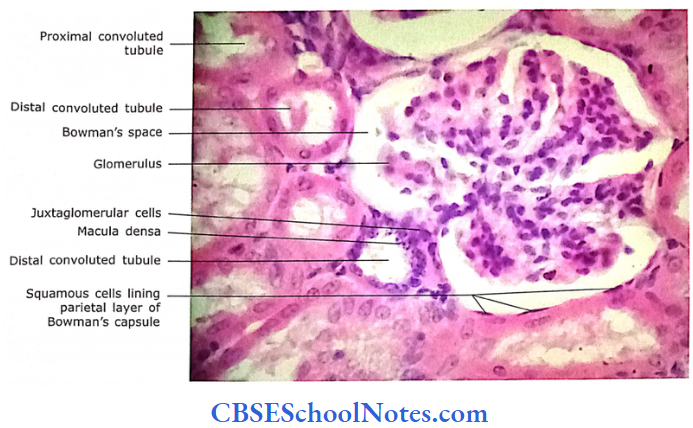
It is also known as the Malpighian corpuscle. It consists of Bowman’s capsule and glomerulus. The Bowman’s cap¬sule has an outer parietal layer and an inner visceral layer The parietal layer is lined by simple squamous epithelium, while the visceral layer is lined by podocytes. The space between the parietal and visceral layer is the urinary space (Bowman’s space), which receives the ultrafiltrate of blood. At the urinary pole, the space between two layers is continuous with the lumen of the proximal convoluted tubule. The squamous epithelium of the parietal layer at this pole becomes continuous with the cuboidal epithelium of the proximal convoluted tubule. The glomerulus is the tuft of capillaries fed by an afferent arteriole and drained by an efferent arteriole. Both these arterioles are present at the vascular pole of the renal corpuscle.
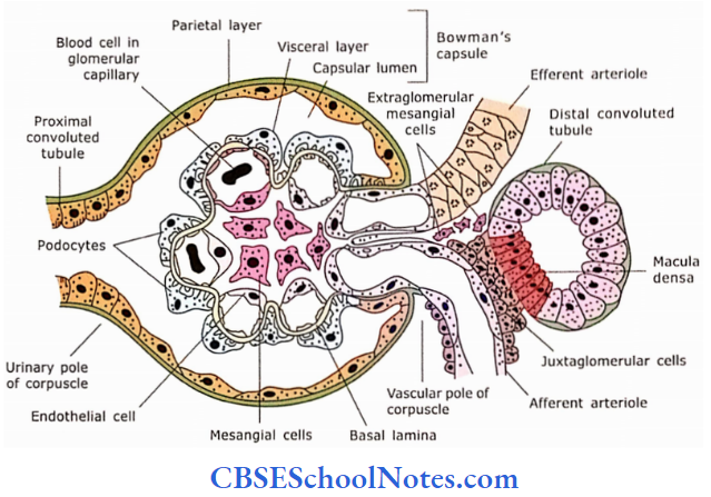
The visceral epithelium of Bowman’s capsule is closely applied to the endothelial lining of capillaries. The cells of the visceral layer become modified and are called Propodocytes and have many radiating processes, which in turn contain secondary processes, called foot processes or pedicels. The foot processes of neighbouring podocytes interdigitate with each other. These foot processes are separated from each other by narrow intercellular spaces that are called filtration slits. The gap of the filtration slit is occupied with a thin membrane called a filtration slit membrane or slit membrane

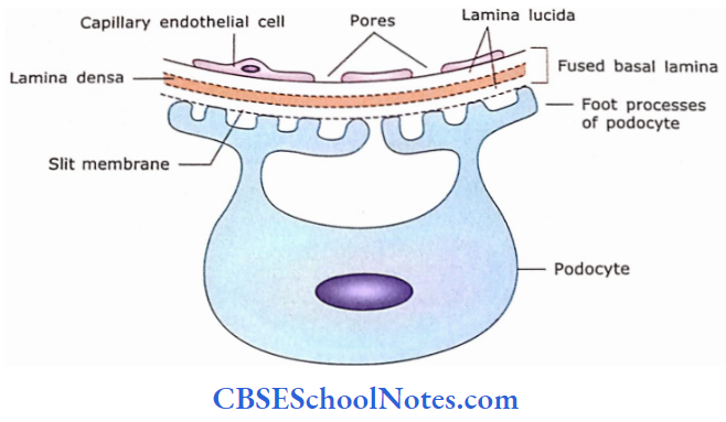
Ultrastructure of Filtration Barrier:
The endothelial cell layer and podocyte cell layer (visceral layer of Bowman’s capsule) share a common fused basal lamina. The foot processes of podocytes are closely applied to the common basal lamina.
The filtration barrier is made up of three components
- Fenestrated Endothelium: The pores of capillary endothelial cells are about 70-90 nm in diameter. These pores are not spanned by a pore diaphragm and allow the passage of molecules up to 70,000 molecular weight. Endothelial cells of glomerular capillaries possess a large number of aquaporin-1 (AQP-1) water channels, which allow fast filtration of water through the endothelium.
- (Glomerular basement Membrane: It consists of fused basal lamina of endothelium and visceral layers of Bowman’s capsule (podocytes). It is made up of type 4 and 8 collagens, proteoglycan and glycoproteins. It forms the major unit of barrier and serves to retain the necessary plasma proteins from leaking out.
- Filtration Slit Membrane (diaphragm): It spans between adjacent foot processes of podocytes and measures 25 nm in width and 4-6 nm in thickness. The slit diaphragm is formed by a transmembrane protein called nephrin. Nephrin proteins emerge from opposite foot processes and form a central density, which has pores. Mutation in the nephrin gene leads to congenital nephritic syndrome which is characterized by proteinuria. This membrane shows the presence of small pores, which prevent the passage of albumin and large molecules from the blood to glomerular filtrate.
Filtration Barrier Remember:
The glomerular filtration barrier is formed by the fenestrated endothelium of glomerular capillaries, glomerular basement membrane and visceral layer of Bowman’s capsule, which consists of epithelial cells that become modified and are called podocytes. The foot processes of neighbouring podocytes interdigitate with each other and contain gaps, which are known as filtration slits. The gap of the filtration slit is occupied with a thin membrane called a filtration slit membrane or slit membrane. This slit membrane acts as a part of the filtration barrier. Thus, the filtration barrier consists of endothelial cells, fused basal lamina and a filtration slit.
Process of Glomerular Ultrafiltration:
Fluid first passes through the pores of capillar}’ endothelial cells then it is filtered by the basal lamina.
- Fluid containing small molecules, ions and macromolecules passes through lamina densa and pores in slit diaphragm of filtration slits.
- If molecules are small (<1.8 nm) and are un¬charged then they pass easily through the slit diaphragm. However, large molecules cannot pass through the slit diaphragm.
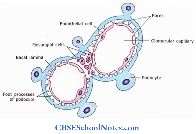
- The large molecules, which are unable to cross the barrier are rapidly removed by the intraglomerular mesangial cells, otherwise, the basal lamina will get clogged with these large molecules. The fluid, which after passing through bar¬riers reaches the Bowman’s space is called the glomerular ultrafiltrate.
Mesangial Cells:
The capillaries of the glomerulus are held together by mesangium (mesangium = between vessels). Mesangial cells are of two different types, i.e., extraglomeru lar and intraglomerular. Extraglomerular cells are located at the vascular pole, while intraglomerular cells are located within the renal corpuscle.
Mesangium is a connective tissue consisting of mesangial cells in an extracellular matrix. These cells are most numerous near the vascular pole of the renal corpuscle. Mesangial cells correspond to the pericyte and may be enclosed by the basal lamina of the glomerular capillaries.
They have many functions:
- Their phagocytic function helps to remove large protein and filtration residues from the glomerular basal lamina. Thus, the integrity of the filter is maintained.
- They participate in the turnover of basal lamina.
- Mesangial cells are contractile, thus regulating the glomerular filtration rates.
- Mesangial cells synthesize and secrete interleukin-1 and platelet-derived growth factor (PDGF).
- The} provides structural support to podocytes.
- Mesangial cells proliferate in certain types of nephropathy.
Juxtaglomerular Apparatus:
This is an apparatus present near the vascular pole of a renal corpuscle and helps in maintaining blood pressure. The macula densa, Juxtaglomerular cells and extra mesangial cells constitute the juxtaglomerular apparatus
- Macula Densa: These are specialized cells at the beginning of the distal convoluted tubule that lie adjacent to afferent and efferent arterioles at the vascular pole of the corpuscle.
- These cells are narrow, columnar and crowded together.
- Cells of macula densa can sense a low sodium concentration of urine in the distal convoluted tubules and help in the release of renin.
- Juxtaglomerular Cells: The smooth muscle cells in the wall of afferent arteriole (and sometimes in efferent arterioles also), which lie close to macula densa, become modified to form juxtaglomerular cells.
- These cells contain secretory granules and no myofilaments.
- They secrete renin hormone, which increases blood pressure. (Renin breaks the angiotensinogen of blood plasma into angiotensin 1, which subsequently is broken down to angiotensin 2 in the lungs. Angiotensin 2 raises the blood pressure by its vasoconstriction activity and controls the glomerular filtration.)
- It also stimulates the synthesis and release of aldosterone, which in turn acts on collecting ducts to increase the reabsorption of sodium and water.
- This leads to a further rise in blood volume and blood pressure.
- Thus, the juxtaglomerular apparatus regulates blood pressure by activating the rennin-angiotensin-aldosterone system.
- Extraglomerular Mesangial Cells: These cells are present in the space between the distal tubule, and afferent and efferent arterioles at the vascular pole of the corpuscle.
- These cells have receptors for angiotensin 2 and may regulate the glomerular filtration rate.
- These cells connect the sensory cells of macula densa with the juxtaglomerular effector cells and transmit the signals through gap junctions.
- They also send signals to the contractile mesangial cells for vasoconstriction within the glomerulus.
Juxtaglomerular apparatus Remember:
The juxtaglomerular apparatus consists of macula dense (which are specialized cells in the beginning of the distal convoluted tubule that lie adjacent to afferent glomerular arterioles), juxtaglomerular cells, (which are smooth muscle cells in the wall of afferent arteriole) and extraglomerular mesangial cells.
The cells of macula densa are specialized to detect the low concentration of sodium and volume of glomerular filtrate in the distal tubule. This leads to the release of renin by juxtaglomerular cells causing the conversion of angiotensinogen to angiotensin 1 which is subsequently converted to angiotensin 2. Angiotensin 2 is a potent vasoconstrictor and helps in the release of aldosterone.
Kidneys Clinical Application
Glomerulonephritis, Filtration Barrier and Proteinuria:
- If kidneys are infected by bacteria, glomeruli are highly affected.
- The urine of a healthy person does not contain protein because the molecules of protein are too large to pass through filtration barrier.
- However, in diseases like glomerulonephritis, the filtration unit (basal lamina of capillaries) may get damaged and large amounts of protein and RBCs can leak into the urine from the blood.
- The presence of protein in urine is known as proteinuria, while the appearance of blood (RBCs) in urine is called haematuria.
- The leakage of protein results in low protein levels in the blood. This causes a collection of fluid in tissue and widespread swelling.
- Proteinuria may also occur in diseases like diabetes mellitus due to damage to the filtration unit of the kidney.
Kidney Failure and Dialysis:
Kidney failure results from the loss of normal function of both kidneys due to a variety of causes, i.e., fall in blood pressure, infection, glomerulonephritis, toxic chemicals, drugs, diabetes mellitus, etc.
- In kidney failure, kidneys are unable to remove waste products and excess water from the blood, thus disrupting the chemical balance of the blood.
- Methods of treatment of kidney failure may include drugs, dialysis or kidney transplant.
- Dialysis is the procedure in which the functions of the kidney (removal of wastes and excess water from the blood) are performed by a machine.
- Each dialysis treatment takes about 3-4 hrs and has to be repeated 3 times a week.
2. Proximal Convoluted Tubule
If starts from the urinary pole of a renal corpuscle and extends up to a thick portion of the descending limb of Henle. This part of the tube is 60 m in diameter and complexly coiled (convoluted). The length of the proximal convoluted tubule is almost double that of the distal convoluted tubule. It is present in the cortex only.
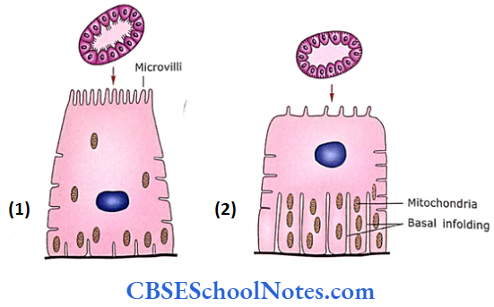
The tube is lined with simple cuboidal or low columnar epithelial cells. These tubules have small uneven lumen (Fig. 17.9 and 17.12). There is the presence of a brush border formed by tall microvilli on the apical surface of cells. Nuclei are round and centrally placed. The cytoplasm stains deeply with eosin. The basal part of the cell may show vertical striations, due to the presence of mitochondria.
Proximal Convoluted Tubule Ultrastructure:
- The electron micrograph of cells of proximal convoluted tubules shows the features that indicate that these cells are involved in absorption and transport.
- The presence of microvilli lateral and basal infoldings of plasma membrane increases the surface area of cell for absorption and transport.
- The presence of mitochondria between basal folds provides for the high-energy requirements needed for active transport.
- Fluid and absorbed substances return to the fenestrated blood capillaries present adjacent to proximal convoluted tubules.
Proximal tubules Functions:
- In proximal tubules, there occurs reabsorption of 80% of salts (Na and Cl), water (85%)
- Most amino acids, ascorbic and lactic acid (100%), filtered proteins, glucose and bicarbonate
- The remaining molecules and fluids are removed in the other portions of the nephron.
3. Loop of Henle
The proximal convoluted tubule continues downward into the medullary ray and medulla as the loop of Henle.
- The histological structure of the thick descending limb of the loop of Henle is similar to that of the proximal convoluted tubule.
- The descending and ascending thin limbs of the loop are about 15 m in diameter and are lined with squamous epithelial cells bearing few microvilli. The cytoplasm is pale staining, nuclei bulge into small lumen.
- The thin limb resembles a venule in cross-section. The histological structure of the thick ascending limb of the loop of Henle is similar to that of the distal convoluted tubule (see below).
Loop of Henle Ultrastructure:
The thin limb of the loop of Henle is lined by squamous epithelium bearing a few’ short microvilli. The presence of very few organelles (including mitochondria) and very few infoldings of plasma membrane indicates that these cells are only involved in the passive transport of fluid and salts.
Loop of Henle Functions:
- The loop of Henle is the essential element in the production of hypertonic urine.
- The thin descending limb is permeable in both water and salt.
- In contrast to this the thin ascending limb is permeable to salt but not to water.
4. Distal Convoluted Tubule
As the distal convoluted tubule is half the length of the proximal convoluted tubule few distal tubules are seen in a microscopic field. The diameter of the distal tubule is less as compared to proximal tubules (15-30 μm). The tubules are lined by cuboidal epithelium.
The cytoplasm of cells stains light eosinophilic. The brush border is not present and the height of cuboidal cells is short (5 μm). These two facts are responsible for the large regular lumen of distal tubules.
Differences between proximal and distal convoluted tubules:
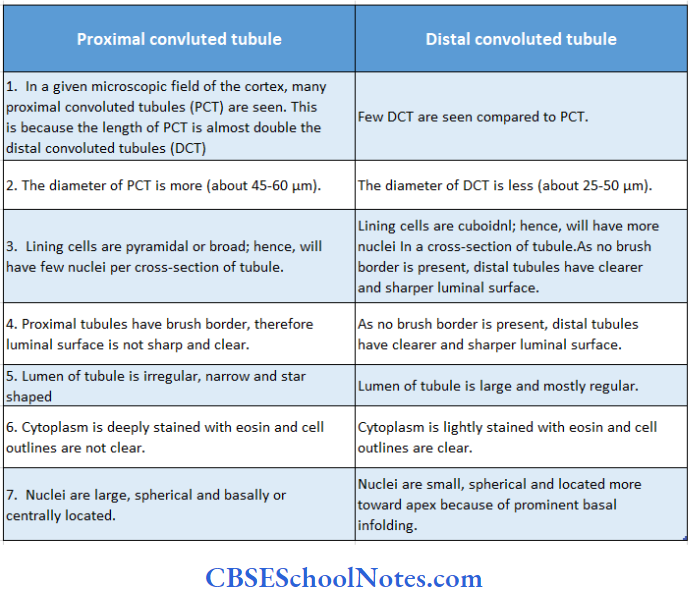
The differences between proximal and distal convoluted tubules are presented in Table
Distal Convoluted Tubule Ultrastructure:
- The cells of distal convoluted tubules show very few short microvilli, but lateral and basal infoldings of the plasma membrane are very prominent.
- The mitochondria are oriented parallel to the long axis of the cell.
- All these features indicate that cells are involved in the active transport of ions.
Distal Convoluted Tubule Functions:
- It is involved in the reabsorption of salt, water and bicarbonate. The distal tubule also secretes potassium and hydrogen ions.
- The distal convoluted tubule is under the control of an antidiuretic hormone, which promotes the reabsorption of water and salts
4. Collecting Tubules
Collecting tubules begin in the cortex and proceed to the medullary ray where they join the larger collecting tubules called as collecting ducts. These ducts in the medulla run toward the apex of the pyramid and join each other to form the duct of Bellini. Collecting tubules are about 40 pm in diameter while ducts are much wider.
Both tubules and ducts are lined by cuboidal to low columnar epithelium. The brush border is not present and cells are lightly stained with eosin. The cell outline is clear and both, tubules and ducts, have a much larger lumen
Collecting Tubules Ultra Structure:
Collecting tubules and ducts are lined by two kinds of cells, i.e., principal and intercalated cells. Most of the lining cells are principal cells, which are wide, low columnar. They have few organelles, lateral and basal infoldings of the plasma membrane and several mitochondria. Intercalated cells are few and they have microvilli and basal infoldings.
Collecting Tubules Functions:
The function is the concentration of urine by sail-free water re-absorption that occurs under the influence of ADH. The result is hypertonic urine.
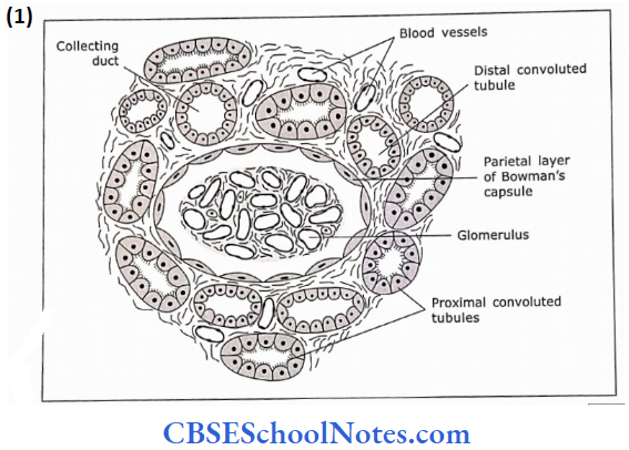
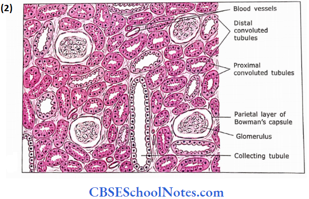
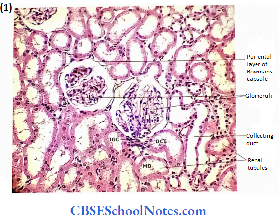
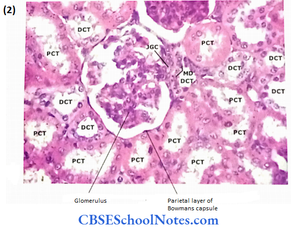
- The light microscopic structure of the cortex of the kidney: A section from the cortex of the kidney shows the glomeruli, proxi¬mal and distal convoluted tubules, blood vessels and col¬lecting tubules.
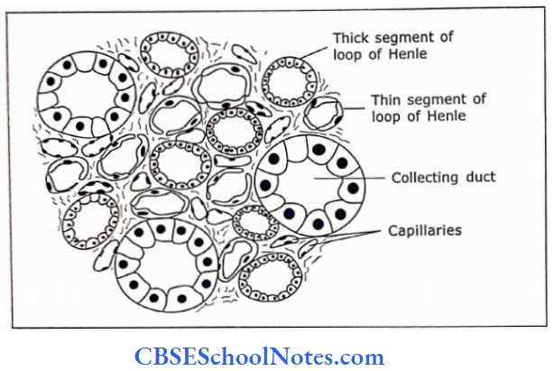
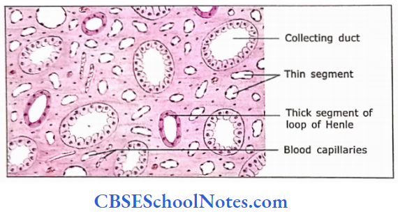
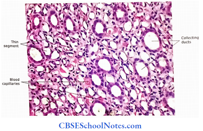
- The light microscopic structure of the medulla of the kidney: A section from the medulla of the kidney shows the thick and thin segment of the loop of Henle, collecting ducts and blood ves¬sels (capillaries).
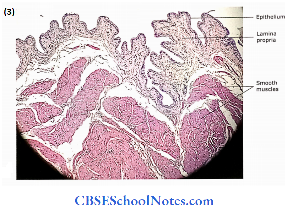
Ureter
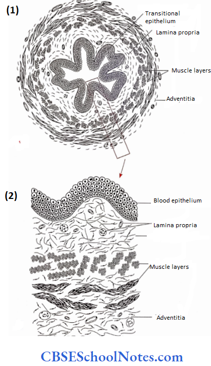
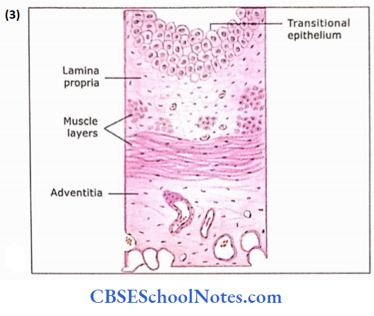
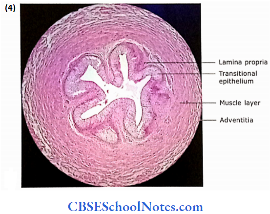
The ureter is a tube with a star-shaped lumen varying in length from 25 to 35 cm. It conducts urine from the renal pelvis to the urinary bladder. The following three layers comprise the wall of the ureter
- Mucosa
- Muscle layer
- Adventitia
The mucosa consists of lining epithelium and lamina pro-pria. The epithelium is transitional and is 4-5 cells thick. The lamina propria is wide and made up of loose connective tissue. Blood vessels and lymphatics are present in it.
The muscle layer consists of an inner longitudinal and outer circular layer of smooth muscle fibres. In the middle and lower partial ureter, a third outer layer of longitudinal smooth muscle is also present. these three layers of muscle arc are not well defined and are difficult to mark off front of each other. The outermost layer (adventitia) is made up of loose connective tissue and contains many blood vessels, nerves and fat cells.
Kidney Stones Clinical Application
Calcium salts and uric acid are excreted In the glomerular filtrate. These salts are less soluble in water The water is reabsorbed from the glomerular filtrate to concentrate the urine Kidney stones occur when urine is saturated with these salts. These salts tryst into stone-like structures. Kidney stones can take years to form These stones are usually formed in the renal pelvis. A small stone may dislodge from the kidney and may pass to the ureter where it may cause severe pain.
Urinary Bladder
Following are the layers in the wall of the urinary bladder
- Mucosa
- Muscle layer
- Serosa/Adventitia
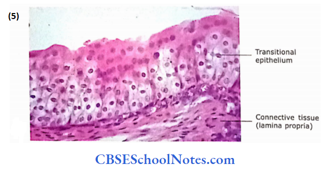
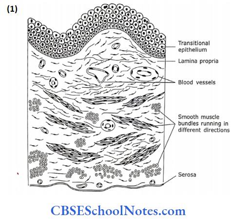
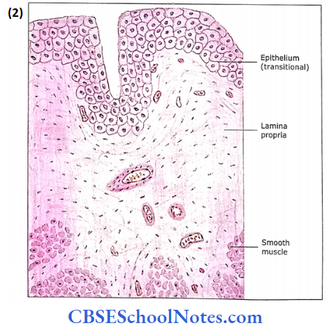
The mucosa is made up of transitional epithelium and lamina propria. The empty bladder shows many mucosal folds and epithelium increases in thickness up to eight cell layers.
- The superficial cell layer takes dark eosinophilic stains due to the presence of plaques. Plaques are modified areas of the plasma membrane.
- These plaques are more rigid and thicker than the rest of the apical plasma membrane (interplaque region). Plaques give attachment to actin filaments on their inner surface. The functional significance of these plaques is not known.
- Probably, these plaques act as osmotic bar¬rier to water and salts.
- When the bladder is filled, the mucosal folds disap¬pear and the epithelium becomes thin to about 3-4 cells. The lamina propria is made up of moderately dense con¬nective tissue. It may occasionally show small lymphatic nodules among the blood vessels and lymphatics.
- The thick muscle coat is made up of smooth muscle fibres running in all directions. Between the bundles of muscle fibres is loose connective tissue.
- Although the three muscle coats, i.e., transverse, longitudinal and oblique are described, these layers are difficult to distinguish. In the region of trigone, the mucosa is thin and directly applied to the muscle layer.
- The superior surface of the bladder is covered by serosa (peritoneum) while all other surfaces are covered with tu¬nica adventitia.
Urinary Bladder Clinical Application
Bladder Tumours and Bladder Stones
- Tumours in the bladder may be either non-cancerous or cancerous.
- The tumour starts growing from the epithelial lining of the bladder and projects into the cavity of the bladder.
- The bladder is also a very common site for the formation of stones.
- Stones are formed because of the crystallization of waste products present in urine.
Urethra
The male urethra is long and consists of prostatic and penile urethra. The male urethra is described along with prostate and penis (see male reproductive system).
The female urethra is short (about 3 cm long) and near the bladder, it is lined by transitional and in the middle portion by pseudostratified columnar epithelium.
- Near the external opening, it is lined by stratified squamous epithelium.
- The submucosa consists of loose connective tissue, which contains many venous plexuses and elastic fibres.
- The muscle coat consists of an inner longitudinal and an outer circular coat of smooth muscle fibres.
- At the terminal end, the urethra is surrounded by skeletal muscle fibres, which constitute the external sphincter.