
The Central Nervous System
The central nervous system consists of the brain and spinal cord. The cut surface of any part of the central nervous system consists of grey matter and white matter.
- The cerebrum and cerebellum have a cortical layer of grey matter on the surface deep to which is white matter. However, in the spinal cord, grey matter is inside, surrounded by white matter.
- Out of the various parts of the brain, we should study only the cerebral cortex and cerebellar cortex which are of histological significance.
Some Important Definitions
- Grey matter: It is a collection of neurons, neuroglial cells, the processes of these cells lying adjacent to the cell body, and blood vessels inside the central nervous system (brain and spinal cord).
- White matter: It consists of bundles of nerve fibers; such as sociated neuroglial cells and blood vessels. The Myelin sheath that surrounds the nerve fibers gives it a white appearance.
- Nuclei: These are islands of grey matter situated deep inside the cerebrum, cerebellum brain stem, and spinal cord For Example., the thalamus, basal nuclei, etc.
- Ganglion: These are collections of nerve cells (grey matter) outside the brain and spinal cord For Example., dorsal root ganglia and sympathetic trunk ganglia.
Cerebral Cortex
Most of the cerebral cortex (except the cortex of hippocampal formation and piriform lobe) is described as having six layers. However, the distinction between these layers is not well-marked.
- These layers are distinguished based on the predominance of cell type and arrangement of fibers.
- The arrangement of fibers can be visualized only by special stains and not by H and E. From superficial to deep, the following are the layers of the cerebral cortex.
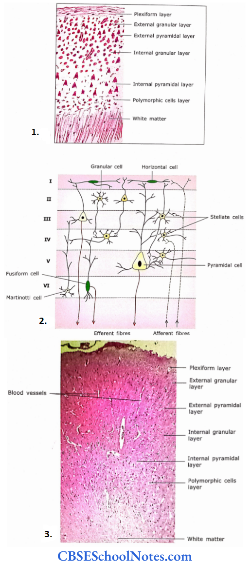
- In a section of the cerebral cortex, it is difficult to distinguish various layers.
- Schematic diagram to show neurons in various layers of the cerebral cortex.
- Section of the cerebral cortex. Six layers of the cerebral cortex are ill-defined as they merge.
Molecularor Plexiform Layer
This layer is situated just beneath the pia mater. It consists predominantly of fibers and neuroglial cells. A few horizontal cells of Cajal are also present.
- The peripheral portion consists largely of fibers, which travel parallel to the surface. In its deeper part lie the horizontal cells of Cajal.
- Their cell body and processes are disposed of horizontally. This layer shows the blood capillaries and many nuclei of neuroglial cells.
External Granular Layer
This layer consists of two types of neurons, i.e., small pyramidal and granular or stellate cells. Their apical dendrites extend in the first layer and the axon ends in the deeper layer.
External Granular Layer Further Details
Pyramidal Neurons
These are triangular and their size ranges from 10 to 120 μm. Their apical dendritic end faces the surface of the cerebral cortex.
- Dendrites take origin from all three angles and synapse with the fibers in other layers. The axon is given off from the base of the cell and extends to deeper layers.
- The large pyramidal cells (Betz cells) are seen in the motor cortex and their axons form pyramidal (corticospinal) fibers.
- The stellate or granular cells are star-shaped small neurons (8 μm). Their processes extend only into neighboring areas.
External Pyramidal Layer
The layer consists of medium pyramidal cells. This layer also consists of a few stellate cells and cells of Martinotti. The axons ofpyramidal cells form association and commissural fibres.
The Martinotti cells are small, triangular, or polygonal cells seen almost in all the layers of the cortex. Their axons travel toward the surface of the cortex.
Internal Granular Layer
This layer consists of densely packed granular cells with a white horizontal fiber layer called the external band of Baillarger. This layer provides connections between neurons of different layers.
Internal Pyramidalor Ganglionic Layer
This layer consists of large pyramidal cells (cells of Betz) and a few cells of Martinotti. The horizontal fibers in the deeper part are called the internal band of Baillarger.
Fusiform Layeror Layer of Polymorphic Cells
This layer predominantly contains spindle-shaped fusiform cells and a few stellate and Martinotti cells. The fusiform cells are located in the deeper layer of the cerebral cortex.
These cells lie perpendicular to the surface with axons coming out from the center of the cell and dendrites from both ends. Deep in the sixth layer of the cerebral cortex lies white matter.
Fusiform Layeror Layer of Polymorphic Cells Remember
The cerebral cortex is composed of 14-16 billion nerve cells approximately. The cortex consists of 6 layers of cells. The two principal neurons are pyramidal cells and granular (stellate) cells. Large-sized pyramidal cells may measure up to 120 μm.
Fusiform Layeror Layer of Polymorphic Cells Clinical Application
Alzheimer’s Disease
The disease is of unknown cause and occurs in old people. To begin with, they suffer from loss of memory, but later all their intellectual capabilities are also lost (dementia).
In this disease, the neurons of the cerebral cortex accumulate late tangled masses of filaments in the cytoplasm and later degenerate. The motor system remains unaffected.
Fusiform Layeror Layer of Polymorphic Cells Further Details
Histologically, one can differentiate motor and sensory cortex. The motor cortex consists predominantly of pyramidal cells in layers 3 and 5. These pyramidal cells are densely packed and large.
On the other hand, the sensory cortex shows very few pyramidal cells in layers 3 and 5 and most of the layers contain small granular cells.
Cerebellar Cortex
The histological structure of the cerebellar cortex is uniform throughout the cerebellum. It consists of three layers, i.e., molecular layer, Purkinje cell layer, and granular layer. The molecular layer is situated just beneath the pia mater.
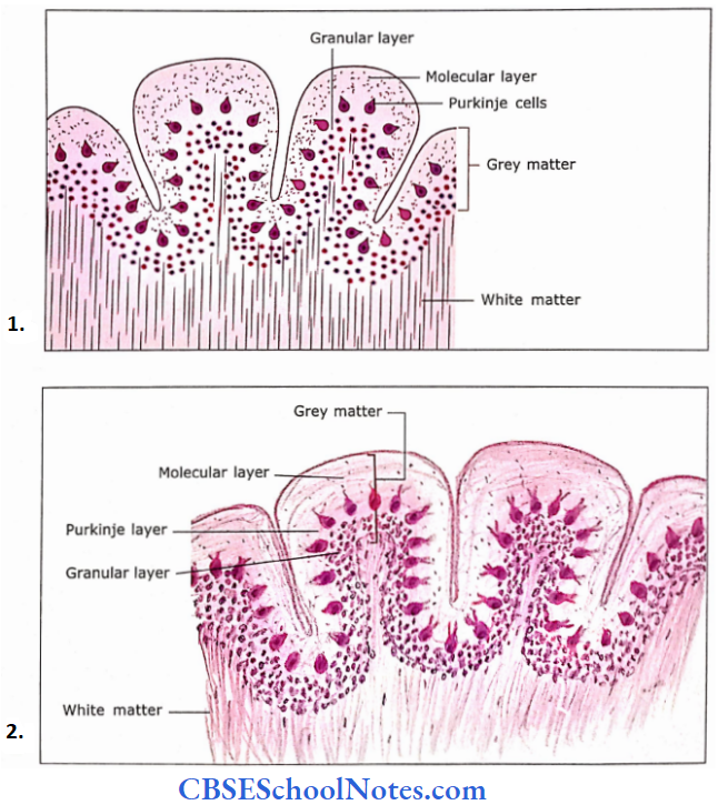
- The structure of two adjacent cerebellar folia shows outer grey and inner white matter.
- The grey matter of the cerebellar cortex consists of three layers, i.e., molecular, Purkinje, and granular layer.
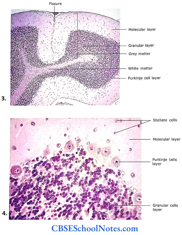
- Photomicrograph of cerebellar cortex at low magnification, cerebellar cortex is folded and presents fissures and folia.
- Photomicrograph of cerebellar cortex showing three layers, i.e., molecular layer, Purkinje cell layer, and granular cell layer.
Molecular Layer
It stains lightly with eosin and is featureless as it consists of a few cells and more myelinated and unmyelinated fibers.
- This layer consists of a few scattered stellate cells in the superficial part and a few basket cells in the deeper part.
- However, the dendritic processes of Purkinje cells and the axonal processes of granular cells (that run parallel to the surface of the cortex) occupy most of the molecular layer.
- The axons of granular cells come in synaptic contact with the dendrites of several Purkinje cells and basket cells. The molecular layer also contains the terminal part of climbing fibers.
Purkinje Cell Layer
These cells are arranged in a single row between molecular and granular layers. Purkinje cell (Golgi type F) is the large pyriform or flask-shaped neuron, that sends numerous dendrites into the molecular layer.
These dendrites synapse with axons of granular cells and climbing fibers. Purkinje cells give a single thin axon, which passes through a granular layer to end in deeper nuclei of the cerebellum.
Granular Layer
The granular layer stains deeply with the hematoxylin because it is densely packed with granular cells. Granular cells are small neurons with round nuclei surrounded by a thin rim of cytoplasm.
- These cells receive impulses from various parts of the CNS through Mossy fibers. The Mossy fibers in the granular layer end as the dilated terminal.
- On which the dendrites of granule cells and axons of Golgi cells (type 2) synapse to form light-stained areas called glomeruli.
- Granular cells send their axons into the molecular layer where they branch in the form of T and come in synaptic contact with dendrites of various Purkinje cells and basket cells.
- At the junction of molecular and granular layers, Golgi type 2 cells are found. Their vesicular nuclei are larger than granule cells. They contain chromophil substances present in the molecular layer.
- However, their axons form synaptic contact with glomeruli in the granular layer. Deep to the granular layer the cerebellar cortex lies in contact with white matter.
Granular Layer Remember
The cerebellar cortex consists of three layers, i.e., the molecular layer, the Purkinje cell layer, and the granular layer. The histological structure of the cerebellar cortex is uniform throughout the cerebellum.
Purkinje cells are large pyriform or flask-shaped neurons arranged in a single row between the molecular and granular layers.
Granular Layer Further Details
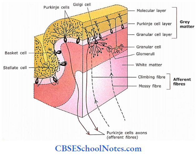
Neuronal Circuit of Cerebellum
- The input to the cerebellum is from different parts of CNS and is in the form of Mossy and climbing fibers
- The climbing fibers ascend to the molecular layer where they form synaptic contact with the dendritic arborization of Purkinje cells.
- The Mossy fibers end in glomeruli and form synaptic contact with granular cells in the granular layer.
- The granular cells convey the impulse received by Mossy fibers to the dendrites of Purkinje cells.
- The output of the cerebellum is in the form of axons of Purkinje cells, which synapse with the intracerebellar nuclei.
- The other neurons like Golgi cells, basket cells, and stellate cells interconnect the intracortical circuit to modify the outgoing impulses.
Spinal Cord
The human spinal cord is about 45 cm long and has cervical, thoracic, and lumbosacral parts. A cross-section of the spinal cord shows a central canal lined by ependyma made up of simple ciliated columnar cells.
- The central canal is surrounded by grey matter, which contains neurons, nerve fibers, neuroglial cells, and blood vessels.
- The grey matter is surrounded by white matter, which consists of bundles of nerve fibers and neuroglial cells. The surface of the spinal cord is covered with pia mater.
Grey Matter
The grey matter of the spinal cord appears roughly in the form of an H. It has anterior and posterior grey columns or horns. The anterior grey column contains large-size multipolar motor neurons.
- The nucleus of the motor cell is a large, spherical, light-staining structure with intensely staining nucleolus. The cytoplasm contains clumps of dark staining basophilic Nissl substance.
- The Nissl substance extends into the dendritic processes neuron but not into the axon. The axons of motor cells form the ventral spinal root.
- The neurons of the posterior grey column are much smaller than the anterior horn cells. Within the grey matter, besides the sensory and motor nerve cells, there are numerous neuroglial cells and blood vessels.
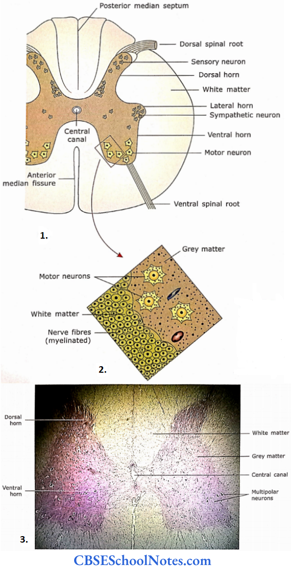
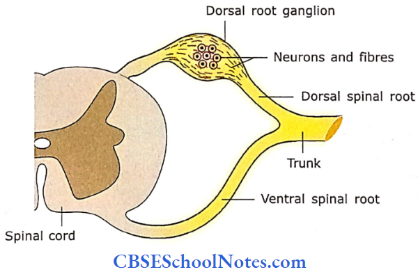
- Drawing to show the arrangement of the grey and white matter of the thoracic spinal cord (transverse section).
- The section of a part of the spinal cord shows the motor neurons in the anterior grey column and fibers in adjacent white matter.
- Section of spinal cord (at low magnification) showing centrally placed grey matter and peripherally placed white matter.
White Matter
The white matter is composed primarily of myelinated nerve fibers, but also neuroglial cells and blood vessels. The nerve fibers are comprised of ascending and descending tracts.
- Nerve fibers are surrounded by a myelin sheath, which in turn is surrounded by a fine connective tissue sheath called epineurium. As the myelin sheath gets dissolved during the preparation of H and E sections.
- The dark-staining axons (in a transverse section of the spinal cord) are surrounded by a clear space, which had been occupied by myelin.
White Matter Remember
A cross-section of the spinal cord shows a central canal surrounded by grey matter. The grey matter is surrounded by white matter. The grey matter of the spinal cord is in the form of an H and has anterior and posterior grey columns or horns.
- The anterior grey column contains large-size multipolar motor neurons, while neurons of the posterior grey column are much smaller than anterior horn cells and are sensory neurons.
- The white matter of the spinal cord consists of ascending and descending nerve fibers, which are mostly myelinated.
White Matter Clinical Application
Multiple Sclerosis
It is the most common disorder of the nervous system affecting young adults. In this condition, myelinated nerve fibers of the brain and spinal cord are progressively damaged due to the destruction of the myelin sheath.
- This affects the sensation, movements, body functions, and balance. Damage to the optic nerve may cause blurred vision. If nerve fibers in the spinal cord are affected.
- It may cause weakness or heaviness in the limbs. Damage to the fibers in the brain stem may affect balance. The demyelination is thought to result from an autoimmune disease with inflammatory features.
Guillain-barre syndrome
ln this disease, there occurs the demyelination of peripheral nerves and motor nerves arising from the ventral roots. The person suffers from muscle weakness and difficulty in respiration.
Peripheral Nervous System
All nervous tissues other than the brain and spinal cord are classified as the peripheral nervous system. The peripheral nervous system consists of nerves (made up of bundles of nerve fibers) and ganglia (collection of neurons and nerve fibers outside CNS).
Nerve
A nerve is defined as the collection of nerve fibers, which may be myelinated and or unmyelinated, and held together by connective tissue.
- A nerve consists of nerve fibers (axons or dendrites), supporting neuroglial cells (Schwann cells) and connective tissue.
- A nerve fiber is first surrounded by Schwann cells (which may form the myelin sheath around it), then it is surrounded by a thin layer of connective tissue called endoneurium.
- The endoneurium is made up of delicate collagen fibers and a few fibroblasts.
- At the light microscopic level, the endoneurium shows the nuclei of fibroblasts, which are difficult to distinguish from the nuclei of Schwann cells.
- Numerous fibers are held together to form a bundle of nerve fibers, which is surrounded by a sheath, called perineurium.
The perineurium is a cellular sheath made up of 3-4 layers of squamous-shaped cells and extracellular material. These cells show basal lamina and are contractile.
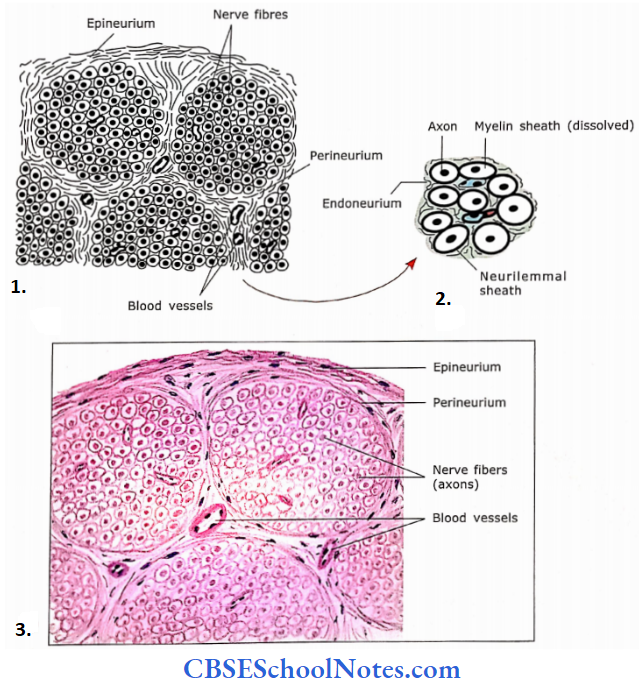
- A part of a transverse section of a nerve showing nerve fibers (axons) arranged in bundles and covered by a connective tissue sheath (perineurium).
- A drawing of the transverse section of a few nerve fibers is shown in an enlarged view.
- Under microscope.
- At low magnification.
- At medium magnification
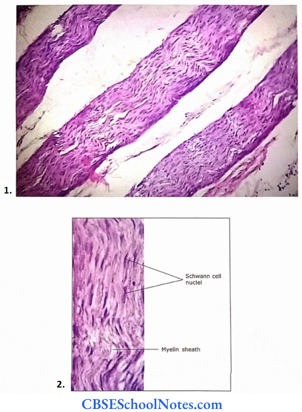
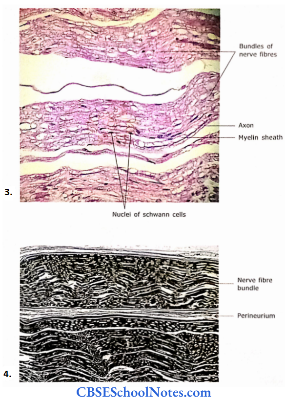
- Photograph of a longitudinal section of nerve at high magnification.
- Micrograph of a longitudinal section of nerve fibers (Sliver Stain).
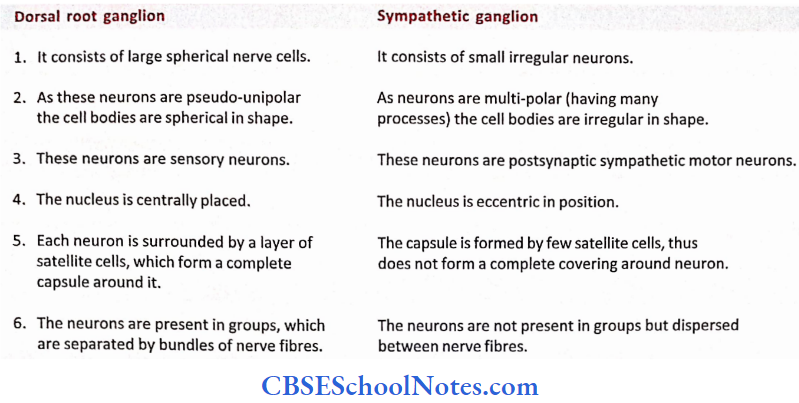
Differences between dorsal root ganglion and sympathetic ganglion
The perineurium forms a semi-permeable barrier. Thus, the cells of perineurium are not in true sense connective tissue cells, which comprise epineurium and endoneurium.
- These cells are more like an epithelioid tissue. Many bundles of nerve fibers are finally covered by a dense connective tissue sheath called epineurium.
- The epineurium not only forms the outermost covering of a nerve, but it also goes inside between perineurial bundles, to bind them together. The epineurium contains blood vessels and adipose tissue.
- The fine branches of blood vessels after penetrating the perineurium reach the endoneurium to supply it and nerve fibers.
Nerve Remember
Peripheral nerves are covered by three different connective tissue sheathes, i.e., epineurium, perineurium, and endoneurium.
Dorsal Root Ganglion
It is also known as sensory ganglion. This ganglion is the collection of sensory neurons on the dorsal root of the spinal nerves.
- Each ganglion is surrounded by a connective tissue capsule, which is the epineurium of the dorsal root. Beneath the capsule, the ganglion contains large cell bodies arranged in groups.
- Also, between and around the groups of neurons there are bundles of myelinated nerve fibers. Most nerve fiber bundles are seen in the central part of the ganglion and groups of nerve cells are seen in the peripheral part.
- The neurons of the dorsal root ganglion have large, spherical bodies with large pale-staining euchromatic nuclei and dark-staining nucleoli. Each neuron is surrounded by the satellite cells.
- The satellite cells, which form a sheath around the neuron, are much smaller compared to neurons. They are flattened or low cuboidal neuroglial cells and form an inner capsule around each neuron.
- The fibroblast and fibers form an outer capsule surrounding the inner capsule formed by satellite cells. The satellite cells prevent unwanted depolarization of sensory neurons.
Dorsal Root Ganglion Remember
The dorsal root (sensory) ganglion is a collection of pseudo-unipolar, rounded sensory neurons on the dorsal root of the spinal nerve.
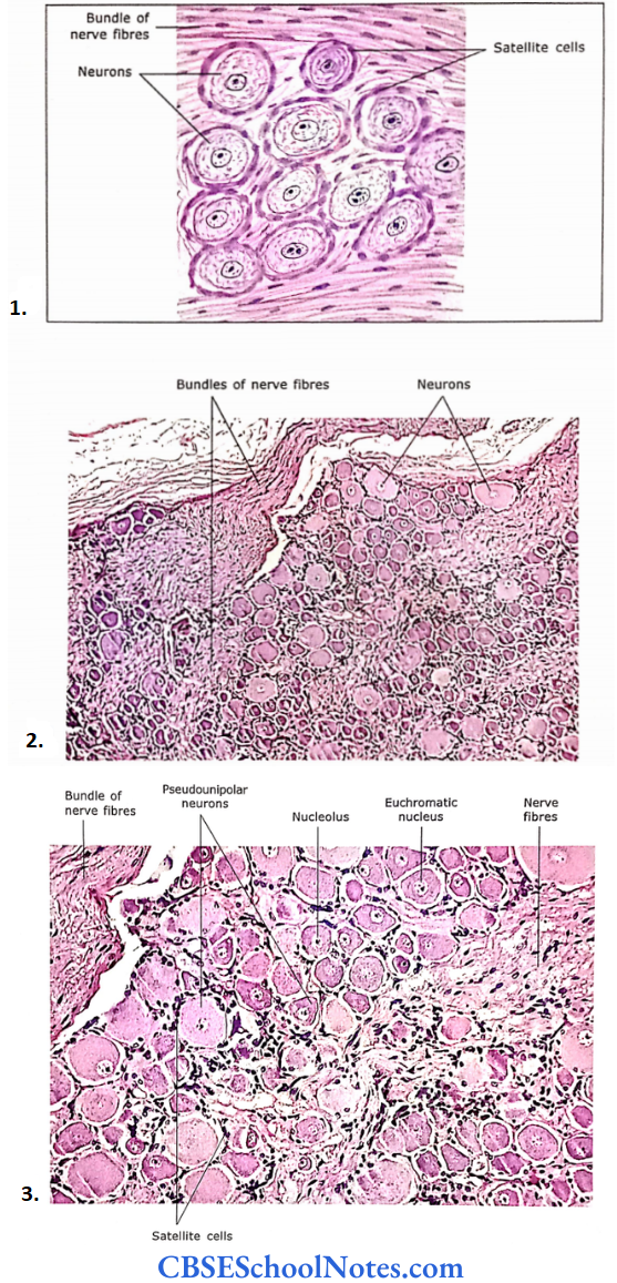
- Section of dorsal root ganglion (spinal ganglion) showing spherical nerve cells with euchromatic nuclei and prominent nucleolus.
- Section of dorsal root ganglion (at low magnification) consisting of large-size pseudo-unipolar neurons.
- Photomicrograph at high magnification.
Sympathetic Trunk Ganglion
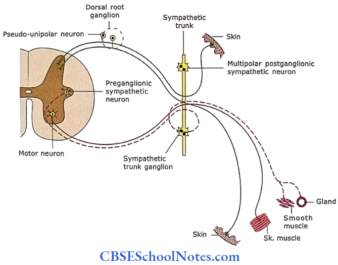
The sympathetic ganglion is the ganglion of the autonomic nervous system and lies along the sympathetic trunk. They contain cell bodies of postsynaptic motor neurons of the autonomic nervous system.
- The ganglion is covered with a thin capsule of connective tissue. It consists of small, irregular neurons dispersed between the nerve fibers. Cells are multipolar and, therefore appear irregular in shape.
- They contain eccentrically placed nuclei with prominent nucleoli. The cytoplasm contains small Nissl bodies. The satellite cells are less in number than in dorsal root ganglion cells.
- In between nerve cells, there is supportive connective tissue, blood vessels, and bundles of nerve fibers (both myelinated preganglionic and unmyelinated postganglionic).
Sympathetic Trunk Ganglion Remember
Autonomic ganglion houses cell bodies of postganglionic autonomic nerves.
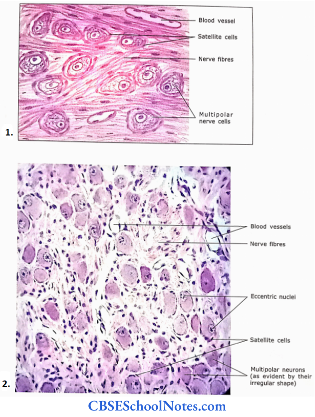
- Section of sympathetic ganglion showing small irregular (multipolar) nerve cells
- Photomicrograph of sympathetic ganglion showing multipolar neurons, scattered between nerve fibers.