
Anatomy Of The Brainstem Pons
The ventral surface is convex and bounded by upper and lower borders. This surface presents transversely running ridges (fibres). Laterally, these ridges come closer to form a bundle, the middle cerebellar peduncle.
The point of junction between the anterior surface of the pons and the middle cerebellar peduncle is marked by the emergence of the trigeminal nerve. The ventral surface also presents a shallow groove in the midline, known as the basilar groove. This groove lodges the ‘basilar artery’.
On the ventral aspect, three cranial nerves (6, 7 and 8) are seen to emerge from the lower border of the pons.
The posterior surface of the pons is formed by the upper part of the floor of the fourth ventricle. The posterior surface of the pons is related to the cerebellum
Read and Learn More Neuroanatomy
Brainstem Pons External Features
Pons is a part of the brainstem, situated between the medulla, below, and the midbrain, above. It lies in front of the cerebellum. It is about 2.5 cm long and presents a ventral and a dorsal surface.
Brainstem Pons Internal Structure
A transverse section passing through the pons is divided into ventral (basilar) and posterior (tegmentum) parts.
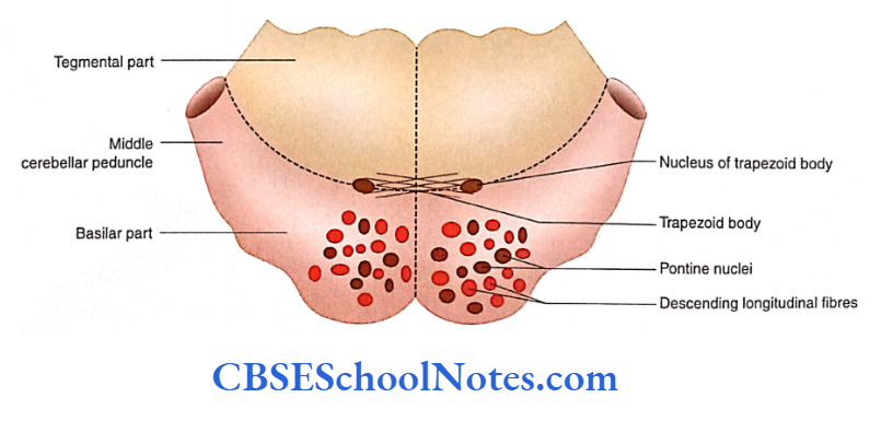
Structure of the Basilar Part
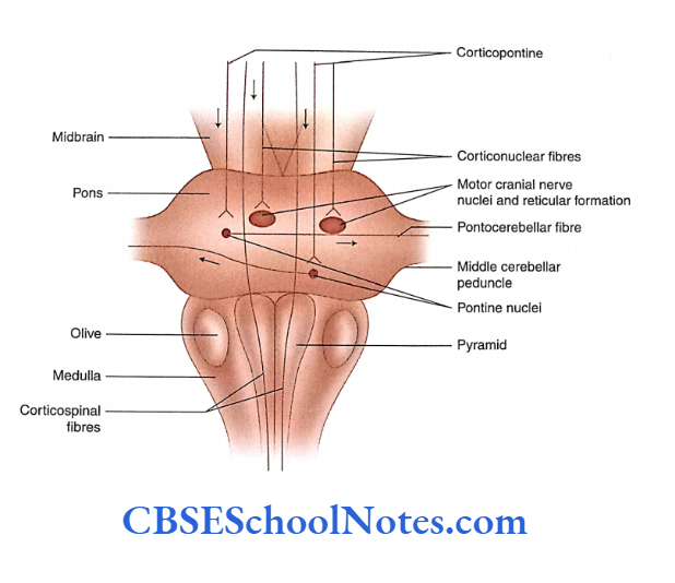
The basilar part of the pons consists of descending longitudinal fibres, transverse pontine fibres and pontine nuclei.
Descending longitudinal fibres: The descending longitudinal fibres consist of corticospinal, corticonuclear and corticopontine fibres.
Pontine nuclei: The pontine nuclei are small masses of grey matter scattered between longitudinal (vertical) and transversely arranged fibres.
Anatomy Of The Brainstem
Corticopontine fibres from various lobes of the cerebral cortex end in pontine nuclei.
Transverse pontine fibres: Transverse pontine fibres are pontocerebellar fibres that run transversely across the midline of the pons.
The pontocerebellar fibres (which form the middle cerebellar peduncle) are a part of the corticopontocerebellar pathway.
Structure of the Tegmental Part
The tegmental (dorsal) part of the pons is the upward continuation of the medullary reticular formation.
The white matter in the tegmentum consists of various ascending and descending tracts.
The structure of the tegmentum is different in the upper and lower parts of the pons. Hence, it is customary to study the internal structure of the tegmental part of pons at two different levels.
- Caudal (lower) Part transverse section passing through the facial colliculus
- Cranial (upper) part, transverse section passing through trigeminal nuclei.
Transverse Section Through The Tegmentum Of Lower Pons
Arrangement of Grey Matter
Anatomy Of The Brainstem
The floor of the fourth ventricle, at this level, is lined by grey matter and shows the presence of two cranial nerve nuclei.
Abducent and Vestibular.
Also seen are the cranial nerve nuclei of the trigeminal (spinal nucleus of the trigeminal nerve) and facial nerve, at deeper levels.
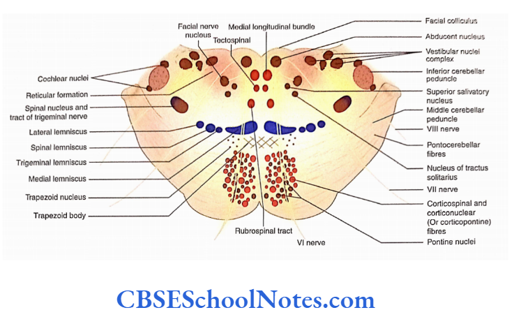
- The abducent nerve nucleus lies beneath the facial colliculus and lateral to the medial longitudinal bundle.
- The facial colliculus is formed due to a complicated loop formed by the fibres of the facial nerve winding around the abducent nucleus.
- The vestibular nuclei complex receives afferent fibres from the vestibular division of the vestibulocochlear nerve.
- There are two cochlear nuclei which are designated as ‘ventral’ and ‘dorsal’. The ventral and dorsal nuclei lie on the ventral and dorsal aspects of the inferior cerebellar peduncle, respectively.
- The motor nucleus of the facial nerve is present in the reticular formation, medial to the nucleus of the spinal tract of the trigeminal nerve.
- The salivatory nuclei are usually divided into superior salivatory, inferior salivatory and lacrimatory nuclei. The nuclei.
- These nuclei send secretomotor fibres via facial and glossopharyngeal nerves to various salivary glands.
- The nucleus of the spinal tract of the trigeminal nerve and the tract is present ventromedial to the inferior cerebellar peduncle and lateral to the nucleus of the facial nerve.
Nucleus of tracts solitarius: At the level ofthe lower pons, this nucleus lies lateral to the superior salivatory nucleus.
- The reticular nuclei are a small collection of grey matter scattered in the network of white fibres (reticular formation).
- The nucleus of the trapezoid body is situated in the the ventral part of the tegmentum.
- This nucleus is placed in the auditory pathway and its fibres constitute the lateral lemniscus.
Arrangement Of White Matter
Trapezoid Body
Brain Stem Parts
The trapezoid body is formed by the fibres of both ventral The trapezoid body is formed by fibres of both ventral.
The fibres of the trapezoid body relay in the trapezoid nucleus and the superior olivary complex. The efferents of these nuclei form the lateral lemniscus (ascending auditory pathways).
Brain Stem Parts
Medial, Trigeminal, Spinal and Lateral Lemnisci
- These lemnisci are situated posterior to the trapezoid body.
- The medial lemniscus is the most medial and now oriented transversely. It was formed by the decussation of internal arcuate fibres in the medulla.
- The trigeminal lemniscus begins to form at this level and is situated lateral to the medial lemniscus. It is formed by the axons arising from the contralateral spinal nucleus of the trigeminal nerve. This tract conveys the exteroceptive impulses (pain, touch, temperature) from the area of supply of the trigeminal nerve (face, nose, mouth, tongue, conjunctiva, etc.
- The spinal lemniscus is formed by the fibres of lateral spinothalamic and spinotectal tracts. It lies lateral to the trigeminal lemniscus.
- The lateral lemniscus begins in the lower half of the pons. Is part of the auditory pathway. It lies most laterally in the lemniscal band, i.e. lateral to the spinal lemniscus.
- The ventral spinocerebellar tract is situated dorsolateral to the lateral lemniscus.
- The spinal nucleus and the tract of the trigeminal nerve are situated lateral to the facial nerve nucleus.
- The lateralmost area of the tegmentum is occupied by the inferior cerebellar peduncle.
Transverse Section Through The Tegmentum of Upper Pons
The transverse section through the upper part of the pons corresponds to the level of trigeminal nuclei. This level Passes through the 1116 motor and Principal Sensory nuclei of the trigeminal nerve.
Brainstem Nuclei
The dorsal part of the tegmentum now contains the cavity of the fourth ventricle, which is bounded dorsolaterally on either side by a superior cerebellar peduncle and roofed by the superior medullary velum.
Arrangement of Grey Matter in Tegmentum
- The following nuclei are seen in the tegmentum.
- The principal (superior) sensory nucleus of the trigeminal nerve: It is situated lateral to the motor nucleus. Below this level, the nucleus is continuous with the spinal nucleus of the trigeminal nerve.
- Motor nucleus of the trigeminal nerve: This nucleus lies medial to the sensory nucleus on the floor
Brainstem Pons Summary
- External features of pons: The pons lie between the medulla and the midbrain and are situated in front of the cerebellum.
- It presents ventral and dorsal surfaces. The ventral surface of the pons is convex and presents a shallow groove in the midline—the basilar groove.
- On each side, the ventral surface is continuous with the middle cerebellar peduncle and shows the attachment of the V cranial nerve.
- The posterior surface of the pons is formed by the upper part of the floor of the fourth ventricle. Three cranial nerves (4, 7 and 8) emerge from the pontomedullary junction.
Brainstem Nuclei
Internal structure of pons: The transverse section passing through pons is divided into basilar and tegmental parts.
- The structure of the basilar part of the pons consists of descending fibres (corticospinal, corticonuclear and corticopontine) and transverse pontine fibres (pontocerebellar).
- The pontine nuclei are small masses of grey matter scattered between longitudinally and transversely arranged fibres.
- The structure of the tegmental part of pons consists of ascending and descending tracts, cranial nerve nuclei of 5 to 8 nerves, and reticular formation.
- The structure of the basilar part of the pons remains constant throughout the pons. However, the structure of the tegmentum differs in the upper and lower parts of the pons.
Transverse section through the lower pons: The tegmentum, at the level of facial colliculus (lower pons), shows the following features:
Arrangement of grey matter in tegmentum: The floor of the fourth ventricle, at this level, shows the presence of abducent and vestibular nerve nuclei.
Brainstem Nuclei
- The ventral dorsal and cochlear nuclei are located on the dorsal and ventral aspects of the inferior cerebellar peduncle, respectively.
- The motor nucleus of the facial nerve is present in the area of reticular formation. The facial nerve follows an unusual course before it comes out at the pontomedullary junction.
- The nucleus of the spinal tract of the trigeminal nerve is located ventromedial to the inferior cerebellar peduncle.
- The nucleus of tractus solitarius and the superior salivatory nucleus is situated ventromedial to the facial nucleus.
Arrangement of white matter: The trapezoid body is seen at the junction of basilar and tegmental parts. Posterior to the trapezoid body, four lemnisci (medial, trigeminal, spinal and lateral) are arranged from the medial to the lateral side.
The medial longitudinal bundle, tectospinal tract and rubrospinal tract are situated at the paramedian position. The inferior cerebellar peduncle is present in the lateralmost area of the tegmentum.
Transverse section through the upper pons
Arrangement of grey matter in tegmentum: The principal (superior) sensory nucleus of the trigeminal nerve is situated lateral to the motor nucleus.
The motor nucleus of the trigeminal nerve lies medial to the sensory nucleus on the floor of the fourth ventricle. The nucleus of the lateral lemniscus is present medial to the lateral lemniscus.
Arrangement of white matter: The medial, trigeminal, spinal and lateral lemnisci are in the same position.
The ventral spinocerebellar tract now forms the superior cerebellar peduncle. The medial longitudinal bundle, tectospinal tract and rubrospinal tracts are seen in the paramedian position.