Bones of the Thoracic Region
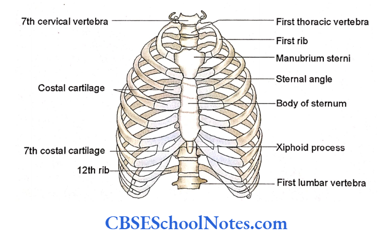
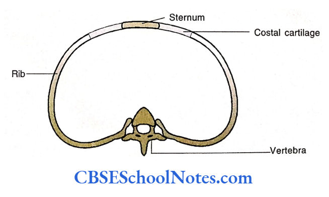
The skeleton of the thoracic region forms a bony cage, which protects the lungs, heart, and some upper abdominal organs.
This cage is formed by the following bones and cartilages:
- Thoracic vertebrae and intervertebral discs.
- Ribs and costal cartilages.
- The sternum.
The thoracic cage is narrower at its superior end and broader at its inferior end. The cage is flattened anteroposteriorly.
Posteriorly, it is formed by 12 thoracic vertebrae and intervertebral discs. The sidewalls of this cage are made up of 12 ribs on each side.
The posterior end of each arched rib articulates with the vertebral column.
Manubriosternal Angle
The anterior end of each rib is attached to the costal cartilage. Through these costal cartilages, ribs gain attachment to the sternum.
The sternum is a flat narrow bone situated on the anterior aspect of the thoracic cage.
Sternum
The sternum is a flat, elongated bone measuring about 6 inches in length. It is situated anteriorly in the wall of the thorax.
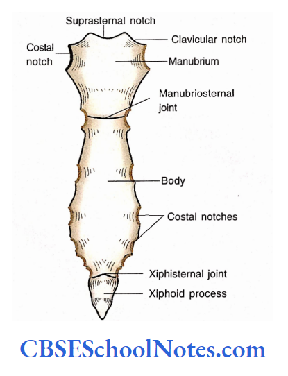
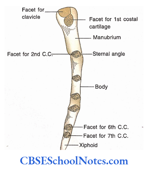
From above downwards, the sternum consists of three portions, i.e., the manubrium, the body, and the xiphoid process.
The body is the middle and largest portion of the sternum. The junction of the manubrium and the upper end of the body forms the manubriosternal joint (the sternal angle).
The junction of the lower end of the body and the xiphoid process forms the xiphisternal joint.
The upper border of the manubrium is concave. It is known as a suprasternal notch.
Manubriosternal Angle
Lateral to the suprasternal notch are clavicular notches that articulate with the medial ends of the clavicles to form sternoclavicular joints.
Read and Learn More Human Osteology Notes
Inferolateral to the clavicular notch there lies a costal notch where the first costal cartilage articulates with the manubrium to form a sternocostal joint. This joint is classified as synchondrosis (primary cartilaginous joint).
The manubrium and the body of the sternum lie in different planes at the manubriosternal joint, therefore the joint projects forwards. This projecting angle is called as angle of Louis or sternal angle.
General Features Manubrium
- The manubrium is the widest and thickest of the three parts of the sternum.
- It is roughly quadrilateral in shape At the sternal angle, the second costal cartilage articulates with the lower end of the manubrium and the upper end of the body.
- As the sternal angle is projecting and subcutaneous it is an easily palpable landmark. This fact is utilized to count the ribs. The rib counting starts with the 2nd rib adjacent to the sternal angle.
- The lower end of the manubrium lies at the level of the lower border of the T4 vertebra.
Body of the Sternum
The body of the sternum is a long and thin plate of bone. Its lateral borders present costal notches for articulation with 2nd to 7th costal cartilages.
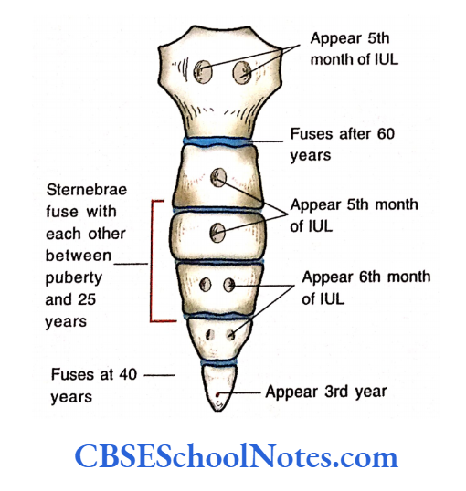
All these joints are synovial. In adults, the body of the sternum is made up of four pieces (sternebrae).
These pieces of bones are joined to each other by primary cartilaginous joints. These joints begin to fuse soon after puberty. In an adult, these fusion sites are seen as three transverse ridges.
Xiphoid Process
The xiphoid process is the smallest, triangular part of the sternum. The upper end of the xiphoid process meets the body of the. sternum to form xiphisternal joint.
The lateral border of the xiphoid process has a demi facet for articulation with the 7th costal cartilage.
The xiphoid process is cartilaginous in young people. It begins to ossify at about the third year of age.
Manubriosternal Angle
It gets completely ossified at about 40 years of age. It fuses with the body of the sternum in old people.
Sternum Particular Features
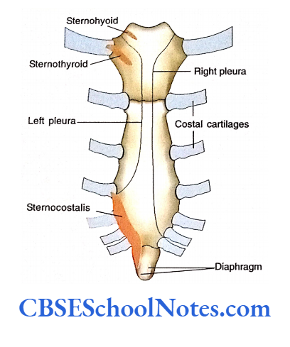
Attachments of Muscles on the Anterior Surface of the Sternum Note the origin of the sternal head of the sternocleidomastoid and pectoralis major muscles and insertion of rectus abdominis with the help.
Muscles attached on the posterior aspect of the sternum. Note the origin of sternohyoid, sternothyroid, and sternocostalis with the help. Also, note the reflection of the pleura on both the sides of sternum.
Sternum Ossification
The sternum develops by the fusion of the right and left cartilaginous plates in the midline. After the fusion, many centers of ossification appear in the cartilaginous model.
Sternum Clinical importance
Fractures
Sternal fractures may occur due to automobile accidents. The sternum is broken into pieces due to crush injuries against the steering wheel.

Sternotomy
For coronary bypass surgery and surgeries on lung, these organs are exposed by cutting the sternum in the midline. A wide gap is exposed between two split parts of the sternum because of the elasticity of the costal cartilage and the flexibility of the ribs. After surgery two splitted parts of the sternum are joined by wire sutures.
Bone marrow biopsy
As the sternum is superficial (subcutaneous) it is best suited for needle biopsy of bone marrow. The bone marrow sample is obtained from the spongy bone of the sternum with the help of a needle. A bone marrow biopsy is needed to diagnose blood abnormalities and for bone marrow transplantation.
Sternum is helpful in the identification of sex To a certain extent the sex of an individual can be identified with the help of the sternum as the female sternum is short and wide as compared to males, Jit, et al. (1980). The determination of the sex of bones is needed in medico-legal cases.
Manubriosternal Angle
Congenital defects of sternum As the sternum develops by the fusion of two halves of the sternum in the midline, if these two pieces fail to fuse then the heart is exposed on the thoracic wall (ectopia cordis).
In this condition, pericardium also fails to form. Sometimes there may be the presence of a foramen in the body of the sternum. This occurs due to faulty ossification.
Ribs
Ribs are curved, flat bones that form the greater part of the thoracic wall.
They are arranged in twelve pairs. The length of the ribs increases from the first to the seventh rib. Thereafter the length gradually decreases from the eighth to the twelfth rib.
Each rib articulates posteriorly with the corresponding vertebra. The anterior ends of ribs (except the eleventh and twelfth ribs) articulate with the sternum through costal cartilages.

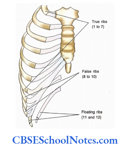
Classification of Ribs
There are three types of ribs:
True Ribs
These ribs are directly attached to the sternum through their costal cartilage. The first seven ribs are true ribs and these ribs are also called vertebrosternal ribs.
False Ribs
These ribs are indirectly connected to the sternum. Their costal cartilages are joined to the costal cartilages of superior ribs. The eighth to the tenth ribs are false ribs and are also called vertebrochondral ribs.
Floating Ribs
The eleventh and twelfth ribs are not connected with the sternum. Their anterior ends are free and covered by rudimentary costal cartilage. They are also known as vertebral ribs or floating ribs.
Ribs may also be classified as typical and atypical ribs. The typical ribs are those, that have the same (common) features. The 3rd to the 9th ribs are typical ribs.
Manubriosternal Angle
The atypical ribs have special features therefore can be differentiated from the rest of the ribs. The atypical ribs are 1st, 2nd, 10th, 11th and 12th ribs.
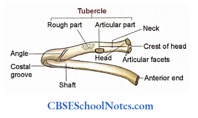
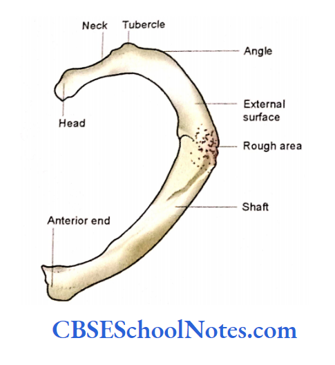
The Typical Ribs
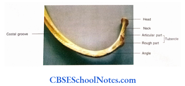
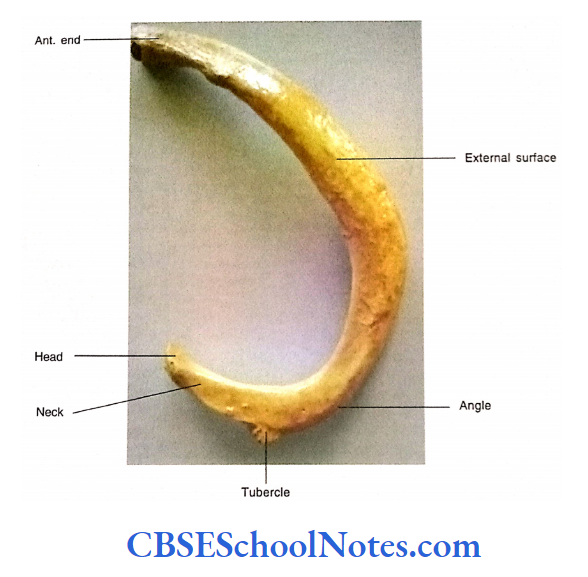
A typical rib consists of a head, neck, tubercle, angle, and shaft. The head is present at the posterior end and bears one or two articular facets.
The tubercle is a knob-like structure present at the junction of the neck and shaft.
The shaft is long and curved. The convex surface is the external surface while the concave surface is the internal surface, which bears a groove called a subcostal groove.
The upper border of the shaft is rounded while the lower border is sharp. The sternal end bears a concave depression for attachment of costal cartilage.
Ribs Side Determination
- The end of the rib having the head, neck, and tubercle (posterior end) should be kept posteriorly.
- The concave curved surface should be kept medially.
- The sharp border (inferior border) of the shaft should be kept downwards.
Thoracic Cage Bones
Ribs Anatomical Position
- The posterior end should be kept near the midline.
- The posterior end is at a higher level as compared to the anterior end.
Ribs General Features
The Posterior or Vertebral End
It includes the head, neck, and tubercle.
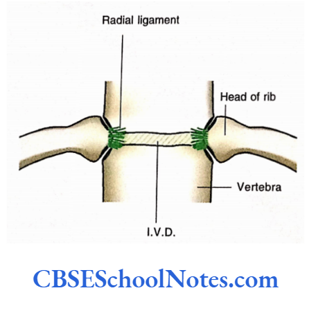
- The head presents two articular facets separated by the crest of the head. The lower facet is large and articulates with the body of the numerically corresponding vertebra.
- The upper small articular facet articulates with the body of the adjacent upper vertebra. The crest of the head is connected to the intervertebral disc by intra-articular ligament.
- The neck is short and has a sharp upper border. It is sometimes called as crest of the neck. The neck has an anterior smooth and a posterior rough surface.
- On the posterior aspect of the rib, just lateral to the neck, there is the presence of an elevation called a tubercle. The tubercle has a medial articular (smooth) and lateral non-articular (rough) part.
The Anterior End
The anterior end of the rib has a cup-shaped depression for articulation with the lateral end of the corresponding costal (smooth) cartilage to form a costochondral joint.
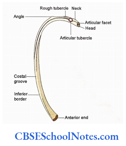
The Shaft
The shaft of a typical rib is thin, flat, and curved. It forms the major part of the rib. It
extends between the tubercle and the anterior end of the rib.
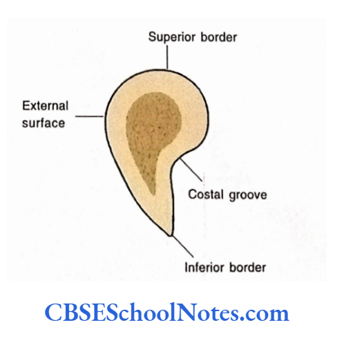
- It has a superior rounded border and a sharp inferior border. It has an outer convex and an inner concave surface The inner surface shows a shallow costal groove just above the inferior border.
- The costal groove is well-defined in the middle part of the shaft.
- A short distance lateral to the tubercle rib bends anteriorly. This is called as posterior angle.
- The rib also shows a twisting, because of which, two ends of the rib cannot touch a horizontal plane simultaneously (when kept on the table.
Ribs Particular Features
Thoracic Cage Bones
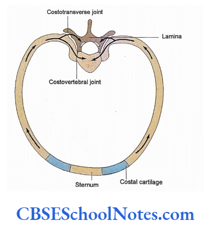
Joints about the Typical Rib
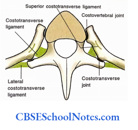
- The head of a typical rib articulates with the adjacent vertebral bodies (with the numerically corresponding body and a body above it) and intervertebral disc to form a costovertebral joint.
- The costotransverse joint is formed between the costal facet of the transverse process and the articular part of the tubercle.
- This joint is supported by various costotransverse ligaments, i.e., the lateral costotransverse, superior costotransverse, and costotransverse ligaments.
The Muscles Attached to the Rib
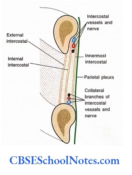
The attachments of external intercostal, The various muscles are also attached on the external surface of the ribs, i.e., pectoralis minor, serratus anterior, latissimus torsi, and back muscles.
The parietal pleura is related to the inner surface of the rib.
Nerves and Vessels Related to the Typical Rib
- The sympathetic trunk descends downwards on the anterior aspect of the heads of the typical ribs.
- The intercostal nerve and vessels lie in the costal groove between the internal intercostals and innermost intercostal muscles.
The Atypical Ribs
The First Rib
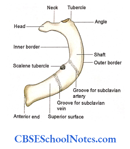
The first rib is the shortest, broadest, curved, and flat rib. As compared to the typical ribs its head has only one articular facet for the body of the first thoracic vertebra.
The posterior angle of the rib lies at the tubercle itself. The shaft shows inner and outer borders thus superior and inferior surfaces.
(This is in contrast to the typical ribs, which show superior and inferior borders and outer and inner surfaces.)
The superior surface of the first rib is rough and shows the presence of two grooves. The inferior surface is smooth and there is the absence of a subcostal groove.
First Rib Side Determination
- The head, neck, and tubercle are located posteriorly near the midline.
- The broad anterior end lies anteriorly.
- The concave inner border lies medially.
- The rough superior surface of the shaft (having grooves) should face superiorly.
Thoracic Cage Bones
First Rib Anatomical Position
- Keep the posterior or vertebral end near the midline.
- The anterior end is at the lower level as compared to the posterior end. In this position, the upper surface should face anterosuperiorly.
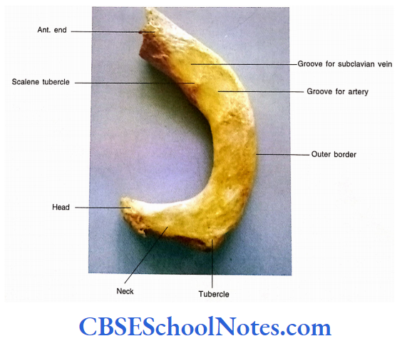
First Rib General Features
First Rib The Posterior End
- The posterior end consists of the head, neck, and tubercle. The head is rounded, small, and bears only one circular articular facet.
- The neck is rounded and extends upwards and posterolaterally.
- The neck shows superior, posterior, inferior, and anterior surfaces.
- The tubercle is large and prominent. The oval articular facet on the tubercle articulates with the transverse process of a first thoracic vertebra.
The Shaft
- The shaft is flat and shows superior and inferior surfaces and outer and inner borders.
- The superior surface of the first rib shows two shallow but wide grooves separated by a faint ridge. This ridge is continuous medially with the scalene tubercle on the inner border.
- The inferior surface is smooth and related to the parietal (costal) pleura.
Particular Features
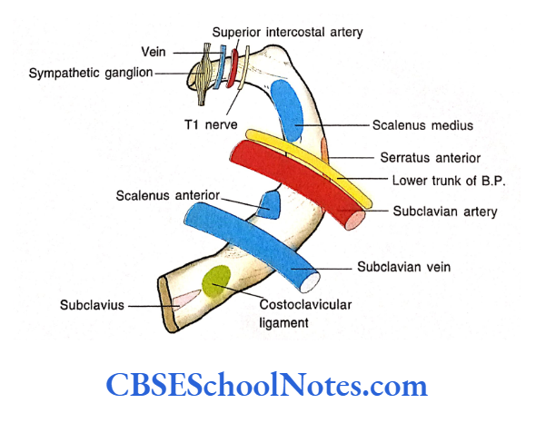
Attachments of the Muscles on the First Rib
- The subclavius muscle arises from the superior surface near its anterior end.
- The scalenus anterior is inserted on the scalene tubercle. The scalenus medius is attached behind the groove for the subclavian artery.
- The intercostal muscles arise from its outer border.
- The first digitation of the serratus anterior arises from its outer border
Relations of the Nerves, Vessels and Ligaments
- The costoclavicular ligament is attached on its superior surface near its anterior end The inner border gives attachment to the suprapleural membrane.
- The groove anterior to the scalene tubercle lodges the subclavian vein.
- The groove posterior to the scalene tubercle lodges the subclavian artery and lover trunk of the brachial plexus.
- The anterior surface of the neck is related from the medial to lateral side to the sympathetic trunk, first posterior intercostal vein, superior intercostal artery, and ascending branch ofthe ventral ramus of the first thoracic nerve.
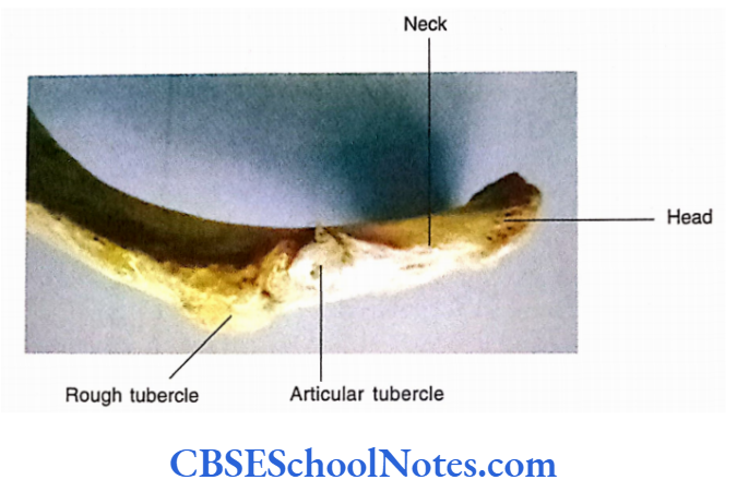
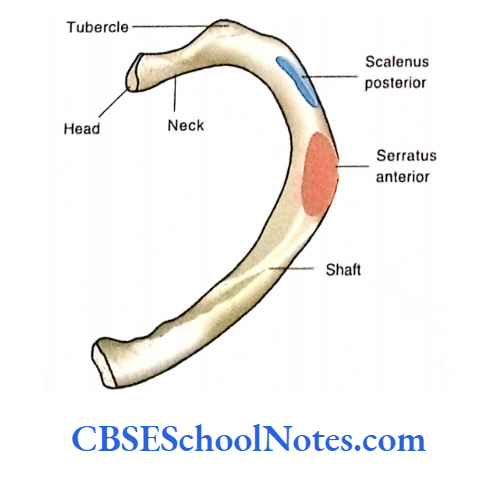
The Second Rib
- The second rib is almost twice the length of the first rib. The shaft has an external surface directed laterally and slightly upwards.
- This surface presents a prominent rough area near the middle of the shaft.
- It has a faint, short costal groove in the posterior part of the internal surface, which is directed downwards and medially.
Thoracic Cage Bones
Second Rib Particular Features
- The prominent rough area on the outer surface, just behind the middle of the shaft, gives origin to the 1st and 2nd digitations of the serratus anterior.
- The outer surface in its posterior part gives insertion to the scalenus posterior muscle.
- The intercostal muscles are attached to its upper and lower borders.
The Tenth Rib
It also presents the head, neck, tubercle, posterior angle, and shaft, like a typical rib.
However, it differs from typical ribs because it presents a single articular facet on its head for articulation with the 10th thoracic vertebral body.
The Eleventh Rib
This rib is short as compared to the tenth rib. It has no neck or tubercle.
Its lateral end is tapering while the vertebral end bears a single articular facet for the body of the eleventh thoracic vertebra.
Thoracic Cage Bones
The Twelfth Rib
- The twelfth rib is shorter than the eleventh.
- It is directed downwards, laterally, and forwards.
- Similar to the eleventh rib it has no neck or tubercle.
- It has no angle and there is also the absence of a subcostal groove.
- It has a single facet on the head for articulation with the body of the 12th thoracic vertebra.
- It has a pointed lateral end, which gives attachment to cartilage.
- This rib presents upper and lower borders and anterior and posterior surfaces.
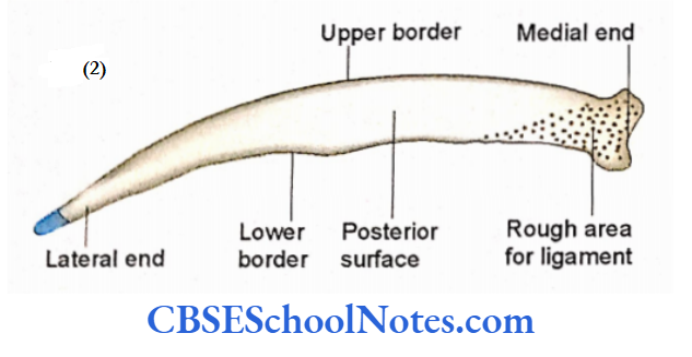
- The anterior surface is smooth and concave and faces slightly upwards.
Particular Features of the 12th Rib
- The medial part of the upper border gives attachment to the intercostal muscles.
- The lateral part of the upper border gives attachment to the diaphragm.
- The quadratus lumborum muscle with its covering (anterior layer of the thoraco¬ lumbar fascia) is attached to the medial half of the anterior surface.
- The transverse abdominis muscle is attached to the lower lateral part of the anterior surface.
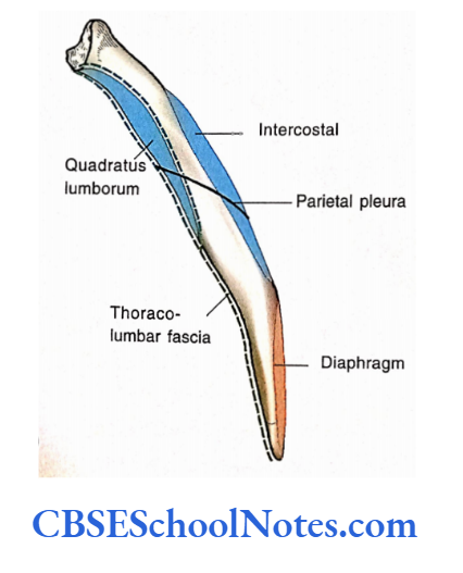

- The lower limit of the parietal (costal) pleura crosses the anterior surface of the rib and quadratus lumborum muscle. Thus costodiaphragmatic recess extends on the anterior aspect of the medial half of the rib.
- The attachments of the muscles on the posterior aspect of the twelfth rib.
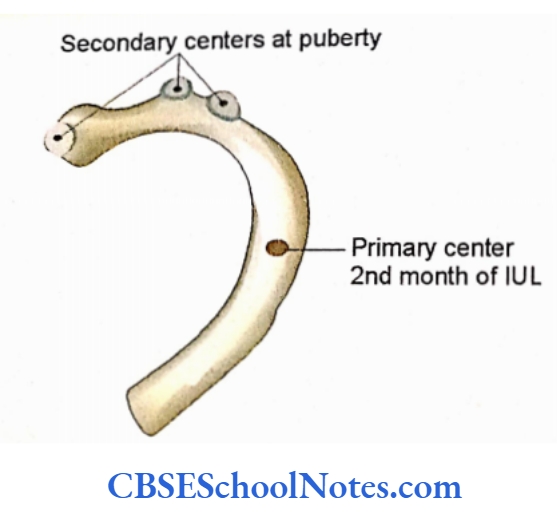
Ossification of Rib
A typical rib is ossified from one primary and three secondary centers (one for the head and two for the tubercle). Primary and secondary centers fuse after 20 years of age.
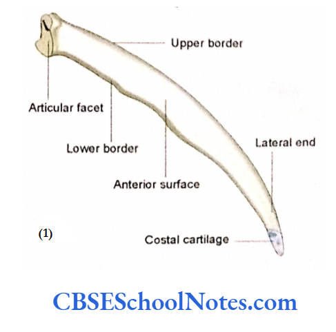
The Costal Cartilages
- The costal cartilages are flattened bars of hyaline cartilage.
- They prolong the ribs anteriorly.
- Each costal cartilage has anterior and posterior surfaces and upper and lower borders.
- The first seven costal cartilages join to the sternum.
- The 8th, 9th, and 10th articulate with the cartilage just superior to them. The 11th and 12th costal cartilages form caps on the anterior end of these ribs.
- The length of the costal cartilage increases from the first to the seventh. The length decreases gradually from the 8th to the 12th.
- The costal cartilage provides elasticity and mobility to the thoracic wall.
- They prevent fractures of the sternum and ribs due to their resilience.
Costal Cartilages Clinical Importance
Fractures of Ribs
- When there occurs a direct injury on the sidewall of the chest, the broken ends of the ribs may tear the pleura and lung. This may lead to the collection of air (pneumothorax) or blood (haemothorax) in the pleural cavity.
- When there is indirect violence, due to compression of the chest against the steering wheel in accidents, ribs are commonly fractured near their angles.
- Ribs are Used for Bone Grafting Similar to the fibula ribs are also used for bone grafting. In this procedure, the periosteum of the rib is incised along its length and a segment of the rib is removed for grafting leaving the periosteum intact. After some time, rib regenerates deep to the periosteum.
Extra Ribs
- Sometimes the number of ribs may increase (than normal 12 pairs) due to the presence of a cervical or lumbar rib. The cervical rib articulates with the 7th cervical vertebra but usually fails to attach to the sternum.
- The anterior end of the cervical rib may attach to the first rib. The cervical rib may compress the lower trunk of the brachial plexus.
- This may lead to pain and numbness in the shoulder and upper limb.
- This rib may also compress the subclavian artery resulting in pain in the upper limb due to poor blood supply to the limb muscles. Lumbar ribs are less common as compared to the cervical ribs.
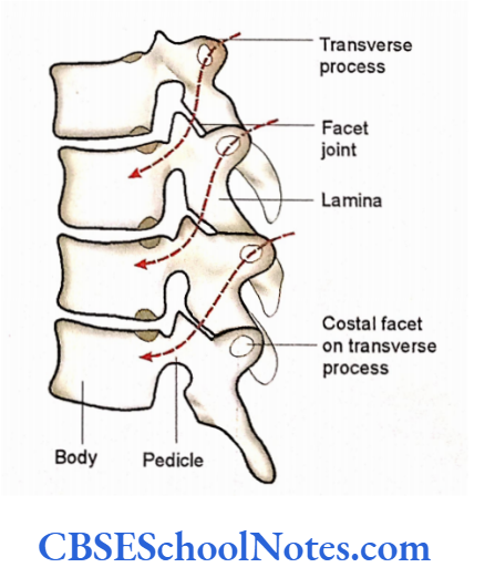
Thoracic Vertebrae
The thoracic part of the vertebral column is formed by the twelve thoracic vertebrae and intervertebral discs. This part of the column is concave anteriorly.
Thoracic vertebrae are larger and stronger than cervical vertebrae.
Compared to the cervical vertebrae they also have longer and larger transverse processes. The following three special features are helpful in the identification of thoracic vertebrae.
- Presence of costal facets or demi-facets on the bodies for articulation with the heads of the ribs.
- Presence of costal facets on their transverse processes for articulation with the tubercle of the ribs (except for Til and T12 vertebrae).
- The spinous processes of T3 to T9 vertebrae are long and slope downwards.
- The spinous process of Til and T12 are shorter, broader, and directed more posteriorly.
- The 2nd to 9th thoracic vertebrae are typical because they bear common bony features. The 1st, 10th 11th, and 12th thoracic vertebrae are atypical.
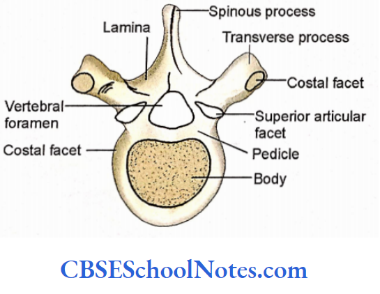
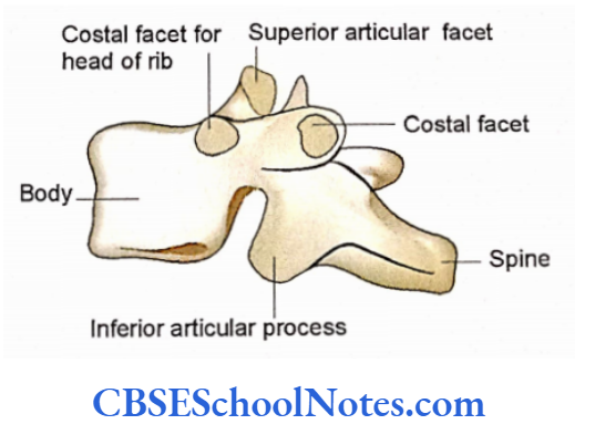
Typical Thoracic Vertebrae
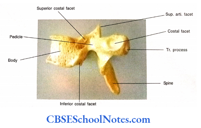
A typical thoracic vertebra is made up of a body and a neural arch (vertebral arch). Each vertebra has seven processes, i.e., four articular processes, two transverse processes, and one spinous process.
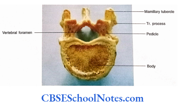
Body
The superior and inferior surfaces of the body of a typical vertebra are heart shaped
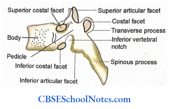
The lateral aspect of the body, close to its upper and lower borders, shows the presence of two costal demi-facets. The upper demi-facet is usually larger and is close to the pedicle.
It articulates with the numerically corresponding rib. The lower costal demi-facet is smaller and lies in front of the inferior vertebral notch and articulates with the lower rib.
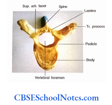
Vertebral Arch
It consists of pedicles and laminae. Pedicles are short and directed backward. Laminae are short, thick, and flat vertical plates of the bone that join in the midline to form the posterior portion of the vertebral arch.
The superior vertebral notch is shallow and present above the pedicle while the inferior vertebral notch is deep and present below the pedicle. The vertebral foramen is small and circular. It is bounded by body, pedicles, and laminae.
Vertebral Arch Processes
The transverse processes are large, club-shaped, and directed laterally and somewhat backward. On their anterior surface, they bear, an oval costal facet for articulation with the tubercle of the rib (costotransverse articulation).
The costal facets on the upper six transverse processes are concave and directed anterolaterally. While the costal facets in the 7th to 10th transverse processes are flat and directed upwards, forwards, and laterally.
The superior articular facets are directed posterolaterally while the inferior articular facets are directed anteromedially. The spinous processes are long and directed downwards
A typical Thoracic Vertebra
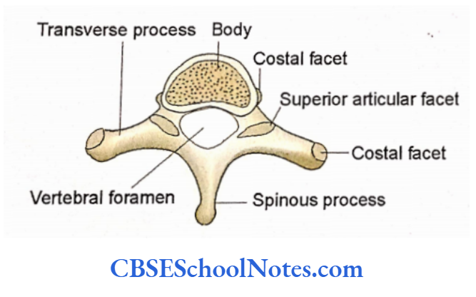
First Thoracic Vertebra
- The following features will help to identify the head of rib J’s first thoracic vertebra:
- The body is like cervical vertebrae, i.e., its anteroposterior diameter is less than its transverse diameter.
- There is the presence of a single circular costal facet on the lateral surface of the body for articulation with the head of the first rib.
- Presence of a small costal demi-facet near its lower border for articulation with the upper demi-facet on the head of the 2nd rib.
- The vertebral foramen is large and triangular (similar to the cervical vertebra).
- The spinous process is long and directed backward
Tenth Thoracic Vertebra
The shape of the body is somewhat like lumbar vertebrae.
Mostly it bears a single circular costal facet for the head of the 10th rib.
It means the head of the 10th rib will not articulate with the body of the 9th thoracic vertebra. Hence the body of the 9th vertebra will have only the upper demi-facet.
The costal facet is present on the anterior aspect of the transverse process for the tubercle of the 10th rib.
Eleventh Thoracic Vertebra
- The body is large and somewhat lumbar type.
- A single circular costal facet on the lateral aspect of the body.
- The transverse process is short and bears no costal facet, as the 11th rib is a floating rib.
- The superior and inferior articular facets are thoracic type. (Sometimes inferior articular facet may be lumbar type).
- The spinous process may be somewhat like a lumbar vertebra.
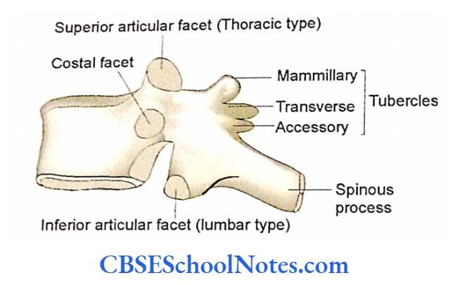
Twelfth Thoracic Vertebra
- The body is large and lumbar-type (kidney-shaped).
- The single circular costal facet for the head of the 12th rib. The facet is present mid-way between the upper and lower borders and tends to encroach on the lateral aspect of the pedicle.
- Similar to the 11th vertebra there is the absence of costal facet on the transverse process.
- The transverse processes of T12 are short (rudimentary) and present three tubercles, i.e., superior, inferior, and lateral. The lateral tubercle represents the true transverse process.
- The inferior articular facets are lumbar-like, i.e., they are convex and face anterolaterally. The superior articular facets are thoracic type, i.e., flat and directed posterolaterally.
- The spinous process is also like a lumbar vertebra, i.e., short, broad, and directed backward.
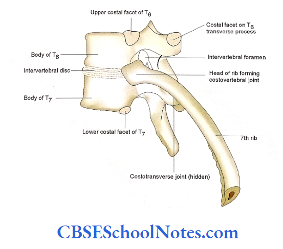
- Articulation of a Typical Rib with the Vertebrae (Costovertebral and Costotransverse Joints)
- A typical rib articulates posteriorly with the bodies of two adjacent vertebrae and the transverse process of the lower vertebra. Students should practice this articulation with the help of two typical thoracic vertebrae and a typical rib.
The Costotransverse Joint
The articular part of the tubercle of the rib presents an oval facet. It articulates with the articular facet on the transverse process of the corresponding vertebra.
This can be further understood by looking at the articulation of the 7th rib. The head of the 7th rib articulates with the body of the 6th thoracic vertebra (with the demi-facet near its lower border) and with the body of 7th thoracic vertebra (with the demi-facet near its upper border).
The head is also connected with the intervertebral disc between the 6th and 7th thoracic vertebrae. The tubercle of the 7th rib also articulates with the transverse process of the 7th thoracic vertebra.
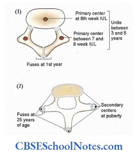
Ossification of Thoracic Vertebra
A typical thoracic vertebra ossifies by three primary centers, i.e., one for the body and one for each neural arch. These centers unite with each other between 3 to 5 years of age At puberty, five secondary centers appear, i.e., one for the spine, one for each transverse process, and two ring-like centers at the margin of the upper and lower surface of the body.
Tuberculosis of Thoracic Vertebrae Spongy bone in the body of thoracic vertebrae is the most common site of tubercular infection. Tubercular bacilli reach the spongy bone through blood circulation and settle there due to sluggish blood flow and restricted mobility of thoracic coloumn.
The infection leads to the destruction of spongy bone and the production of “Cold pus”. As the chronic injection of tubercle bacilli does not produce the local signs of heat, pain, and redness, it is called a cold abscess. Due to the destruction of spongy bone body is collapsed resulting in the production of kyphosis.
Scoliosis and Kyphosis
The thoracic coloumn is the most common site where scoliosis and kyphosis are produced (See Further Details). Kyphosis is most commonly seen in old age due to weakness of spongy bones of vertebral bodies (Osteoporosis).
Hemivertebra
Sometimes the body of the vertebra ossifies by two primary centers, one for each lateral half of the body. If one of these centers fails to ossify then only half of the vertebral body is formed. This leads to the lateral bending of the column on one side.