
Autonomic Nervous System
The autonomic nervous system controls and coordinates the internal environment of the body. This system has two divisions:
- Sympathetic
- Parasympathetic
Both these divisions are complementary to each other. They function in coordination and adjust the body unconsciously to maintain the internal environment.
Most organs of our body receive innervations from both sympathetic and parasympathetic fibers. In general, a nerve impulse from one division stimulates the organ to increase the activity while the other will have the opposite action, i.e. decreases the activity.
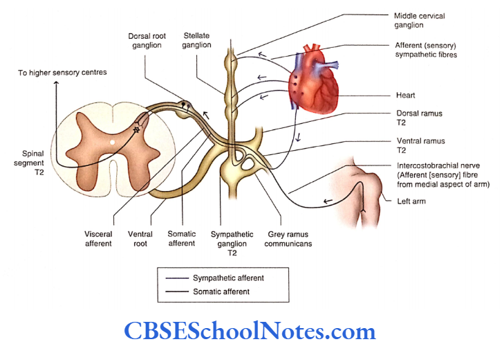
For example, stimulation of sympathetic nerves increases the heart rate while stimulation of parasympathetic innervations decreases the heart rate.
Read and Learn More Neuroanatomy
The ANS consists of both afferent (sensory) and efferent (Motor) components and an integration center.
Autonomic Sensory (Visceral Afferent) Component The sensations of the autonomic system are transmitted from the viscera to the CNS through somatosensory fibers.
The cell bodies of sensory neurons are located in the dorsal root ganglion of spinal nerves and the sensory ganglia of some cranial nerves.
Somatosensory neurons are also responsible for visceromotor reflex activities. Though these sensations hardly reach the level of consciousness, the sensations of nausea and pain in
the heart is perceived.
Integration and Control of the Autonomic Nervous System. The autonomic activities are integrated at higher levels. These centers are situated in the reticular formation of the brainstem, hypothalamus, limbic cortex, and prefrontal cortex.
The hypothalamus is considered the higher center (control and integration) of ANS. It controls both sympathetic and parasympathetic divisions of ANS.
The neurons of sympathetic and parasympathetic nuclei in the brainstem and spinal cord are connected with the nuclei of the hypothalamus through the reticular formation.
Autonomic Motor (Visceral Efferent) Component. The autonomic activities regulate body temperature, heart rate, respiration, blood pressure, gastrointestinal motility, and secretion from glands.
Thus, ANS consists of motor (efferent) fibers that innervate smooth muscle, cardiac muscle, and glands.
The effector cells of different organ systems are not under voluntary control; they work mostly at an unconscious level.
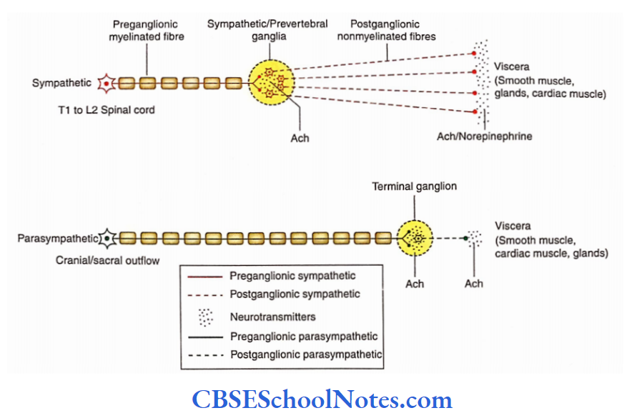
Both sympathetic and parasympathetic motor pathways consist of two motor neurons that conduct the impulses from the CNS to the effector organ.
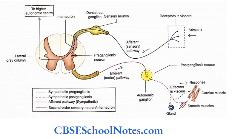
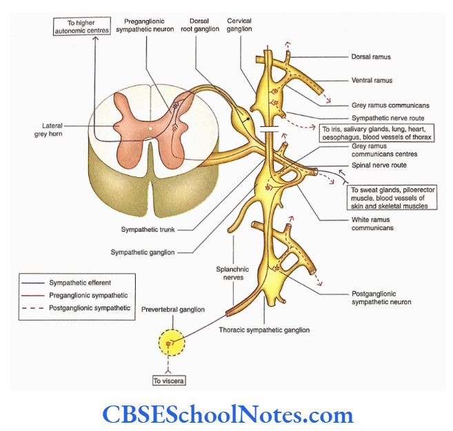
The cell bodies of the first motor neurons are located in the grey matter of the CNS (brain and spinal cord). These are called preganglionic or presynaptic neurons.
The preganglionic neurons send their myelinated axonal processes to synapse with the second motor neurons in the pathway.
The axons of presynaptic neurons come out of the CNS as part of either the spinal or the cranial nerve.
The cell bodies of second motor neurons are located in the autonomic ganglia outside the CNS.
These are called post-synaptic or post-ganglionic neurons because presynaptic neurons make synaptic contact with them.
The axons of post-synaptic neurons are unmyelinated and terminate on the effector organs (smooth muscle, glands, or cardiac muscle).
Sympathetic Division Of Autonomic Nervous System
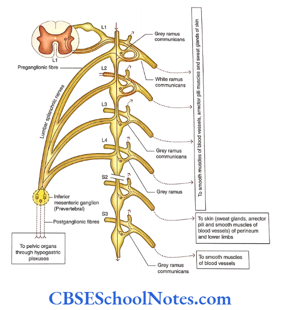
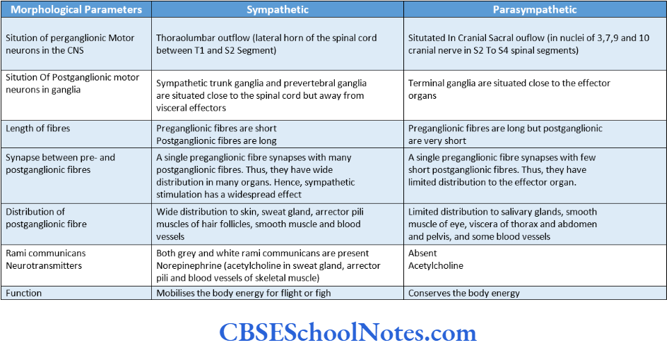
The sympathetic division of the autonomic nervous system is also called thoracolumbar outflow as the preganglionic nerve cells are situated in the thoracic and upper lumbar segments of the spinal cord.
The cell bodies of presynaptic neurons of the sympathetic division of ANS are located in the lateral grey column (horn) of the spinal cord between the T1 and L2 spinal segments.
The cell bodies of postsynaptic neurons are located in two groups:
- Paravertebral ganglion (sympathetic trunk ganglia) and
- Pacvcrtebral ganglion.
- Sympathetic trunk ganglia lie in a vertical row on either side of the vertebral column extending from the base of the skull to the coccyx.
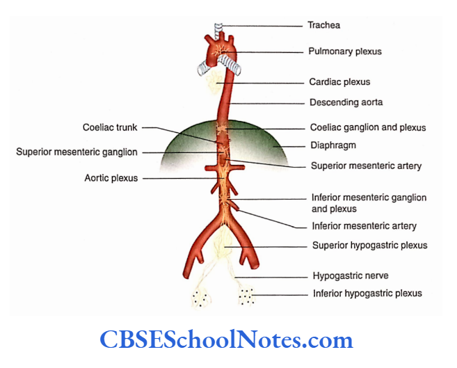
- On the other hand, prevertebral ganglia are situated anterior to the vertebrae and close to the origin of the main branches of the abdominal aorta.
- Examples of prevertebral ganglia are celiac ganglion, superior mesenteric ganglion, and inferior mesenteric ganglion close to the beginning of arteries of the same name.
- The axons of presynaptic neurons leave the spinal cord through ventral roots and enter the ventral rami of spinal nerves (T1 to L2).
- These axons are myelinated fibers. They leave the ventral ramus as a slender branch called white ramus communicantes and enter the sympathetic trunk.
- The presynaptic fibers of white ramus communicantes, after entering the sympathetic trunk, may synapse with the postsynaptic neurons present in the sympathetic ganglion at the same level.
- These fibers may also ascend or descend in the sympathetic trunk for a few levels before forming synaptic connections.
- It may also happen that these fibers pass through the sympathetic trunk without synapsing and join the splanchnic nerve to reach the prevertebral ganglia and synapse with the post-ganglionic neurons there.
The splanchnic nerves of the thorax are as follows
- Greater (T5 to T9)
- Lesser (T10 and Til)
- Least (T12)
The splanchnic nerves of the thorax are greater (T5 to T9), lesser (T10 and Til), and least (T12) while lumbar splanchnic nerves take origin from LI to L4 sympathetic ganglia.
The post-synaptic neurons of sympathetic ganglia send their axons (post-synaptic sympathetic fibers) to the adjacent ventral rami of spinal nerves.
These axons are non-myelinated and are called gray rami communicantes. From the ventral rami, they go to all branches of the spinal nerve including the dorsal rami.
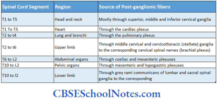
The post-ganglionic fibers from paravertebral sympathetic ganglia (sympathetic trunk ganglia) supply the blood vessels (vasomotor), sweat glands, and smooth muscle of hair follicles (erector pili) in the skin of limbs and body wall.
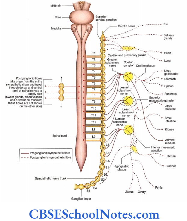
The post-ganglionic fibers from T1 to T5 supply thoracic viscera (heart, lung, trachea, and esophagus) via cardiac and pulmonary plexus.
The post-ganglionic fibers from prevertebral sympathetic ganglia (coeliac, superior and inferior mesenteric, and hypogastric ganglia, and plexuses) supply the abdominal and pelvic viscera (liver, kidney, stomach, intestine, rectum, colon, urinary bladder, genital organ, etc.)
The post-ganglionic fibers from the prevertebral sympathetic ganglia follow the course of various arteries.
(Students should note that the prevertebral ganglia consist of post-ganglionic sympathetic neurons only. However; these plexuses consist of both sympathetic and parasympathetic fibres.)
The sympathetic nervous activities are widely diffused activities that affect the whole body. The sympathetic reaction deals with emergencies or emotional stress.
The sympathetic reaction leads to the dilatation of the pupil, pale face (due to vasoconstriction in the skin), dry mouth, increased heart rate, and raised blood pressure.
The peristaltic movements of the intestine are suppressed and sphincters are closed. The blood vessels of skeletal muscles, heart, and brain dilate to supply more blood to these vital organs.
Parasympathetic Division Of Autonomic Nervous System
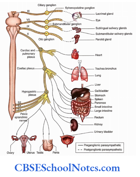
The parasympathetic division of ANS is also known as craniosacral outflow as preganglionic neurons are situated in the brain and sacral segment of the spinal cord.
Cranial outflow: The cell bodies of the cranial part of parasympathetic preganglionic neurons are situated in the brain and axons come out along with cranial nerves 3, 7, 9, and 10.
Sacral outflow: The sacral parts of preganglionic neurons are situated in the lateral grey column (horn) of spinal cord segments S2 to S4.
Their axons come out of the spinal cord through the ventral rami. The fibers from ventral rami then travel into pelvic splanchnic nerves.
The axons of pre-ganglionic neurons are very long, as they reach up to the effector organs. Close to the organs or within the substance of the organ, there is the presence of ganglia called terminal ganglia.
The terminal ganglia consist of post-synaptic neurons. Pre-synaptic axons form synaptic contacts with these neurons.
The terminal ganglia consist of post-synaptic neurons. Pre-synaptic axons form synaptic contacts with these post-synaptic neurons.
As the terminal ganglia are close to the organ supplied by the parasympathetic nerve, the axons of post-synaptic neurons are very short. They innervate the smooth muscles and glands in the wall of an organ.
The terminal ganglia associated with cranial outflow are ciliary (3 cranial nerve), pterygopalatine ganglia, submandibular ganglia (4 cranial nerve), and otic ganglia (9 cranial nerve).
The post-ganglionic fibers from these ganglia supply the eye, salivary glands, and other structures of the head and neck.
The pre-ganglionic parasympathetic fibers in the vagus nerve extend to many terminal ganglia in the thorax and abdomen. The post-ganglionic fibers supply the heart and lungs in the thorax (through cardiac and pulmonary plexuses). They supply the liver, gall bladder, stomach pancreas, small intestine, and part of the large intestine pancreas, small intestine, and part of the large intestine in the abdomen (through coeliac and superior mesenteric plexuses).
The postganglionic fibers from sacral outflow supply smooth muscle and glands in the wall of the colon, ureter, urinary bladder, and reproductive organs (through the hypogastric plexus).
Sympathetic And Parasympathetic Systems A Comparison
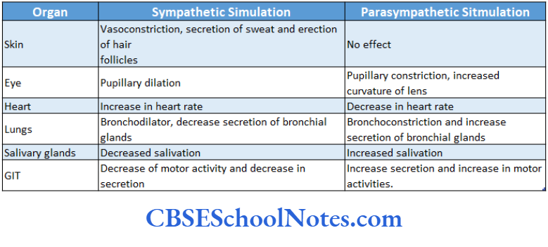
Most of the organs in the body receive both types of motor innervations (sympathetic and parasympathetic). These two types of motor innervations usually have opposite ctions (antagonistic); that is, if one type of innervation increases the activity (excitation) of viscera, then the other type will decrease (inhibition) the activity.
For example, sympathetic stimulation leads to decreased mobility of the intestine and contraction of sphincters. On the other hand, parasympathetic stimulation leads to increased motility of the intestine and relaxation of sphincters.
Sympathetic overactivity causes dilatation of the pupil while parasympathetic overactivity leads to its constriction.
In parasympathetic nerves, the neurotransmitter at pre- and post-synaptic nerve terminals is acetylcholine.
Acetylcholine is liberated at the sympathetic preganglionic nerve terminals while norepinephrine is liberated at postganglionic nerve terminals.
The autonomic innervations of some important organs of the body are described in brief in the following text
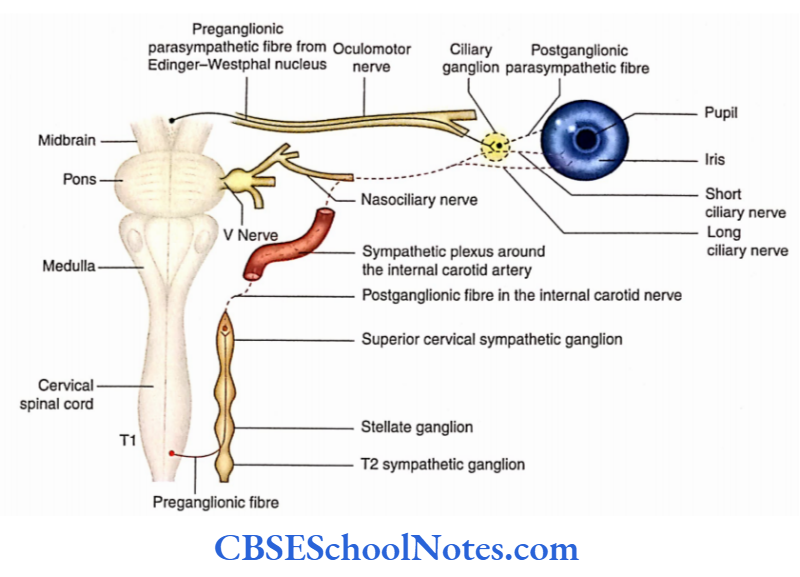
1. Eyeball
- Sympathetic innervation—from the Tl spinal cord segment
- Parasympathetic innervation—from the Edinger- Westphal nucleus
- Stimulation of sympathetic innervation causes dilatation of the pupil and relaxation of ciliary muscles.
- Stimulation of parasympathetic innervation causes constriction of pupil and ciliary muscle.
- Horner’s syndrome (which occurs due to deinnervations of sympathetic nerves of the head and neck) has the following characteristics: Constriction of a pupil, partial ptosis, absence of secretion on the face, flushing of the face, and enophthalmos.
2. Submandibular and sublingual salivary glands
- Sympathetic innervation—from Tl and T2 spinal segments
- Parasympathetic innervation—from the superior salivatory nucleus of the facial nerve
3. Lacrimal gland
- Sympathetic innervation—from Tl and T2 spinal segments
- Parasympathetic innervation—from the lacrimatory nucleus of the facial nerve.
4. Parotid gland
- Sympathetic innervation—from T1 and T2 spinal segments
- Parasympathetic innervation—from the inferior salivatory nucleus of the glossopharyngeal nerve
5. Heart
- Sympathetic innervation—from T1 to T5 spinal segments
- Parasympathetic innervation—from the dorsal nucleus of the vagus nerve.
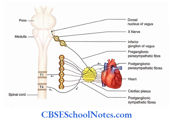
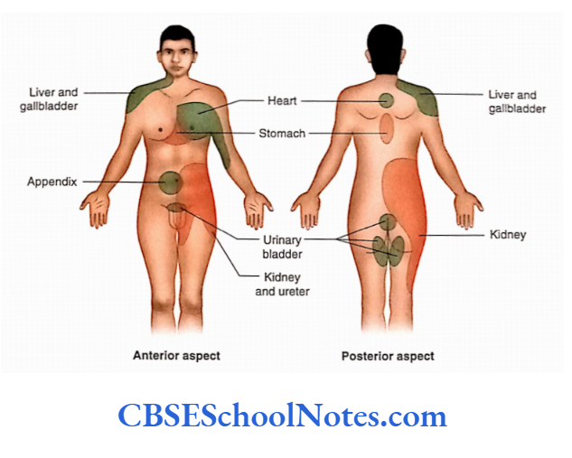
The pain of a heart attack is felt over the middle of the sternum, left shoulder, jaw, and medial aspect of the left arm.
This is because these areas are supplied by the same segments of the spinal coral as the heart (T1 to T5).
6. Lungs
- Sympathetic innervation—from T2 to T5 spinal segments
- The pain of a heart attack is felt over the middle of the sternum, left shoulder, jaw, and medial aspect of the left arm.
- This is because these areas are supplied by the same segments of the spinal coral as the heart (T1 to T5).
7. Gastrointestinal tract
- Sympathetic innervation—from T5 to L2 spinal segments
- Parasympathetic innervation—from the dorsal nucleus of the vagus and from the S2 to S4 spinal segment
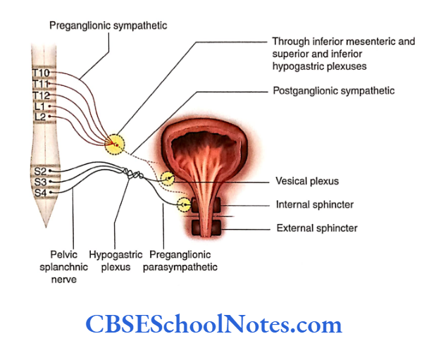
8. Urinary bladder
- Sympathetic innervation—from T10 to L2 spinal segments
- Parasympathetic innervation—from S2 to S4 spinal segments
- Sensory innervation—from T10 to L2 and S2 to S4
9. Arteries of the upper limb
- Sympathetic innervation—from T2 to T8
- Arteries of the lower limb
- Sympathetic innervation-from T10 to L2 spinal cord segments
- Autonomic innervations for the erection of the penis
- Parasympathetic innervation—S2 to S4 spinal segments
- Autonomic innervations for ejaculation from the penis
- Sympathetic innervations from the LI segment.
Localisation Of Visceral Pain
Viscera are usually insensitive to touch, heat, and cutting (cutting of the intestine in a conscious person does not elicit visceral pain).
But if receptors are stimulated in large areas (diffuse stimulation) due to inadequate blood supply, collection of metabolites, distension (stretch), and spasm, then visceral pain can be very severe. When the kidney stone obstructs and distends the ureter, it causes severe pain.
The visceral pain is poorly localized and dull because receptors are stimulated in a large area. The pain may be felt in the viscera itself or it may be felt just deep in the skin that overlies the viscera. However, in many cases, pain cord segments may also be felt in a surface area of the skin far from the stimulated organ. This phenomenon is called refereed pain.
Pain originating from a particular viscus is usually felt at a distance from the site of the visceral organ involved. This may be because afferent fibers from the skin (dermatome) and viscera enter the same segment of the spinal cord.
The first-order neurons of the afferent fibers of both visceral and somatic are situated in the dorsal root ganglia of the spinal nerve.
Probably, both the fibers synapse with the common (same) second-order neuron of the dorsal grey horn of the spinal cord.
The axons of the second-order neuron reach higher centers (via the thalamus) which probably fail to recognize the source of pain (skin or viscera).
Functions Of Autonomic Nervous System
- Both sympathetic and parasympathetic systems of ANS innervate almost all the organs of the body. Thus, their action on one particular organ is antagonistic to each other.
- If one system stimulates (excites) the organ, the other system depresses (inhibits) it. This is because their postganglionic neurons secrete different neurotransmitters
- (Ach for parasympathetic and noradrenaline in case of sympathetic) and their effector receptors for parasympathetic and adrenergic receptors for sympathetic).
- As the hypothalamus is the higher center of ANS it controls and balances the sympathetic and parasympathetic activities.
- However, students should also note that organs such as sweat glands, arrector pili muscles, kidneys, adrenal medulla, and many blood vessels are innervated only by the sympathetic system of the autonomic nerves.
- As there is no parasympathetic innervation in these organs, they do not face opposition. Thus, sympathetic stimulation leads to action while its inhibition leads to cessation of action.
- For example, when sympathetic fibers release norepinephrine into the smooth muscles of blood vessels, blood pressure rises due to the contraction of smooth muscles by mediation of the al receptor.
- Meanwhile, the epinephrine of circulating blood acts on the same smooth muscles by mediation of P2 receptors in the cell membrane leading to vasodilatation and a fall in blood pressure. Parasympathetic fibers do not terminate on many blood vessels.
Enteric Nervous System
The enteric nervous system (ENS) is defined as the system of neurons that is found within the wall of the GIT, gallbladder, and pancreas.
The entire length of GIT is supplied by sympathetic and parasympathetic parts of ANS.
The ANS forms the following two different nerve plexuses in the gut wall:
Submucosal plexus (plexus of Meissner): Situated in the subamucosa
Myenteric plexus (plexus of Auerbach): Situated in between circular and longitudinal muscle coats
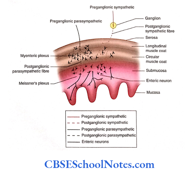
These plexuses consist of the following:
- Parasympathetic preganglionic fibers terminate on postganglionic parasympathetic neurons situated in the myenteric plexus.
- Postganglionic sympathetic and parasympathetic fibers forming a network in Meissner and myenteric plexuses
- Sensory and motor enteric neurons and their processes consist of 100 million neurons in the plexuses of GIT.
All these innervate the mucosa, submucosa, muscle coats, and blood vessels. These regulate the secretion from the mucosal gland and motility of GIT.
The gallbladder and pancreas also have ganglia and nerve plexus. The ENS was previously considered a part of ANS.
However, now it is considered a system separate from the ANS. The neurons of the ENS arise from neural crest; thus, they differ from the origin of sympathetic and parasympathetic neurons.
Functions of the Enteric Nervous System
- The functions of ENS include both sensory and motor.
- The sensory neurons of ENS monitor the stretching of the walls of the intestine and chemical changes within the gastrointestinal tract.
- The motor neurons of ENS control the contraction of the smooth muscle, secretion of the gastrointestinal gland, and endocrine cells associated with GIT.
Autonomic Nervous System Summary
- The autonomic nervous system (ANS) innervates the cardiac muscle, smooth muscle (present in the wall of viscera and blood vessels), and glands.
- The ANS works by forming the reflex arc which is organized in afferent (sensory) pathways, integrating higher centers and efferent (motor) pathways.
- Visceral sensation hardly reaches the level of consciousness. The cell bodies of sensory neurons are situated in the sensory ganglia of cranial nerves and the dorsal root ganglia of spinal nerves.
- The visceral activities are integrated at higher levels, especially in the hypothalamus.
- Efferent pathways convey motor impulses from the CNS to the cardiac muscle, smooth muscles, and glands.
Two neurons are involved in this pathway:
- Preganglionic and
- Postganglionic neurons.
The visceral motor signals reach various organs through two major subdivisions of the ANS:
- Sympathetic and
- Parasympathetic nervous systems.
Sympathetic nervous system
- The preganglionic motor neurons are situated in the lateral horn of the grey matter of the spinal cord from T1 to L2 segments.
- The postganglionic motor neurons of this division are situated in the sympathetic trunk ganglia and prevertebral ganglia.
- The preganglionic fibers reach the sympathetic trunk ganglion through white rami communicans while the postganglionic fibers after coming out through the sympathetic ganglia join the ventral ramus through grey rami communicans.
- Some preganglionic sympathetic fibers, which do not relay in the sympathetic chain (paravertebral), may relay in the prevertebral ganglion through splanchnic nerves.
Parasympathetic nervous system
- The parasympathetic nervous system consists of cranial and ‘sacral’ outflow.
- The preganglionic parasympathetic fibers of both cranial and sacral outflow end in ‘terminal ganglia’ containing postganglionic parasympathetic neurons.
- The terminal ganglia associated with cranial outflow are the ciliary, pterygopalatine, submandibular, and otic ganglia. The terminal ganglia of the vagus and sacral outflow are situated in the wall of the viscera and are unnamed.
- The actions of sympathetic and parasympathetic parts of ANS on any particular organ are antagonistic to each other.
Enteric nervous system
- The enteric nervous system (ENS) consists of motor, sensory, and interneurons, which are found within the walls of GIT.
- The functions of ENS include both sensory and motor.
Autonomic Nervous System Multiple Choice Questions
Question 1. The following structures are innervated by ANS except
- Smooth muscles of viscera
- Smooth muscles of blood vessels
- Cardiac muscles
- Glands
- Articular capsule of joints
Answer: 5. Articular capsule of joints
Question 2. Which of the following facts about visceral sensations are true?
- They hardly reach the level of consciousness
- Sensations of nausea and retrosternal pain are perceived
- The sensation of distension of the bladder and bowel reaches the level of consciousness
- Pain sensation from the viscera is perceived as secondary to a lack of oxygen supply
- All of the above
Answer: 3. Sensation of distension of bladder and bowel reach the level of consciousness
Question 3. Which of the following facts about the autonomic motor pathway is false?
- Visceral motor signals reach organs through the sympathetic and parasympathetic nervous systems
- Both sympathetic and parasympathetic pathways consist of two neurons—preganglionic and postganglionic
- Preganglionic neurons are situated in the CNS while postganglionic in the peripheral ganglion
- The axons of preganglionic neurons are myelinated and synapse with postganglionic neurons
- None of the above
Answer: 5. None of the above
Question 4. Which ofthe following statements about the sympathetic nervous system is false?
- Preganglionic sympathetic neurons are located in the lateral horn of the spinal cord from T1 to L2 and from S2 to S4 spinal segments
- Postganglionic sympathetic neurons are located in the sympathetic trunk (sympathetic chain) ganglia
- They are also located in prevertebral ganglia
- Prevertebral sympathetic ganglia are situated in front of the vertebral column
Answer: 1. Preganglionic sympathetic neurons are located in the lateral horn of the spinal cord from T1 to L2 and from S2 to S4 spinal segments
Question 5. Prevertebral sympathetic ganglia consist of the following except
- Pulmonary ganglion
- Superior mesenteric ganglio
- Inferior mesenteric ganglion
- Celiac ganglion
Answer: 1. Pulmonary ganglion
Question 6. Which of the following statements about splanchnic nerves is false?
- They arise from the thoracic sympathetic chain
- They are medically directed branches of the sympathetic trunk
- They contain postganglionic myelinated nerve fibers
- There are three splanchnic nerves—greater, lesser, and least splanchnic nerves
- These splanchnic nerves reach the prevertebral ganglion
Answer: 3. There are three splanchnic nerves—greater, lesser, and least splanchnic nerves
Question 7. The following facts about the distribution of sympathetic fibres are true except
- Preganglionic sympathetic fibers travel in the ventral spinal nerve root
- They then travel in the ventral ramus of spinal nerves
- They soon leave the ventral ramus to join the sympathetic trunk through the white ramus communicans to synapse with the postganglionic neurons
- The axons of postganglionic neurons join the ventral ramus again through grey ramus communicans
- Grey ramus communicans contain myelinated fibers of postganglionic neurons
Answer: 5. Grey ramus communicans contain myelinated fibers of postganglionic neurons
Question 8. Following is the location of preganglionic motor neurons of the parasympathetic nervous system except
- Few cranial nerve nuclei in the brainstem
- Lateral horn of spinal cord between S2 and S4 spinal segments
- Terminal ganglia of the parasympathetic nervous system
- Pterygopalatine ganglion
Answer: 3. Terminal ganglia of the parasympathetic nervous system
Question 9. Which of the following statements is false?
- Acetylcholine (ACh) is liberated at the terminals of preganglionic parasympathetic neurons
- ACh is liberated at the terminals of preganglionic sympathetic neurons
- Adrenaline is liberated at the terminal of postganglionic parasympathetic neurons
- ACh/norepinephrine is liberated at the terminals of
postganglionic sympathetic neurons
Answer: 3. Adrenaline is liberated at the terminal of postganglionic parasympathetic neurons Part of the thalamus, subthalamus, middle and posterior parts of the hypothalamus, part of the midbrain