Structure Of DNA And RNA Notes

Structure of DNA and RNA

The chromosomes are principally constituted of nucleic acids and are structurally supported by protein molecules. However, it is only the nucleic acids that build-up the genes and are linked to the transmission of a trait.

There are two types of nucleic acids.

  • Deoxyribonucleic acid (DNA): The bulk of the cellular DNA is found in the nucleus as chromosomes. Circular strands of DNA are also found in the mitochondria. Human genes are made up of only of DNA>
  • Ribonucleic acid (RNA): RNA is largely found in the nucleolus within the nucleus. Other locations where RNA is found are the ribosomes and the cytoplasm. RNAs work as functional intermediaries between genes and their final products, the proteins.

Structure And Packaging of DNA

Structure of DNA (Chemical):

Three different types of chemical compounds compose the DNA.

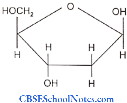
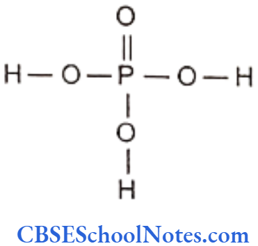
  • Sugar molecule – It is called deoxyribose and is a 5 carbon pentose sugar.
  • Phosphoric acid.
  • Nitrogenous bases.

These are of 4 types:
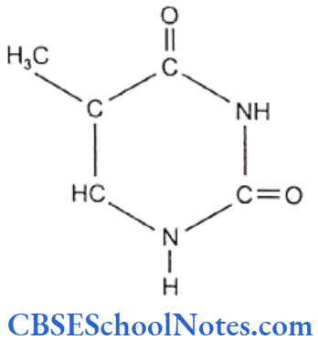
  • Adenine – (A)
  • Thymine – (T)
  • Cytosine – (C)
  • Guanine – (G)

Adenine and Guanine are classified as Purines while Cytosine and Thymine as Pyramidines.

Read and Learn More Genetics in Dentistry Notes

Genetics In Dentistry Structure of DNA and RNA Deoxyribose molecule

Genetics In Dentistry Structure of DNA and RNA Phosphoric acid

Genetics In Dentistry Structure of DNA and RNA Molecular structure of thymine

Structure of DNA (Molecular):

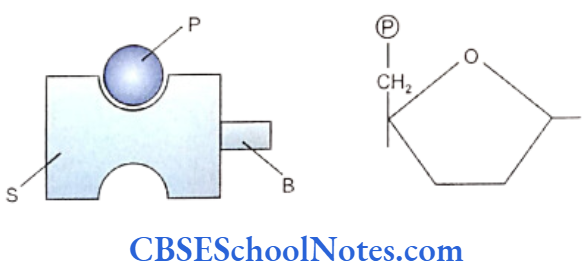
  • DNA exists in the form of a long polymer which is formed by linkage of a series of nucleotide molecules like in a chain.
  • A nucleotide molecule is formed of one molecule of deoxyribose sugar, one molecule of phosphoric acid and one nitrogenous base attached on the sides of the deoxyribose. As there are four varieties of nitrogenous bases, there are four types of nucleotides in the DNA.
  • The phosphate molecule in a nucleotide is attached to the fifth carbon atom of the sugar (deoxyribose) and the nitrogenous base to the first carbon atom of the sugar molecule.
  • The third carbon atom of the deoxyribose of the next nucletide is attached to the phosphate molecule of a nucleotide. Hence, the sugar and the phosphate molecules are arranged in a linear fashion to form a polynucleotide chain. The nitrogenous base attached to sugar molecule is directed at right angle to the long axis of a single polynucleotide chain.
  • All polynucleotide chains have marked ends. It can be noticed that at the upper end of the chain the 5th carbon atom of the sugar molecule of the last nucleotide just terminates in a phosphate. This end is called as 5′ or 5’P terminus.
  • The other end of the chain ends in sugar molecule or a nucleotide whose 3rd carbon atom is free and not linked to the phosphate of any nucleotide and bears an OH group (hydroxyl group) instead. This end of polynucleotide chain is called 3′ end or 3′ OH terminus.

Genetics In Dentistry Structure of DNA and RNA Schematic diagram

Genetics In Dentistry Structure of DNA and RNA The schematic diagram of a part of polynucleotide chain

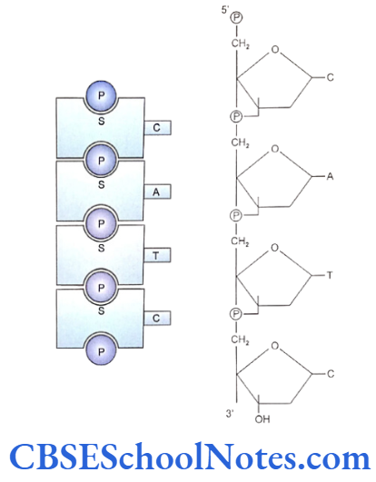
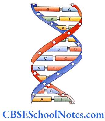
  • Waston and Crick in 1953 worked out the DNA helix model as been made up of two such polynucleotide chains which lie side by side but run in opposite directions. One chain runs from its 5′-3′ direction whereas the other in 3′-5′ direction.

The nitrogen bases face towards the inside of the skeleton formed by the two strands of the nucleotide chain.

  • The 3′ end of the DNA strand is called the “head” end and the 5′ ends its “tail”.
  • The two chains are held together by two types of hydrogen bonds between the nitrogenous bases.

Genetics In Dentistry Structure of DNA and RNA Schematic diagram of antiparallel polypeptide chains

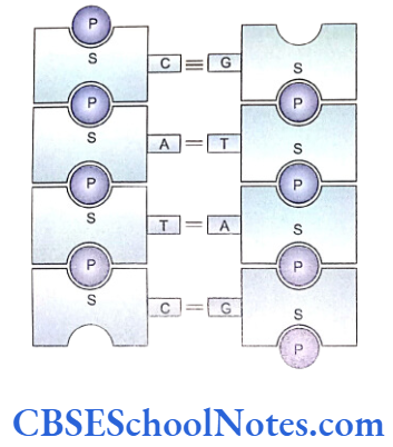
  • Pairing between two nitrogenous bases is predetermined and constant, i.e. Adenine(A) always pairs with Thymine (T) and Cytosine(C) with Guanine(G). This specific pairing is due to the fact that these molecules are complementary and the combination of the specific bases facilities stable hydrogen bonds between them. Nucleotides A and T s hare two hydrogen bonds while C and G are joined by three bonds.
  • As a consequence of specific base pairing, two strands of DNA are complementary to each other. It means that if the sequence of bases on one chain is A T G C A, then correspondingly, the exactly opposite region on other chain will have the sequence T A C G T that can thus anneal together.
  • The double helix of a DNA molecule is formed as the two complementary chains (polynucleotide chains) twist around each other.

A single and complete 360° turn of the helix measures about 3.4 nm along the long axis and contains 10 pairs of nucleotides. The distance between two adjacent nucleotides. The distance between two adjacent nucleotides is 0.34 nm. The diameter of helix is about 3 nm.

One turn of helix measure about 3.4nm and contains 10 pairs of nucleotides.

Genetics In Dentistry Structure of DNA and RNA The DNA double helix

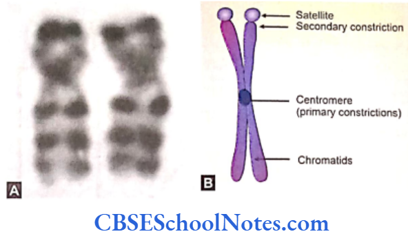
Packaging of DNA in a Chromosome

A chromosome is composed of a double helix of DNA and histone proteins. The average length of the DNA filament of a single chromosome can extend upto 50 mm but the chromosome is only 5 microns in length when maximally condensed in the metaphase. Thus there is about 10,000 times reduction in length. This is due to the fact that in a metaphase chromosome filament of DNA undergoes several orders of coiling or condensation.

  • The primary or first order coiling is due to turning of the DNA double helix on itself.
  • These primarily coiled DNA double helix then wind around histone complexes (histone beads). This secondary coiling of DNA filaments around histone beads forms structures called nucleosomes. The DNA filaments wind twice around each histone bead and contain approximately 146 nucleotide pairs. Nucleosomes are attached to one another forming long chains.
  • The nucleosomes arrange in a spiral to form a closely stacked thick structure; the chromatin filament.
  • Chromatin filament coil again to form chromatin loops.

Genetics In Dentistry Structure of DNA and RNA DNA packs tightly into metaphase chromosomes

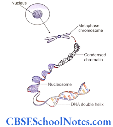
  • Additional coiling of the loops on themselves to give the shape of a chromosome as visible during the metaphase of cell division.

These successive degrees of coiling gives rise to the solenoid model of chromosomal structure.

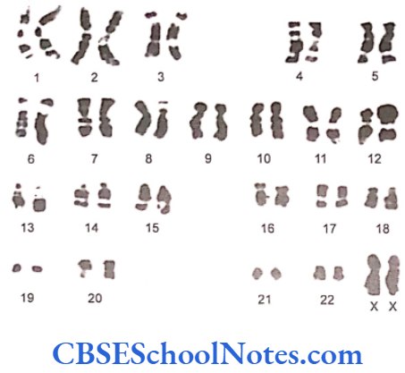
On straightening a strand of DNA taken from a typical human chromosome, it measures about 5 cm in length. It may have about half a billion to 3 billion nucleotides. If we arrange all the molecules of DNA present in the 46 human chromosomes end-to-end, they would measure about 2 meters or 6 ft in length.

Human body consists of approximately 1014 cells and if all the DNA of an individual is joined end-to-end, the total length of DNA would measure approximately 2 x 10¹⁴m or 2 x 10¹¹ km. This length would be good enough to go from the earth to the sum and back for about 500 times.

Replication Of DNA

Nondividing cells remain in the interphase stage of the cell cycle. Cell division begins with the doubling or duplication of the DNA content of each chromosome. This event of DNA replication is also called the synthetic phase and results in the formation of two sister chromatids in each chromosome.

DNA replication is followed by the prophase, metaphase, anaphase and telophases of mitosis or meiosis that include distribution of chromosomes and cytoplasm to the daughter cells. The double helix model of Watson and Crick ideally explains the events during replication.

Genetics In Dentistry Structure of DNA and RNA The process of replicaton

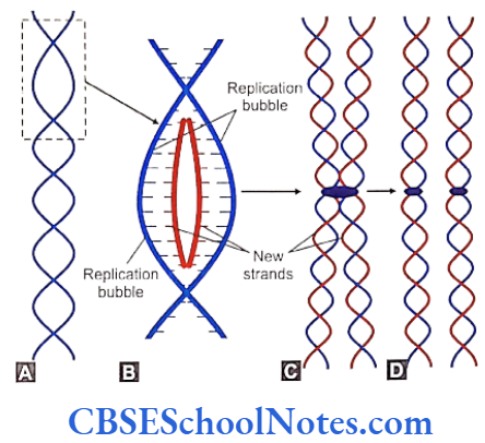
  • The tightly coiled DNA filament gets uncoiled during s (synthesis) phase of cell division. The two strands of DNA molecules are separated (denatured) by specific enzymes on breaking the hydrogen bonds between nitrogenous bases. The two separated strands of polynucleotide chains are complementary to each other.
  • Origin of replication (ori) are sites along a DNA strand at which replications being. The double stranded DNA gets denatured at these sites and the replication begins on both the strands but in opposite directions. Due to replication, bubble-shaped structures pop up long the chromosome at multiple points simultaneously, called replication bubbles.
  • The human genome doubles in approximately 9 hours in a cell with about 100 bubbles being active in each chromosome, each bubble apart by about 40000 nucleotide pairs.
  • The region in each bubble at which parental DNA strand is progressively separated with the help of enzymes looks like the alphabet Y. The stem of the Y is formed by the double stranded DNA whereas the two arms of the Y are made-up of the dentured single strands. This region of on the chromosome is called the replication fork. The total replication time is reduced as each chromosome replicates by many thousands origin sites.

At each replication fork about 10 to 100 nucleotide pairs are added per second. A chromosome usually takes 15 to 30 minutes to replicate. Because all the chromosomes of a cell do not replicated simultaneously, complete replication of all chromosomes of a cell takes 8 to 10 hours.

  • As specified by the rules of base pairing, each nucleotide of an old chain attracts is complementary nucleotide that attach through hydrogen bonds with their complementary nucleotides on the old chain.

The growing end of a replicating new DNA strand elongate with the addition of one nucleotide at a time

  • The phosphate components link the sugar radicals of neighboring nucleotides to each other. Thus a new chain is formed opposite to the old polynucleotide chain. The new chain grows only at its 3′ end.
  • the genetic information is conserved and transmitted unchanged to each daughter cell as the new strand is identical to the old template strand.
  • As the newly synthesized DNA double helix contains an original or old strand (that is said to be conserved as it comes from the parent) and a newly constituted complementary strand, this method of DNA replication is described as semiconservative.

Mitochondrial DNA

In addition to the nucleus, the mitochondria also contain DNA. Mithochondrial DNA, similar to the nuclear DNA, is double-stranded but arranged as circular structures. It consists of about 16.6 kb nucleotide base pairs and codes for 37 genes with 22 genes for tRNAs, 2 for rRNA and 13 genes for enzymes responsible for oxidative phosphorylation.

Oxidative phosphorylation enzymes are involved in energy production. Therefore mitochondrial abnormalities are associated with the loss of coupling between oxidation and phosphorylation. Presentations of mitochondrial disorders are variable because of the phenomenon of heteroplasmy. The characteristics and examples of mitochondrial disorders.

Mithochondrial of sperm are not transmitted into the oocyte during fertilization and the entire mitochondrial complement in the zygote is derived exclusively from the mother. Thus mitochondrial DNA abnormalities are transmitted only through females and follow maternal pattern of inheritance. Both sexes are equally affected.

mtDNA acts as excellent genetic markers for tracing human ancestry as they do not undergo genetic recombinations during gametogenesis, similar to what happens with the Y-chromosomes. It is established that about 1 change per mitochondria lineage occurs in every 3800 years at a constant rate. This fact helps us to estimate that modern human population originated somewhere in the Sub-Saharan Africa approximately 130,000 years ago and migrated to various parts of the world.

They first moved out of Africa tot he Middle-East about 100,000 years age and from there to the east and south Asia (67,000 years age). The journey continued to Australia and to Europe anout 40,000 years ago. From East Asia migration went on further to North America (about 20,000 years back) and from there to South America about 13,000 years ago.

Structure Of Ribonucleic Acid (RNA)

Both the nucleolus and cytoplasm contain RNA molecules. RNAs work as functional intermediaries between genes and their final products, the proteins. RNA is not concerned with inheritance in human beings. It is synthesized by reading DNA template molecules with the help of ribosomes.

There are three types of RNAs.

  • Messenger RNA (mRNA)
  • Ribosomal RNA (rRNA)
  • Transfer RNA (tRNA).

Messenger RNA (mRNA)

The nucleus is the site for messenger RNA (mRNA) synthesis. It is single stranded product of transcription. mRNA is formed at transcription bubbles with arrangement of nucleotides on the template strand that is read from its 3′ to 5′ end. mRNA itself, thought, is synthesized from its 5′ to the 3′ end. It thus carries all the genetic information present on a particular segment of the DNA strand in the form of sequence of base arrangements.

However, there is no thymine in mRNA and has a uracil molecule instead. Several hundred to several thousand nucleotides arranged in a single strand compose a messenger RNA molecule. mRNA comes out through nuclear pores into the cytoplasm after its formation in the nucleus. Soon it gets attached to ribosomes outside the nuclear envelope.

The protein synthesizing apparatus of the cell utilizes the genetic information on the mRNA for translation of proteins. mRNA population constitutes about 10% of the total RNA present in a cell. The life span of mRNA varies from few hours to few days.

Ribosomal RNA (rRNA)

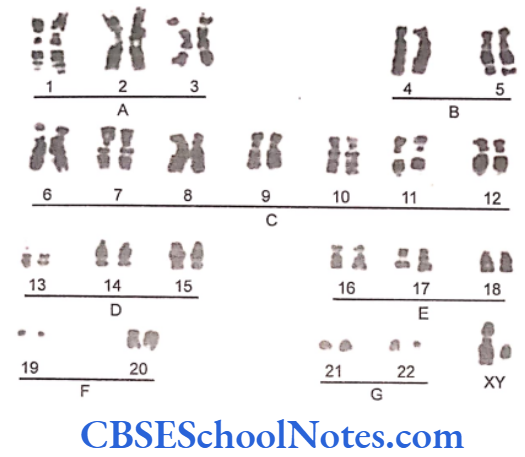
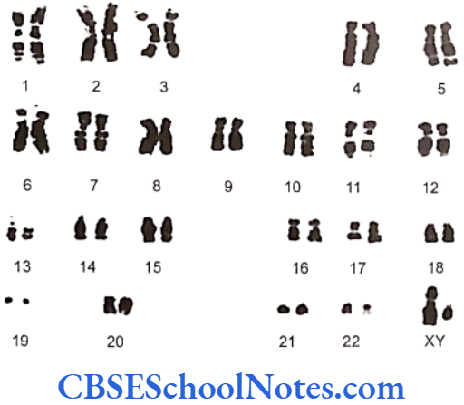
About 80% of the total RNA present in the cell is contributed by rRNA. As implied, rRNA occur in ribosomes. The part of the DNA which codes for rRNA is associated with formation of the nucleolus and is called the nucleolar organizer. DNA loops of chromosomes 13, 14, 15, 21 and 22 contain genes for ribosomal RNA and constitute the nucleolus. rRNA is produced inside the nucleus.

Two subunits, a large and a small, make-up the ribosome. The rRNA molecule occurs as three different dimensions; the 28s, 18s and 5s units. The large ribosome subunit contains the 28s and 5s molecules. The 18s molecules are present in small ribosomal subunits.

Ribosomal RNAs in the ribosome initiate as well as maintain the process of protein formation (translation) by interaction with the mRNA strands as they pass through the ribosomes.

Transfer RNA (tRNA)

Consisting of about 75 to 80 nucleotides, a tRNA molecule is single stranded and is synthesized at particular regions of the genome. The tRNA molecule isbent in the middle of the polynucleotide chain and forms two arms on its sides named clover leaf model for obvious similarly with the structure.

Genetics In Dentistry Structure of DNA and RNA Clover leaf model of tRNA

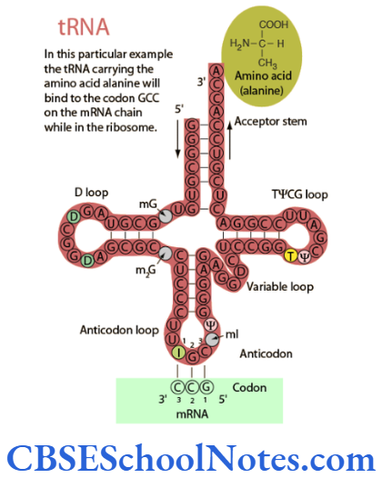
A specific amino acid is designated to each tRNA molecule and hence 20 types of tRNA exist in the cytoplasm. A tRNA with its amino acid (amino-acyl tRNA) is transported to the ribosome where it docks and pairs on the mRNA molecule after being correctly recognized for such a base pairing. Protein synthesis and chain elongation occur with the sequential assembly of the amino acids by the tRNA on the mRNA molecule. Four different special sites are present in the tRNA molecule.

  • Recognition site – Recognizes the appropriate amino acids to be attached with the help of specific amino-acid sequences.
  • Codon recognition site – A 3 base sequence site that is complementary to a sequence of three bases (codon) on the mRNA molecule. Base pairing between tRNA and mRNA happens at these sites after tRNA molecule lands on the mRNA.
  • Amino acid attachment side – This sites attach specific amino acids after their correct identification.
  • Ribosomal recognition site – This site facilities tRNA to recognize their specific positions inside the ribosome.

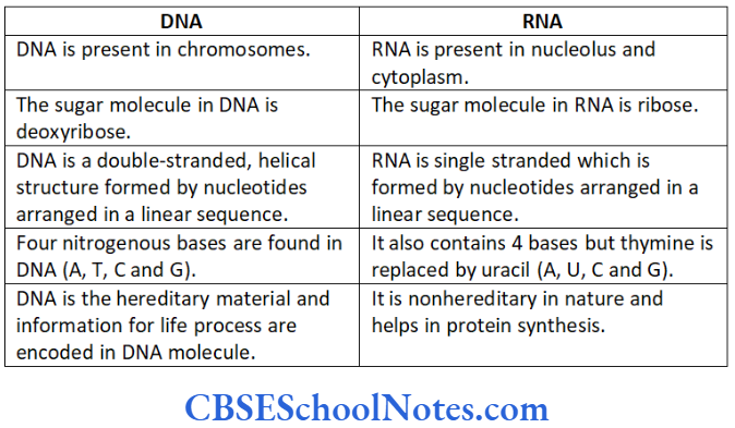
Following are the difference between DNA and RNA molecules.

Genetics In Dentistry Structure of DNA and RNA Differences between DNA and RNA molecules

Structure Of DNA And RNA Summary

  • DNA
    • Eukaryotic genes are composed of DNA molecules and are responsible for inheritance of characters. DNA is present in nucleus (chromosomes) and mitochondria.
    • DNA is in the form of a long sequence that is formed by adding up of nucleotide molecules as in a chain. (A nucleotide molecule itself is formed of a single molecule of deoxyribose sugar, a single molecule of phosphate and single nitrogenous base).
    • The DNA molecule is made up of two hightly coiled and condensed polynucleotide chains (double helix) which lie side by side but runs in opposite directions (antiparallel).
    • There is strict and definite pattern of pairing between the bases of the two parallel running DNA strands.
    • During cell division chromosomes (and the DNA) duplicate themselves by the process of replication.
    • The process of replication generates a new strand of DNA (semiconservative) against each old and complementary template strand.
  • RNA
    • RNA does not constitute eukaryotic genes and therefore is not a heriditary material.
    • RNA is abundant in the nucleolus as well as in the cytoplasm.
    • The sugar molecule in RNA is ribose and nitrogenous bases are A, G, C and U.
    • There are three different types of RNAs (mRNA, rRNA and tRNA) which play an important role in synthesis of proteins.

Classification Of Human Chromosomes Notes

Chromosomes And Their Classification

It was the beginning of the 20th century that the importance of Mendel’s findings was beginning to get appreciated. This was due to simultaneous understanding of several aspects of the cell division and the structure of the chromosome.

The first account of mitosis was accounted by A Schneider in 1873 followed by W Fleming in 1879 who described the migration of individual chromosomes into the daughter cells after the detachment of the sister chromatids.

Subsequently Brenden showed haploid (half) number of chromosomes in the gametes and restoration of the diploid number of chromosomes in the somatic cells after fertilization.

It was 1902 when Walter S Sutton and Theodore Boueri came up with the ‘chromosome theory of heredity’ that claimed that Mendel’s pair of ‘hereditary factors’, were in fact, physically located on the chromosomes.

According to Mendel each trait was represented by a pair of factors. The presence or the absence of one or both the factors determined the expression of that particular trait in an individual.

The emerging concepts of gametogenesis and fertilization further explained Mendel’s observations, calculations and foresight.

Read and Learn More Genetics in Dentistry Notes

Classification Of Human Chromosomes Notes

Introduction To Human Chromosomes

The human chromosomes are nuclear structures. They look like a net that is spread across the nucleus in a nondividing cell (interphase). The strands of the net are called chromatin and are chiefly made up of Deoxyribonucleic acid (DNA) and histone proteins; stained dark with basic dyes.

Certain areas in the net look thick, coiled and condensed and are called heterochromatin whereas certain other areas resemble thin and lightly stained threads termed the euchromatin.

The euchromatin is active during the functioning of the cell. The chromosomes get fully coiled and look like separate and individual thick rod like entities only during cell division.

The chromatin net structure is restored once the cell has completed its mitotic or meiotic phases of cell division.

The Number of Human Chromosomes

  • The number of chromosomes is always specific and constant for each species.
  • In each of human somatic cell there are 46 chromosomes referred to as the diploid set and designated as 2n.
  • The 46 chromosomes can be grouped into 23 pairs of chromosome; each pair different from the other. The constituent partners in a group are very similar to each other.
  • During the process of formation of sperm or ovum (gametogenesis), the number of chromosomes is reduced to half, i.e. to 23 or to haploid (n) state with one chromosome from each pair of the diploid set migrating to the gametes.
  • With fertilization the haploid sets (n/23) of the sperm and the ova fuse to restore the diploid set (2n/46) in the first cell of the embryo.
    • Chromosomal Classification into Autosomes and Sex Chromosomes
    • The complement of 46 chromosomes in each human cell is classified into 44 autosomes (22 pairs) and 2 sex chromosomes (1 pair).
    • One member of each pair of autosomes and sex chromosome is contributed by either the father (paternal) or the mother (maternal).
    • The sex chromosomes are of two different types; the X and Y chromosomes.
    • The chromosomal constitution of the females of the human race is 44 autosomes and two X chromosomes (44 + XX), forming a homomorphic pair of sex chromosomes.
    • The chromosomal organization in human males is 44 autosomes and a pair of dissimilar sex chromosome, (44 + XY), i.e. one X and one Y chromosome, forming a heteromorphic pair of sex chromosomes.
    • The Y chromosome is always contributed by the male parent via its Y chromosome-containing-gamete.

Chromosomal Size and Shape

The Chromosomes remain extended and uncoiled in the interphase stage of cell cycle with the length of the chromatin, if measured, extending a few meters.

The chromosomes coil and condense maximally during the metaphase stage of cell division when the average size of the chromosomes is about 5μm. It is during the cell division that we can, in fact, visualize individual chromosomes.

Chromosomes look like an entangled mesh of chromatin thread when in the interphase. Prior to the oneset of cell division and progressive thickening of individual chromosome, each chromosome undergoes duplication of its DNA content and appears like two closely placed free strands attached together roughly near their waists.

This event is called the phase of DNA replication. Subsequently during the later stages of cell division, as the chromosomes get condensed further; each of them looking like a thick rod (in metaphase) or like the letters J or V (in anaphase).

Chromosomal Structure

Each metaphase chromosome comprises of two identical components (after DNA replication). These two symmetrical halves are called sister chromatids and they are attached together at a constricted region that stains lightly and is called the centromere.

The centromere defines the primary constriction of the chromosome and divides the chromosome into a short arm (p) and a long arm(q). Centromeres play a pivotal role during the movement of chromosomes during cell division.

Genetics In Dentistry Chromosomes And Their Classification Structure of a chromosome as seen under the microscope during metaphase

Certain chromosomes usually carry an additional secondary constriction in one or both the chromatids. These constrictions are linked to the formation of the nucleolus and hence referred to as the nucleolar organizing region.

The secondary constriction may lie at the distal end of a chromatid giving rise to a small fragment of chromosome at the extreme end of the chromosome called the satellite.

The centromere (primary constriction) is situated anywhere along the length of the chromosome. The level of the constriction and consequently the lengths of the p and q arms are different for different chromosomes but specific for a particular chromosome.

The location of the centromere, the length of the chromosome and the existence of satellites are taken as parameters to classify as well as to identify chromosomes.

Classification Of Chromosomes And Analysis

 Classifications are used to identify chromosomes.

  • Standard (Denver) classification:

    • Chromosomes are classified into seven groups in an arrangement in descending order of their lengths. Groups are designated alphabetically from groups A to G. The longer female sex Chromosome X is included in the group C and the smaller male sex chromosome Y is included in group G.

Genetics In Dentistry Chromosomes And Their Classification Photograph of karyotype

  • Classification based on the position of the centromere:
    • Metacentric: Centromere located near the middle of the chromosome; the length of p = q.
    • Submetacentric: Centromere located slightly away from the middle; the length of p < q.
    • Acrocentric: Centromere located very near to the end; the length of p << q.
    • Telocentric: Centromere located at one end of the chromosome; effectively having only a single arm.

Genetics In Dentistry Chromosomes And Their Classification Classification of chromosomes

  • The Paris nomenclature: This classification entails banding techniques (special staining) and therefore is more accurate in identification of chromosomes. The arms of the chromosomes are divided into short segments and designated numbers 1, 2 and 3 beginning from the centromere and proceeding distally. Each of these small segments or regions is subdivided into Z banded regions. Thus not only a particular chromosome and a segment in its arm can be accurately identified in this classification; small structural anomalies can also be detected within small regions in the segments.

Chromosomal Analysis

Chromosomal analysis is an accurate tool to investigate several clinical conditions to arrive at a precise diagnosis. It may be indicated in cases of congenital malformation, mental retardation, repeated abortion, sex determination, prenatal diagnosis and other analytical purposes.

The chromosomal make-up of an individual is called as his or her karyotype. Karyotype is essentially a photomicrograph of an individual’s chromosomes arranged according to the standard classification. Diagrammatic representation of karyotype is called as ideogram. A karyotype is done to:

  • Identify and number the chromosomes
  • Detect numerical and structural anomalies of chromosomes.

Technique of Karyotping (Chromosomal Preparation)

The procedure to obtain a karyotype of an individual is called Karyotyping. The metaphase chromosomes from somatic cell are prepared and photographed. Photographs of individual chromosomes are cut and arranged as per the Standard Classification.

Rapidly dividing cells are used to yield the chromosomes. The cells are usually obtained from sources like peripheral blood lymphocytes (most commonly used), skin fibroblasts, bone marrow cells, chorionic villi and amniotic fluid cells. The sequential steps followed and described below.

About 5 ml of venous blood is collected in a heparinized vial under sterile conditions and then the Lymphocytes are separated from the red cell population with the help if a centrifuge.

A culture via is prepared that contains culture media and fetal calf serum for nourishment of the lymphocytes. Phytohemagglutinin in the vial stimulates cell division. Antibiotics are added to the medium to prevent infection.

Genetics In Dentistry Chromosomes And Their Classification Flow chart of steps in the preparation of a karyotype

The white cell suspension is then put in the culture vial. The vial is incubated for three days at 37°C.

Colchicin stops the formation of mitotic spindles and arrests cell division in metaphase. The chromosomes are maximally condensed and easily visible at this stage.

The dividing lymphocytes are separated off with a centrifuge 2 hours after the colchicin is added.

The cells are subsequently treated with hypotonic saline. This causes the cells to swell and become turgid.

The cells are then fixed by adding a mixture of glacial acetic acid and methanol.

When the cells get suspended in the fixative, they are dropped on chilled slides from a height. This causes the cell wall to disintegrate thereby allowing the chromosomes to spread in a limited area of cell rupture. This is called the metaphase spread.

These slides are stained and microphotographed. The Karyotype of an individual is obtained after the images of chromosomes are cut from the photograph and arranged. Karyotypes of male and female sexes.

Banding of Chromosomes

Banding techniques allow precise analysis of chromosomes. Bands are obtained with the help of several staining methods.

Genetics In Dentistry Chromosomes And Their Classification Male karyotype

Genetics In Dentistry Chromosomes And Their Classification Female karyotype

G-banding

Unique pattern of light and dark bands are obtained on the chromosomes after treating the slides with trypsin that denatures the chromosome proteins and then staining the cells with Giemsa solution.

Q-banding

The method involves staining of chromosomes with quinacrine mustard. The pattern of banding is similar to the G-banding but the slides can only be visualized under ultraviolet fluorescent microscope.

R-banding

R-bandings are the reverse banding as seen in G-banding. The slides are preheated before staining with the Giemsa solution.

C-banding

Both the primary as well as the secondary constrictions are stained with this method.

Fluorescent in situ Hybridization (FISH)

This technique is based on the principle of DNA hybridization. A radiolabelled single stranded DNA probe is manufactured having a known and desired sequence of nucleotides. This probe gets annealed to the complementary target sequence on the interphase or metaphase chromosomes. These probes can be localized on a nitrocellulose filter by autoradiography. The technique is now widely used as it is accurate and rapid.

Various types of FISH available are:

Centromeric Probe

These probes are helpful for identification of chromosomes. Each chromosome has highly repetitive and specific DNA sequences in and around the centromere. The probes are designed to identify a particular chromosome accurately.

Chromosome Specific Unique Probe

These probes are designed to anneal onto very precise segments of the chromosome that bears unique sequences of DNA. They are useful even to detect sub-microscopic deletions or duplication.

Whole Chromosome Paint Probe

Entire chromosomes are visualized with this technique.

Multicolor Spectral Karyotyping

This technique allows observing all the chromosomes simultaneously. A multicolor spread is obtained after all the chromosomes are painted or fluoresced to get a multicolor karyotype. Special Karyotyping (SKY) detects chromosomal deletions and translocations.

Sex Chromatin

Interphase nuclei in the female exhibits a dark stained mass of heterochromatin just beneath the nuclear membrane. This mass of chromatin material is called the sex chromatin or Barr body. Sex chromatin is observed only in females and is absent in males. It is thus a tool for determination of sex in humans.

The identification of Barr body can be done rapidly after isolating epithelial cells from the skin, vagina and oral cavity or from blood cells. Typically, the buccal mucosa is scraped and put on a slide and evenly spread. The cells are then fixed in alcohol. The slides are observed under high magnification after staining with any basic dye. Chromatin positive cells usually denote a female sex

Genetics In Dentistry Chromosomes And Their Classification Spectral karyotyping of a normal female

Human female polymorphonuclear white cells also show a small drumstick like structure at one end of the nucleus. This drumstick body is absent in males. The Barr body technique is not a very suitable method for determination of sex. Karyotyping is a more acceptable and accurate method for the purpose.

Barr Body and the Drumstick are Features of the Female Nuclei

The distinct relation between sex chromatin and sex chromosomes was worked out by Ohano, Kaplan and Kinosita in 1959. They observed that the sex chromatin was derived from one of the two X chromosomes in females. In females one of the X chromosomes became the condensed and inactive heterochromatin (Barr body) whereas turned into euchromatin, active in cellular metabolism.

Genetics In Dentistry Chromosomes And Their Classification Two different cells from a female

Lyon’s Hypothesis

The process of inactivation of one X chromosome is called Lyonization after Mary F Lyon. In 1962 she demonstrated that during the early stages of embryogenesis at about 15th or 16th day of development, one of the X chromosomes convert into a coiled and inactive heterochromatin structure; the Barr body.

Features of Lyonization

  • One of the two X chromosomes becomes inactive.
  • The inactivation occurs at about 5000 cell stage in early embryonic life.
  • The X chromosome in a female cell is randomly selected for inactivation. Thus is some cells the maternally derived X chromosomes are inactivated whereas in the rest the inactivated X chromosomes are of paternal origin.
  • Therefore the cell populations in a female represents a mosaic pattern with respect to having a cluster of cells with active paternally derived X chromosomal genes and also a set of active X chromosomal genes of maternal origin, in the same individual.
  • During cell division the Barr body uncoils and participates in the division and shows late replication.
  • After cell division the same X chromosome gets inactivated again. This pattern continues in all subsequent cell divisions.
  • Barr bodies may number more that one but the number of Barr bodies is always one less than the total number of X chromosomes in the cell.

Thus Barr bodies in the following situations are as follows:

Normal male(XY) exhibit no Barr body, normal female(XX) – One Barr body, Turner syndrome (X0) no Barr body, klinefelter syndrome (XXY) – One Barr body and triple X syndrome (XXX) – two Barr bodies.

Thus at any time point a somatic cell contains only a single active X chromosome and the other X chromosome (s), if present, shows up ad Barr body/ bodies.

The Importance of X Inactivation

At its onset embryogenesis in the females requires active participation of both the X chromosomes. Thereafter one of the X chromosomes is randomly inactivated in subsequent course of development. The presence of only a single active X chromosome in either a male or a female cell is sufficient to maintain the protein levels expressed by the genes on the X chromosome.

The presence of an extra active X chromosome causes the dose of the gene products to be double and are eventually deleterious or fatal. Nature has thus evolved a mechanism of inactivation of an X-chromosome for the regulation of dose of its genes. This mechanism is called dosage compensation.

The Y chromosomes never from Barr bodies though at times they may be more than one in number in certain abnormal situations. This is because the Y chromosome has very few genes and has very negligible influence on the phenotype. Hence the Y chromosomes are not subjected to dosage compensation.

Classification Of Human Chromosomes Summary

  • The human chromosomes are 46 in number comprising 22 pairs of autosomes and a pair of female (XX) and male (XY) sex chromosomes.
  • Chromosomes are visualized during cell division. Metaphase chromosomes are thick rod like. The centromere forms the primary constriction. Chromosomes are classified according to the location of the centromere into metacentric, submetacentric, acrocentric and telocentric.
  • Karyotype denotes the chromosomal make-up of an individual.
  • Chromosomal spreads are prepared by arresting cell division in metaphase. Special staining is used for banding the segments of chromosomes in a pattern to identify and detect structural alterations in chromosomes.
  • Specific and precise detection of microdeletions, translocation and identification is done by using modern techniques like SKY and FISH.
  • Barr body is formed by the inactivation of one X chromosome if they are more than one in number in a cell. Barr bodies and drumsticks are used to determine the sex of an individual.