Meninges And Cerebrospinal Fluid
- The delicate nervous tissue of the central nervous system is protected by structures such as bones, meninges, and cerebrospinal fluid (CSF).
- Meninges are connective tissue membranes, which cover the brain and the spinal cord. The CSF surrounding the brain and the spinal cord acts as a cushion.
- The meninges surrounding the brain are called cranial meninges and those surrounding the spinal cord are called spinal meninges.
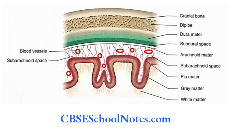
- Meninges are of three different types: From without inwards they are named as Dura mater, Arachnoid mater, and Pia mater.
Read and Learn More Neuroanatomy
Cranial Meninges
Dura Mater
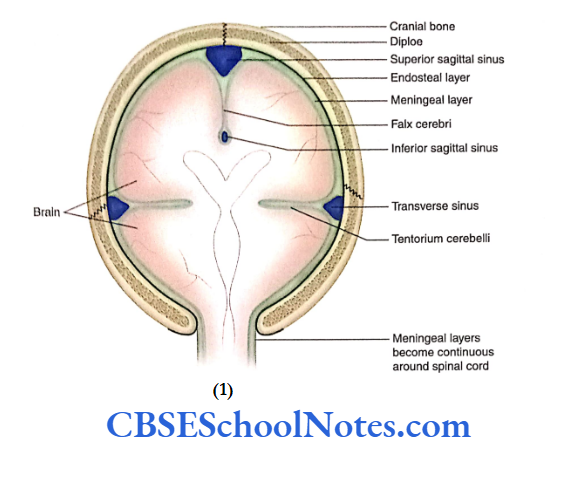
The dura covering the brain is known as cerebral dura. The cerebral dura is made up of an outer endosteal layer and an inner meningeal layer. The endosteal layer of the dura mater is nothing but the endocranium or the inner periosteum.
The meningeal layer of the dura mater is the membranous layer. It covers the brain and then becomes continuous with the dura mater covering the spinal cord.
The two layers of dura mater are tightly fused except in a few places. At these places, the meningeal layer separates from the endosteal layer to form a double-layered fold or partition.
These folds of dura mater extend between the major parts of the brain. As the meningeal layer separates from the endosteal layer, a triangular space is formed.
This space encloses the dural venous sinus. Here, the internal surface of the dura mater is smooth, shining, and lined by endothelial cells.
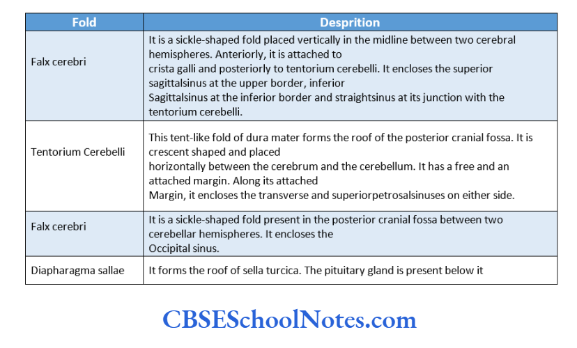
Folds of Dura Mater
The folds of the dura mater play an important role in supporting the brain tissue.
The following folds or septa of the dura mater are formed in the cranial cavity due to duplication of the meningeal layer of the dura.
Dural Venous Sinuses
- As mentioned earlier, the dural venous sinuses are formed due to the separation of meningeal and endosteal layers.
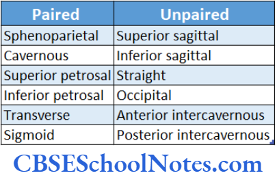
- The dural venous sinuses formed between the layers of the dura mater can be paired or unpaired.
- The dural venous sinuses drain the blood from some cranial bones, meninges, and brain. They ultimately pour the blood into the internal jugular veins.
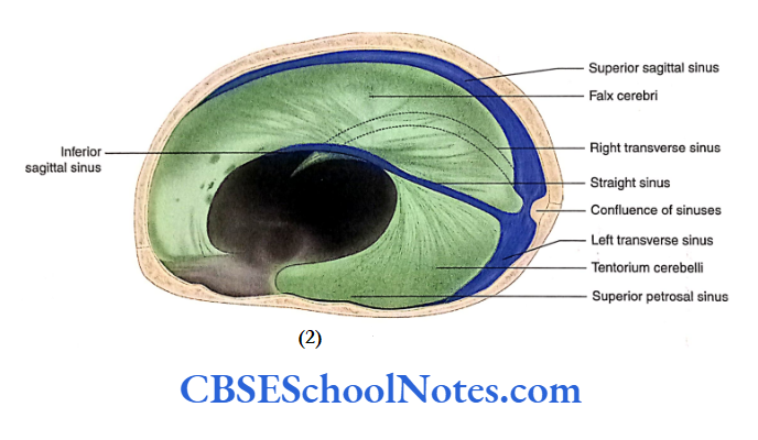
Superior Sagittal Sinus
- The superior sagittal sinus runs at the superior border of the falx cerebri. It begins at the crista galli and is formed by venous blood that is drained from the frontal sinus and veins of the nose.
- This sinus drains superior cerebral veins and becomes continuous with the right transverse sinus at the internal occipital protuberance.
- The inferior sagittal sinus is present at the inferior border of the falx cerebri and drains the falx and medial surface of the cerebral hemisphere. Posteriorly, it becomes continuous with the straight sinus.
Straight Sinus
- The straight sinus is present at the junction of the falx and tentorium.
- The union of the inferior sagittal sinus and the great cerebral vein forms the straight sinus. At the internal occipital protuberance, it becomes continuous with the left transverse sinus.
Transverse Sinus
- The transverse sinus is present on each side of the attached margin of tentorium cerebelli. This sinus extends from the internal occipital protuberance to the base of the petrous temporal bone.
- The right transverse sinus is formed by the continuation of the superior sagittal sinus while the left transverse sinus is formed by the continuation of the straight sinus.
- The transverse sinuses receive blood from the veins of the occipital lobe of the cerebrum and cerebellum and ultimately drain into the right and left sigmoid sinuses.
The sigmoid sinus is situated behind the base of the petrous temporal bone.
This S-shaped sinus is the continuation of the transverse sinus. It passes through the jugular foramen to form the internal jugular vein.
Cavernous Sinus
The right and left cavernous sinuses are situated on either side of the body of the sphenoid. These are formed due to the separation of the endosteal and meningeal layers of the dura mater. These layers are lined by endothelium.
Extension: From the superior orbital fissure anteriorly to the apex of the petrous temporal bone posteriorly.
Tributaries: Superior and inferior ophthalmic veins, cerebral veins, sphenoparietal sinus and frontal trunk of the middle meningeal vein.
Drainage: Into superior and inferior petrosal sinuses and to the basilar plexus of veins.
Communication: With the facial vein through the superior ophthalmic vein, with the pterygoid plexus through the emissary’s veins and with the internal vertebral plexus through the basilar venous plexus
Thrombosis of Cavernous Sinus
- Sometimes, infection may reach the cavernous sinus from the dangerous area of the face and scalp through deep facial and ophthalmic veins.
- This septic thrombosis of the cavernous sinus compresses nerves passing through its lateral wall, and this produces the corresponding symptoms.
- Cavernous thrombosis also causes pain in the eye and swelling of the eyelids.
Nerve Supply of Dura
- The branches of the trigeminal nerve supply the dura mater of the anterior and middle cranial fossae.
- The dura of the posterior cranial fossa is supplied by the vagus nerve and the meningeal branches of Cl to C3 spinal nerves.
Arterial Supply of Dura
Several branches of the following arteries supply the dura mater:
External carotid: Branches of the middle meningeal, ascending pharyngeal and occipital arteries.
Internal carotid
Subclavian: Vertebral branch of the subclavian artery
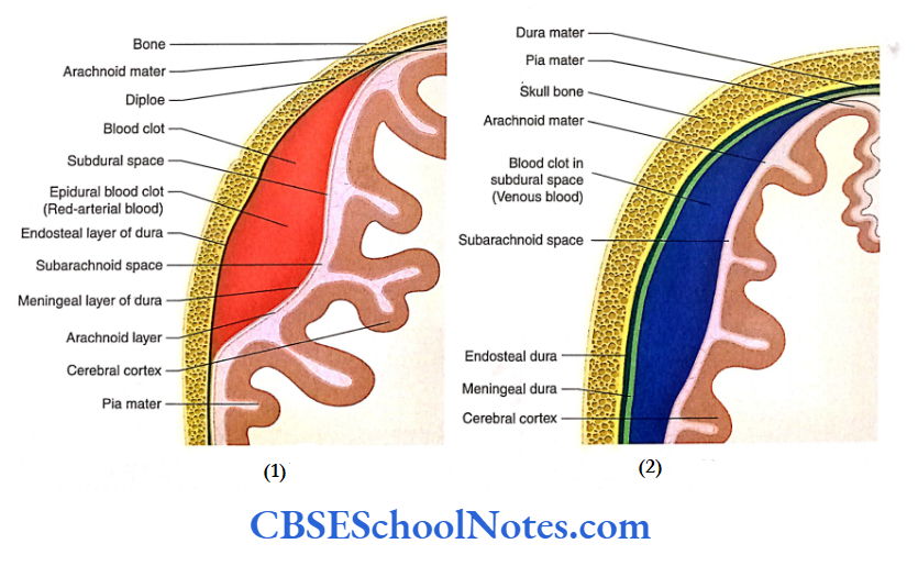
Extradural (Epidural) Haemorrhage
- Epidural haemorrhage occurs due to the rupture of meningeal vessels running between the endosteal and meningeal layers of the dura mater.
- The tear in the meningeal vessels is usually secondary to the fracture of the skull.
- The most common artery affected is the anterior branch of the middle meningeal artery and vein, which lies in the area of the pterion.
Subdural Haemorrhage
- The superior cerebral veins open into the superior sagittal sinus. Just before their opening in the sinus, they run for a short distance in the subdural space.
- Trauma to the head (forceful movement of the brain within the cranial cavity) may tear the superior cerebral vein(s). This results in the collection of blood in subdural space.
Arachnoid Mater
- Deep to dura mater, there lies a delicate, thin and almost transparent membrane known as arachnoid mater.
- It is separated from the dura mater by a capillary space which is called subdural space- it contains a thin film of fluid.
The subarachnoid space lies beneath the arachnoid mater, between the arachnoid and pia mater.
It is filled with CSF. The CSF acts as a buffer, which distributes and equalises the pressure on the surface of the brain.
A meshwork or filaments (trabeculae) extends through the fluid-filled subarachnoid space between the arachnoid and pia mater.
The arteries, veins and cranial nerves, while entering or leaving the brain, lie in the subarachnoid space.
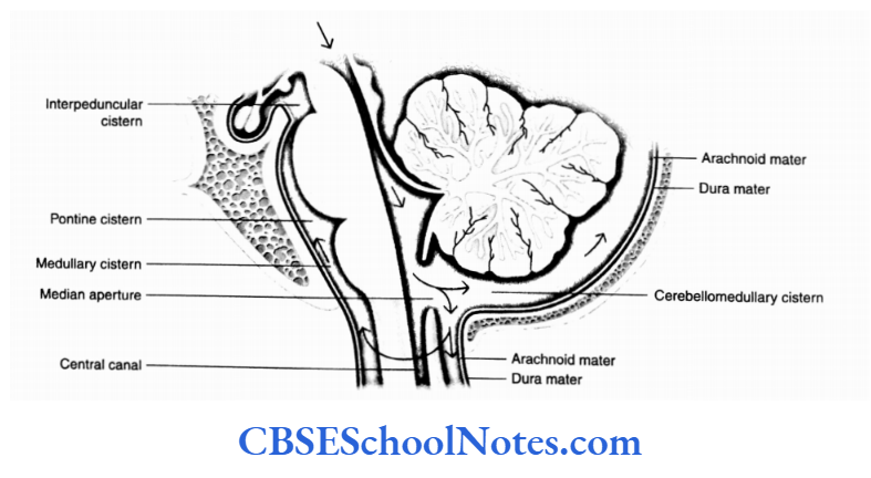
Subarachnoid Cisterns
The arachnoid mater forms bridges over sulci and other irregularities on the surface of the brain. In some situations, on the surface of the brain, the pia and arachnoid mater are widely separated from each other to form subarachnoid cisterns. Large cisterns are formed around the brainstem and cerebellum.
Cisterna Magna
Cisterna magna is the largest cistern and is also known as the cerebellomedullary cistern. It lies in the angle between the cerebellum and the medulla oblongata.
This cistern is continuous above the fourth ventricle through its median aperture and below the subarachnoid space of the spinal cord.
Cisterna Pontis
Cisterna pots lie anterior to the pons and medulla and contain vertebral and basilar arteries.
Cistnrna Intetrpeduncularis
Cisterna interpeduncularis lies between two cerebral
peduncles. It contains the circle of Willis.
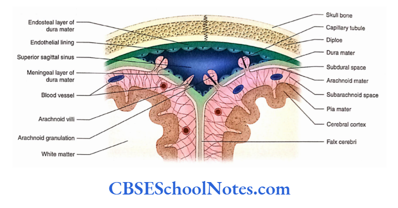
Arachnoid Villi and Granulations
Arachnoid villi arc minute finger-like elevations of arachnoid mater that project into the dural venous sinuses (especially the superior sagittal sinus) through apertures in dura mater. These act as channels of communication between the subarachnoid space and the dural venous sinus.
However, these capillaries (tubules) act as a valve or a one-way communicating channel for the escape of CSF into the venous blood.
The CSF from the subarachnoid space passes into the bloodstream of dural venous sinuses through these villi.
As the age advances, the arachnoid villi become large and globular in shape. These arachnoid villi are now known as arachnoid granulation.
Pia Mater
The pia mater is a delicate, thin membrane adherent to the surface of the brain, i.e. covering the gyri and sulci. It is made up of flattened mesothelial cells. There exists a microscopic subpial space between the pia and the brain.
The blood vessels present in the subarachnoid space anastomose with the neighbouring vessels on the surface of the pia before penetrating the pia mater. Thereafter, the blood vessels pass into the substance of the brain as end arteries.
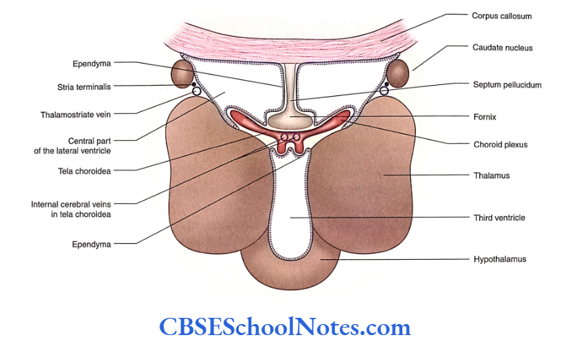
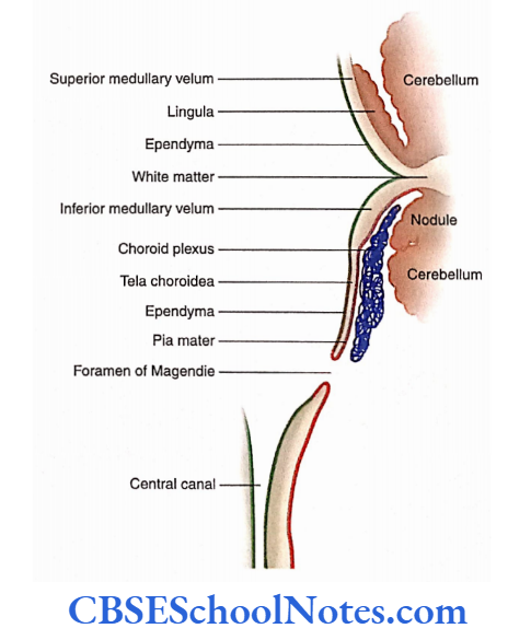
At certain places, the wall of the ventricles of the brain is thin and only lined by ependyma. In this situation, the fold of the pia mater along with the blood capillaries invaginates into the ventricular cavities.
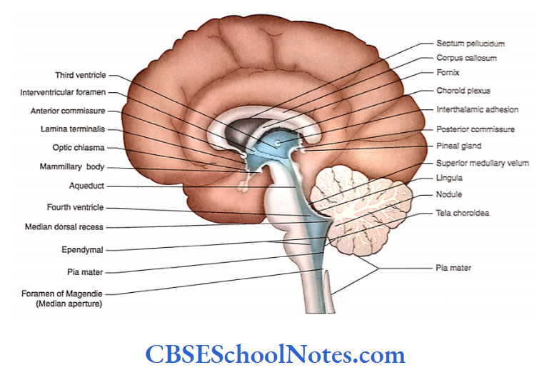
This invaginating vascular tuft is known as the choroid plexus of the ventricles. It is made up of (from outside to inside) ependyma, pia mater and blood vessels.
The pia mater of the choroid plexus is called tela choroidea. In the ventricles, the choroid plexus forms the CSF.
Meninges of the Spinal Cord
- Similar to the cranial meninges, the spinal meninges also consist of the spinal dura mater, arachnoid mater and pia mater.
- These tubular membranes cover the spinal cord and extend into the vertebral canal from the foramen magnum to the level of the second sacral vertebra.
Spinal Dura Mater
- The spinal dura mater is a tough fibrous membrane, which is continuous with the cranial dura at the level of the foramen magnum.
- Below, it narrows at the lower border of the S2 vertebra and covers the thin filum terminale. A the level of the coccyx, it blends with the periosteum covering the posterior aspect of the coccyx.
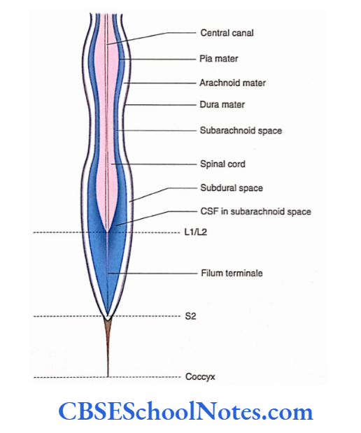
Arachnoid Mater
The arachnoid mater is the continuation of the cranial arachnoid mater. Similar to the dura, it also extends from the foramen magnum to the S2 vertebra where it blends with
the filum terminale.
Pia Mater
- The pia mater closely covers the spinal cord. Above, it is in continuation with the pia mater covering the brain. At the lower end of the spinal cord (conus medullaris), it covers the filum terminale.
- On each side of the spinal cord, the pia mater is present in the form of a fold. This fold is attached between the origin of dorsal and ventral spinal roots. It is known as ligamentum denticulatum.
Cerebrospinal Fluid
- The CSF is a clear, colourless liquid containing a small amount of protein, glucose and potassium and a large amount of sodium chloride.
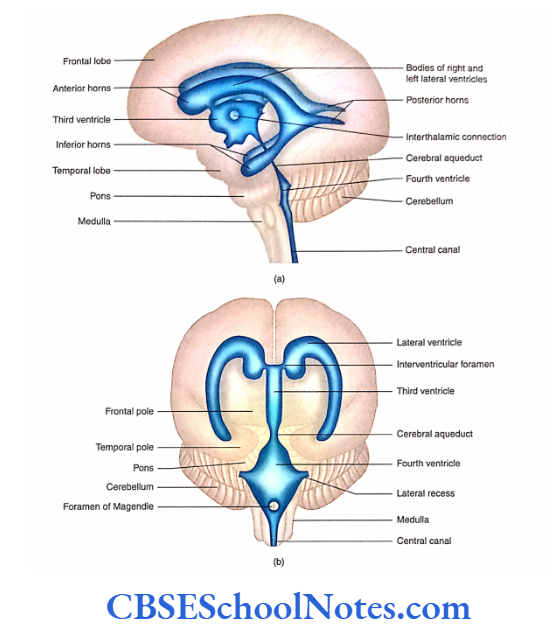
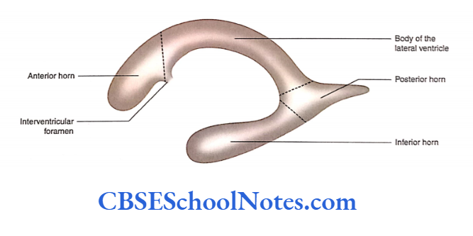
- The CSF is present in the ventricles and subarachnoid space surrounding the brain and the spinal cord.
- It protects the brain and the spinal cord from physical injuries and carries oxygen and nutrients from blood to neurons and neuroglia.
Formation of the Cerebrospinal Fluid
The CSF is produced by choroid plexuses of the lateral, third and fourth ventricles. The net production of CSF is about 400-500 mL/day.
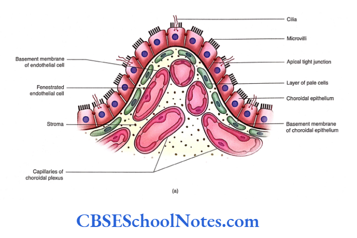
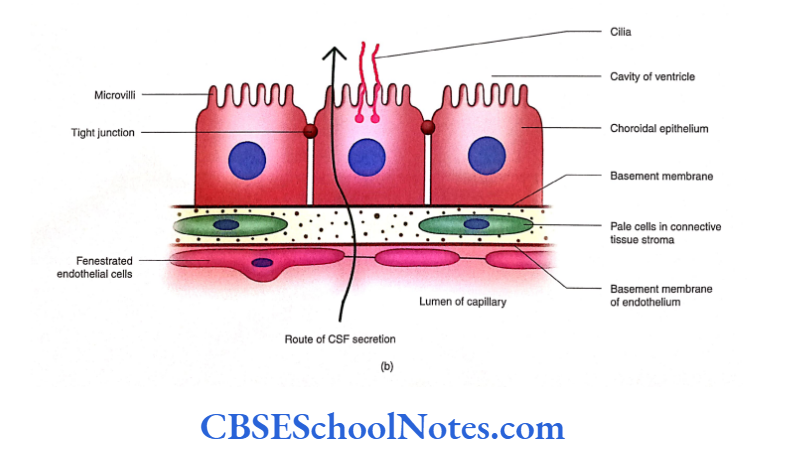
Blood-CSF Barrier
The choroid plexues are made up of a single layer of cuboidal epithelium (modified ependyma) enclosing an extensive capillary network embedded in the connective tissue stroma. Thus, the blood-CSF barrier is formed by the following structures:
- Endothelial cells which are fenestrated
- Basement membranes of endothelial cells
- Layer of pale cells and their processes (derived from pia mater)
- The basement membrane of the choroidal epithelium
- Choroidal epithelium (modified ependymal cells) with tight junctions
The CSF is formed due to the passage of materials through these barriers and also by active secretions from the choroidal epithelium.
The barrier protects the brain and spinal cord from potentially harmful blood-borne substances.
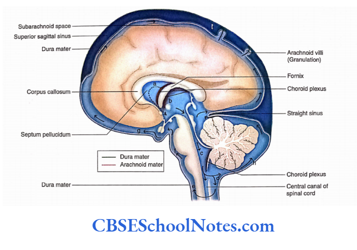
Circulation and Absorption of the Cerebrospinal Fluid
- After being produced by the choroid plexus, the CSF circulates through the ventricles ofthe brain, subarachnoid space and central canal of the spinal cord.
- The movement of the vertebral column and the pulsation of arteries present in the subarachnoid space assist the movement of CSF.
- The CSF is reabsorbed into the venous blood through arachnoid villi and granulation. Ependyma, arachnoid capillaries and lymphatics of meninges also absorb some CSF.
Functions of the Cerebrospinal Fluid
- The CSF surrounds the brain and the spinal cord; therefore, it serves as a cushion between the delicate nervous tissue and the surrounding cranial and vertebral bones.
- The CSF provides a medium for the exchange of nutrients and waste products between the nervous tissue and blood.
- By providing a medium for the exchange of nutrients and removal of waste products, the CSF maintains intracranial pressure.
- Certain hormones are transported by the CSF; for example, the secretion of the pineal gland is carried by the CSF to the pituitary gland.
Hydrocephalus
Hydrocephalus is defined as excessive collection of CSF leading to an increase in CSF pressure. High CSF pressure causes atrophic changes in the brain substance.
Hydrocephalus may be caused due to the following reasons:
Blockage in the normal circulation of CSF
In rare cases, it may also be due to excessive production of CSF as in the case of a tumour of choroid plexus Lumbar Puncture (Spinal Tap)
The CSF can be obtained for biochemical analyses by a procedure called lumbar puncture.
Lumbar puncture is sometimes also used to inject drugs into the subarachnoid space, for example, spinal anaesthesia.
The CSF is obtained by inserting a long needle into the lumbar subarachnoid space in the midline between the third and fourth lumbar spine.
Apart from lumbar puncture, CSF can also be obtained from cisternal puncture.
Summary
- The three meninges—dura mater, arachnoid mater and pia mater—are connective tissue membranes, which cover the brain and the spinal cord.
- Arachnoid and pia are collectively called leptomeninges.
- The cerebral dura mater consists of two layers:
- Endosteal and Meningeal.
- At places, the meningeal layer separates from the endosteal to form a double-layered fold known as ‘dural folds’. These folds or septa lie between the major parts of the brain, for example, falx cerebri and tentorium cerebelli.
- At places, the separation of meningeal and endosteal layers of dura encloses a triangular space. This space encloses
the ‘dural venous sinus’. - The extradural haemorrhage occurs between the endosteal and meningeal layers of the dura mater.
- Superior sagittal, cavernous and sigmoid sinuses are common sites of thrombosis.
- The arachnoid mater is a delicate, thin and transparent membrane.
- The cerebellomedullary cistern is the largest cistern, which lies at the angle between the cerebellum and the medulla oblongata.
- Arachnoid villi and granulations are minute projections of arachnoid mater into dural venous sinuses.
- The spinal cord is also covered by three meninges:
- Dura, Arachnoid and Pia mater
- The dura and arachnoid mater extend from the foramen magnum to the S2 vertebral level where they blend with the filum terminale.
- The pia mater closely covers the spinal cord, rootlets and filum terminale.
- CSF is produced in choroid plexuses, which are present in the ventricles of the brain.
- The fold of the pia mater along with blood capillaries invaginates into ventricular cavities. This invaginating vascular tuft is known as the choroid plexus of the ventricles.
- A blood-CSF barrier is formed by choroidal epithelium, a layer of pale cells and fenestrated capillary epithelium.
- The movement of CSF, in the spinal subarachnoid space, is assisted by the pulsation of the arteries which are situated around the spinal cord. The movement of CSF is also assisted by the movement of the vertebral column.
Multiple Choice Questions
Question 1. The delicate nervous tissue of the CNS is protected by the following structures except
- Bones
- Meninges
- Ligamentum denticulatum
- Cerebrospinal fluid
Answer: 3. Ligamentum denticulatum
Question 2. Which of the following is known as leptomeninges?
- Dura mater
- Arachnoid mater
- Dura and arachnoid mater
- Arachnoid and pia mater
- Pia mater
Answer: 4. Arachnoid and pia mater
Question 3. Which are the structures drained by dural venous sinuses?
- Cranial bones
- Brain
- Meninges
- All of the above
- Only b and c
Answer: 4. Only b and c
Question 4. The following are the paired dural venous sinuses except
- Cavernous
- Sigmoid
- Occipital
- Sphenoparietal
- Inferior petrosal
Answer: 3. Sphenoparietal
Question 5. The following facts about extradural haemorrhage are correct except
- Most commonly, it occurs due to rupture of the middle meningeal artery
- It is usually secondary to a fracture of the skull in the area of the pterion
- Blood collects between the endosteal and meningeal layers of dura mater
- The shape of the blood clot, in the CT scan, is biconcave
- The blood clot presses the lateral surface of the cerebrum
Answer: 4. The shape of the blood clot, in the CT scan, is biconcave
Question 6. Which are the subarachnoid cisterns surrounding the brainstem and cerebellum?
- Cerebellomedullary cistern
- Pontine cistern
- Medullary cistern
- Interpeduncular cistern
- All of the above
Answer: 5. All of the above