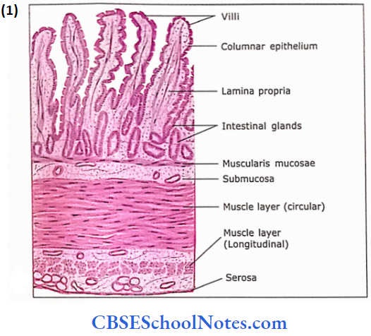
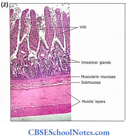
General Histology Of The Alimentary Canal
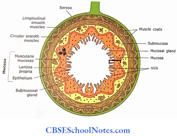
The alimentary canal, from the oesophagus to the anal canal, may be identified by its tubular nature and also by the division of its wall into four layers.
These layers from deep to superficial are :
- Mucosa
- Submucosa
- Muscle layer
- Serosa
1. Mucosa
This is the innermost layer of the alimentary canal and com-prises lining epithelium, lamina propria and muscularis mucosae.
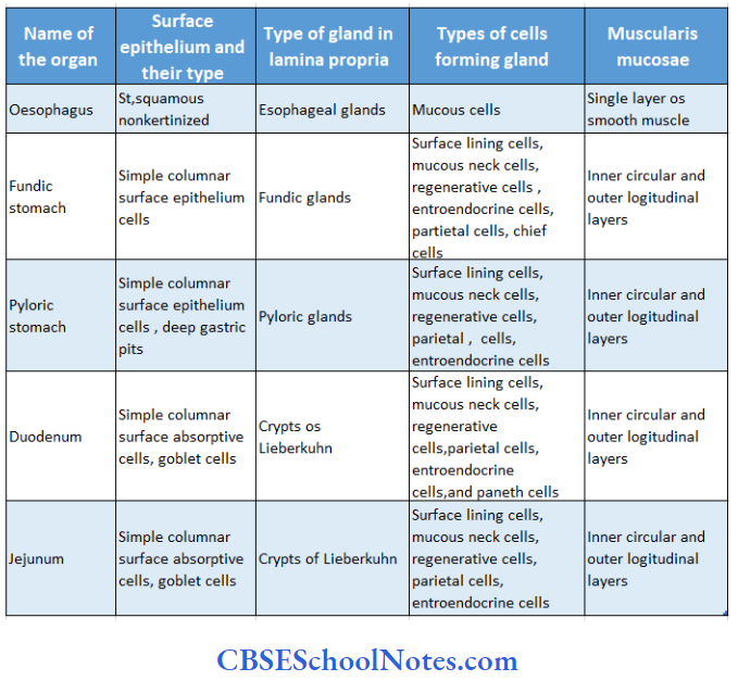
- Epithelium: Different types of epithelium line different parts of the alimentary canal, i.e., the oesophagus and anal canal are lined by protective epithelium (stratified squamous); the intestine and stomach are lined by simple columnar, which is absorptive and secretory.
- Lamina propria: This is a layer of loose connective tis¬sue, which supports the epithelium. It contains the blood and lymphatic vessels through which nutrients are absorbed.
- Lamina propria also accommodates glands like gastric glands in the stomach and crypts of Lieberkuhn (intestinal glands) in the mucosa of the intestine. These glands develop from the invagination of luminal epithelium.
- The lamina propria also contains “mucosa-associated lymphoid tissue.” i.e.. lymphocytes and macrophages.
- These lymphocytes and macrophages are defensive as the alimentary canal is subjected to the entry of foreign substances like bacteria and other microbes.
- These lymphoid tissues may be in the form of lymphatic nodules (as in the appendix) or the form of diffuse tissue,
- Muscularis mucosae:
- This consists of a thin layer of smooth muscle cells. These are arranged as inner circular and outer longitudinal layers. The muscularis mucosae can change the shape of the mucosa, which helps in absorption and secretion.
Lamina propria Remember:
Lamina propria contains the blood and lymphatic vessels through which nutrients are absorbed. It also contains glands (intestinal glands) and lymphoid tissue. The secretory function of mucosa provides lubrication and delivers digestive enzymes and hormones. Lymphoid tissues are defensive and protect the intestine from foreign substances.
2. Submucosa:
It consists of moderately dense, irregular connective tissue rich in collagen and elastic fibres.
- The submucosa contains blood vessels, lymphatic vessels, nerves and Meissner’s plexus.
- In some parts of the alimentary tract (oesophagus and duodenum), the submucosa contains glands that pour their secretion into the lumen of the gut through ducts.
- This layer may also contain scattered lymphoid nodules, particularly in the large intestine.
3. Muscle Layer:
In most of the parts of the alimentary tract, this layer is made up of an outer sheet of longitudinally arranged smooth muscle fibres and an inner sheet of circularly arranged fibres.
- This layer also contains myenteric plexus (Auerbach’s plexus) between circular and longitudinal sheets of muscle fibres.
- This plexus mostly controls the motility of the tract.
4. Serosa Adventitia:
The serosa is the superficial layer on the parts of the alimentary tract present in the abdominal cavity.
- It is a visceral peritoneum composed of connective tissue and simple squamous epithelium.
- The oesophagus part of the alimentary tract, which is outside the abdominal cavity, has a superficial layer called adventitia that consists of areolar connective tissue.
Serosa Remember:
Contraction of the muscle layer mixes and propels the contents of the digestive tract. The serosa or adventitia is the outermost layer of the alimentary canal.
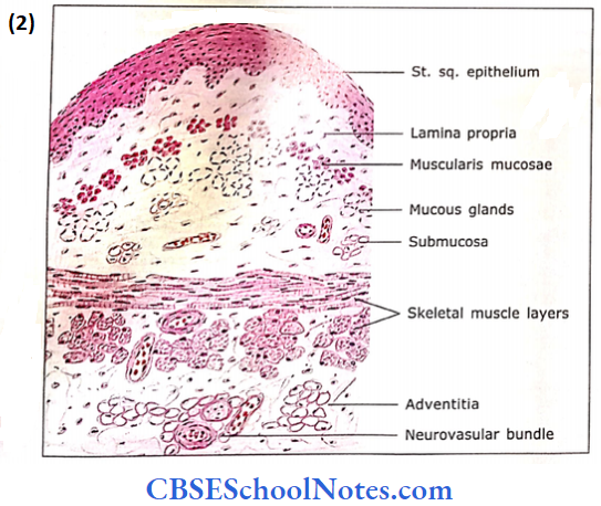
Oesophagus
The oesophagus is a straight muscular tube extending from the pharynx in the neck to the stomach in the abdomen. Most of it lies in the thoracic cavity.

Its wall is made up of four layers:
- Mucosa
- Submucosa
- Muscle layer
- Serosa
1. Mucosa
- Epithelium: It consists of thick non-keratinized strati¬fied squamous epithelium.
- Lamina propria: It consists of a thin layer of loose con¬nective tissue
- Muscularis mucosae: It is unusual in two respects; firstly, it is thicker compared to other parts of the alimentary canal. Secondly, it is single-layered and formed by longitudi¬nally running smooth muscle fibres. Sometimes, the mucosa of the oesophagus may contain mucous glands in its uppermost (near the laryngopharynx) and lower (near the stomach) regions.
2. Submucosa
It is a wide layer of irregular, moderately dense connective tissue composed of bundles of collagen and elastic fibres.
- Due to the presence of elastic fibres, the submucosa is thrown into folds.
- Hence, the lumen of the oesophagus appears star-shaped. The submucosa contains blood vessels and branched tubuloalveolar mucous glands.
- Ducts arising from these glands run through lamina propria to open into the lumen of the oesophagus.
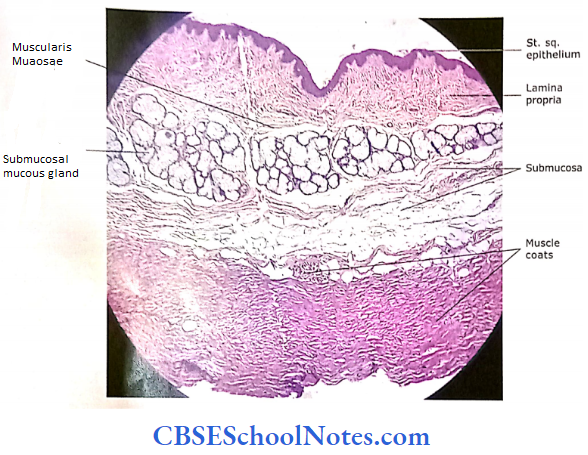
3. Muscle Layer
The muscle layer is different in different parts of the oesophagus.
- In the upper third portion, the muscle is skeletal. It consists of both skeletal and smooth muscles in the middle portion.
- In the lower third, it consists of purely smooth muscle.
These muscles are arranged in two layers, i.e… an outer longitudinal and an inner circular. A well-formed myenteric plexus of nerve fibres and ganglion cells exists between the two muscle layers. It controls the peristaltic movements.
4. Serosa/Adventitia
The superficial layer, adventitia, consists of loose areolar connective tissue, which merges with the connective tissue of surrounding structures. The adventitia surrounds most of the oesophagus, except for the lowest 1 inch where it is covered by serosa.
Oesophagus Functions
- The oesophagus has neither digestive nor absorptive functions.
- It is involved in the transport of food and water from phar¬ynx to the stomach. The bolus of the food travels in the oesophagus at the rate of 50 mm per second.
- It secretes mucus that lubricates the lumen of the oesopha¬gus to facilitate the transport of food.
Oesophagus Remember:
The glands of the mucosa and submucosa of the oesophagus secrete the mucus to facilitate the transport of food and protect the mucosa of the oesophagus. The muscle layer in the upper part of the oesophagus consists of skeletal muscle, while in the lower part it consists of smooth muscle. However, in the intermediate part, it is mixed.
Stomach
Based on histological structure, the stomach is divided into three distinct regions
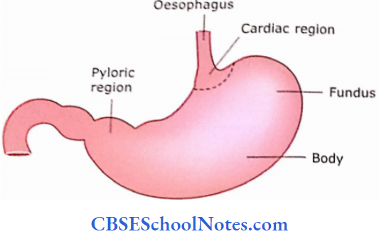
- Cardiac region: This part of the stomach is about 2-3 cm wide at the junction of the oesophagus.
- Fundic region (or body): This region consists of fun¬dus and body.
- Pyloric region: This region connects the stomach to the duodenum
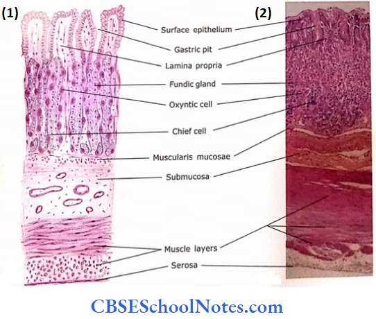
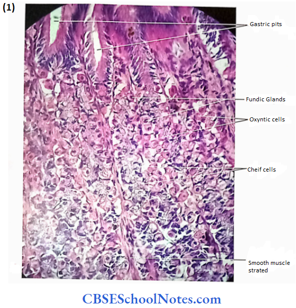
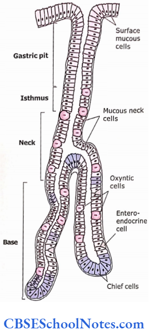
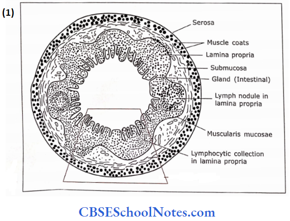
In an empty stomach, the mucosa and submucosa are thrown into longitudinal folds called rugae. These rugae disappear when the stomach is distended with food. The epithelial lining of gastric mucosa contains many depressions called gastric pits.
From the bottom of each gastric pit, several tubular gastric glands extend into the lamina propria A section through the wall of the stomach shows the usual four layers, i.e., mucosa, submucosa, muscle layer and serosa
1. Cardiac Region
1. Mucosa:
- The epithelial lining, at the cardio-oesophageal junction, changes from stratified squamous epithelium to simple columnar epithelium. The columnar epithelium of the cardiac region shows basal, oval nuclei.
- These cells are involved in the secretion of mucus and arc called mucous surface cells.
- The goblet cells are not present in this epithelium. The lamina propria contains small tubular glands that open in gastric pits.
- These glands are lightly stained with ll&E and arc called mucus-secreting cardiac glands. The muscularis mucosae contain smooth muscle fibres arranged in two layers.
2. Submucosa:
The submucosa is a broad layer of connective tissue. It consists of blood vessels and nerve plexus (Meissner’s plexus).
3. Muscle Layer:
It is composed of three thickened layers of smooth muscle bundles inner oblique, middle circular and outer longitudinal. The inner oblique layer is not always apparent. The myenteric plexus may be seen in connective tissue between muscle layers.
4. Serosa:
Serosa is our most covering formed by simple squamous epithelium resting on a layer of connective tissue.
2. Fundic or Body Region
Following lour layers mo observed in this region.
1. Mucosa:
Lining Epithelium:
- The epithelium, which lines the surface and the gastric pits, is a simple columnar mucous epithelium.
- These cells are involved in the secretion of mucus. The mucus protects the epithelial lining from damage due to the presence of acid in the stomach.
- These cells are replaced every few days (3 5) with the proliferating cells of gastric pits.
Lamina propria:
Simple tubular glands arc present in lamina propria, which extend from the bottom of the gastric pit to the muscularis mucosae. They are called gastric glands or fundic glands and several may open at the bottom of a gastric pit. The isthmus is a small segment between the gastric pit and the fundic gland.
The cells of the isthmus divide and migrate upwards and downwards. These cells give rise to the cells of the surface epithelium and various cells of the gastric gland. In H&E sections.
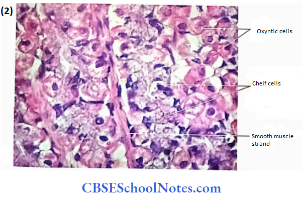
The following types of cells can be identified in fundic glands:
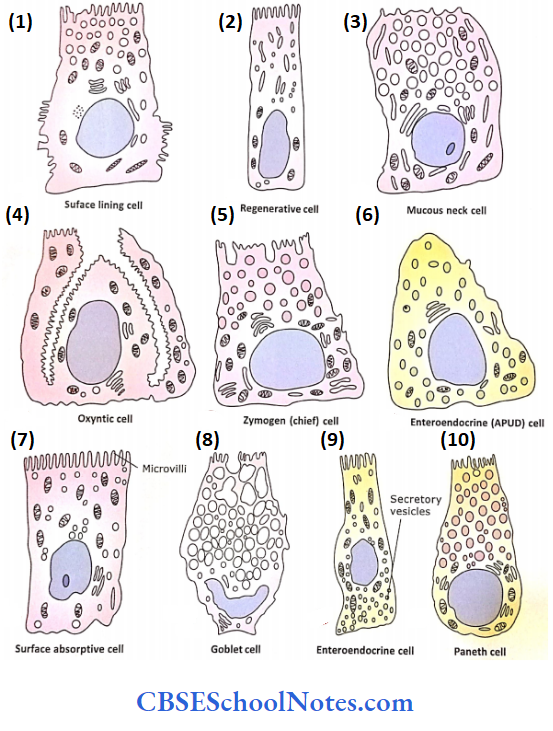
- Mucous neck cells:
- These cells are located just below the gastric pit. They are columnar and have mucinogen granules in apical cytoplasm, while nuclei arc basally situated.
- They produce mucus of a different nature than that of surface epithelium.
- They produce soluble mucus compared to insoluble mucus produced by surface mucous cells. The parietal cells are seen located with mucous neck cells.
- Mucous neck cells also contain few regenerative (stem) cells. These cells proliferate to replace all of the specialized cells lining the gastric gland, gastric pit and surface. The mucous neck cells are replaced every 5-7 days.
- Parietal or oxyntic cells:
- They produce HCl and intrinsic factors. These cells are mostly found in the upper half of gastric glands.
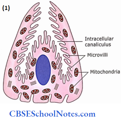
- The parietal cells are large, rounded or pyramidal. These cells are wedged against the basement membrane.
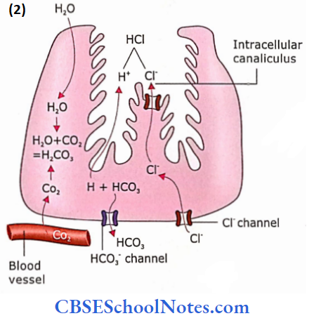
- In H&E preparation, they are stained dark pink. The electron micrographs, show the presence of a deep infolding of the apical membrane forming an “intracellular canaliculus” into which many microvilli. The cytoplasm contains tubulovesicular structures under the microvilli and many mitochondria.
- The tubule-vesicular structures are involved in HCI secretion.
- The synthesis of HCl takes place by the combination of Cl ions inside the intracellular canaliculi (outside cell). Both H+ and Cl– ions are secreted by parietal cells. The life span of parietal cells is 150-200 days.
HCI Remember:
HCI is produced outside the parietal cell, be, in the lumen of the intercellular canaliculus.
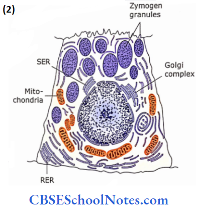
- Chief or zymogenic cells:
- Most of these cells are located in the lower third of gastric glands.
- They usually take basophilic stains and thus are easily differentiated from parietal cells, which are strongly eosinophilic.
- The chief cells contain a rough endoplasmic reticulum near the base, secretory granules near their apex and a small Golgi apparatus.
- These cells are typical protein-secreting cells and secrete pepsinogen, which is converted to a proteolytic enzyme pepsin in an acid environment.
 0
0
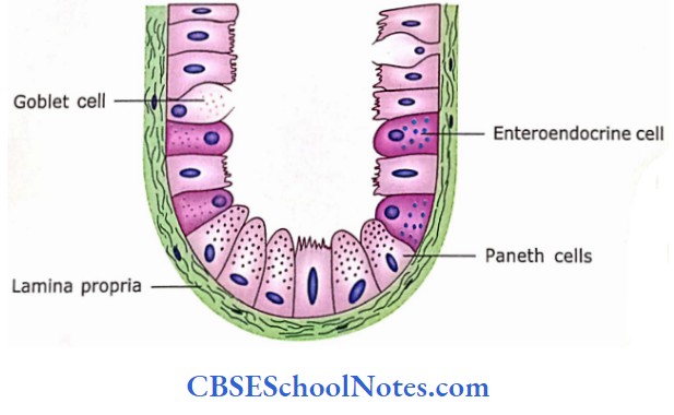
- Enteroendocrine and APUD cells:
- These cells are located in the basal portion of gastric glands.
- In the H&E section, the cells appear unstained. They contain secretory granules, which stain only in special preparations.
- They are thought to produce serotonin, histamine and gastrin.
- As these cells are endocrine cells, they release their products into the blood vessels.
Muscularis mucosae:
It consists of two thin layers of smooth muscles, i.e., outer longitudinal and inner circular.
Lamina propria Remember:
Lamina propria of the fundic or body region contains fundic (gastric) glands, which secrete gastric juice of the stom¬ach. Gastric juice contains hydrochloric acid (HCI) and intrinsic factors, pepsin and mucus. Parietal cells produce HCI and gastric intrinsic factor, while chief cells produce pepsinogen, reninin and gastric lipase. Mucus neck cells produce mucus.
2. Submucosa:
The submucosa is the same as described under general histol¬ogy of the alimentary canal.
3. Muscle Layer:
It has three layers of smooth muscle, i.e., in the outermost layer muscle fibres are oriented longitudinally, in the middle layer Fibres are circular and in the inner layer are obliquely arranged. The outer longitudinal layer is not very complete.
4. Serosa:
It is the outermost layer of the stomach and consists of loose connective tissue covered by mesothelium.
Fundic Or Body region Functions
- Mixing, partial digestion and storage of food.
- Production of gastric juice, i.e., pepsin, HC1, renin, in¬trinsic factor, hormones and mucus.
- Absorption of water and salt (slight). Alcohol and aspi¬rin enter the lamina propria by damaging surface epithelium.
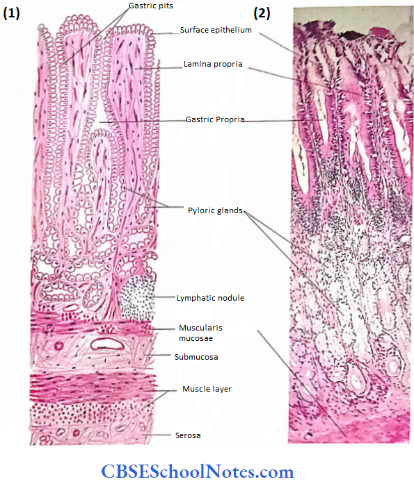
3. Pyloric Region
1. Mucosa:
- The surface epithelium is the same as present in the fundic part of the stomach. The pyloric glands occupy the lamina propria (Fig. 14.8 A and B).
- The gastric pits of these glands are deeper and extend through half the thickness of the mucosa. These glands are short, tortuous and branched. They are lined by one-cell
- Type, which is similar to mucous neck cells. They produce mucus and gastrin. Muscularis mucosae is same as in fundic part.
2. Muscle Layer: The circular muscle is much thickened and forms a pyloric sphincter.
3. Submucosa and Serosa: Same as in the fundic part.
Pyloric Region Clinical Application
Pernicious Anaemia:
The parietal cells also serve as the source of intrinsic factors. The intrinsic factor is a must to absorb vitamin Br from the small intestine. In the absence of parietal cells (due to disease or surgery), the vitamin Bn is not absorbed, which leads to a condition known as pernicious anaemia. Parietal cells may also decrease in old age. The pernicious anaemia is treated by giving injections of vitamin B12.
Small Intestine
The small intestine is about 5 m in length and consists of three parts, i.e., duodenum (about 12 cm in length), jejunum (2 m) and ileum (3 m). This long tube extends from the stomach to the colon and is highly coiled. The major function of the small intestine is digestion and absorption. Most of the nutrients are absorbed by the small intestine.
Intestine Remember:
The absorptive surface area of the intestine is increased by the formation of plicae circularis, villi and microvilli. The plicae circularis (valves of Kerckring) are permanent mucosal folds. These structures do not disappear on distension as they contain the core of the submucosa. Besides increasing the absorptive surface area, they also decrease the velocity of movement of contents.
1. Duodenum
It consists of four layers, i.e.,
- Mucosa
- Submucosa
- Muscle layer and
- Serosa/adventitia.
1. Mucosa:
- In the duodenum, the mucosa and submucosa are thrown into circular folds called plicae circularis. These mucosal folds are permanent structures and do not disappear on distension. This is because they contain the core of submucosa.
- The function of plicae circularis is to slow the passage of contents and to increase the surface area of mucosa.
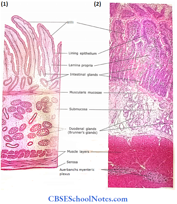
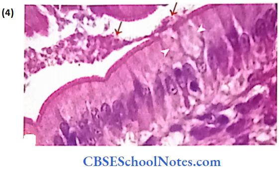
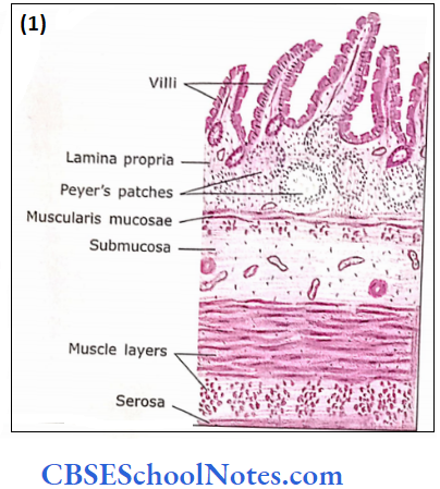
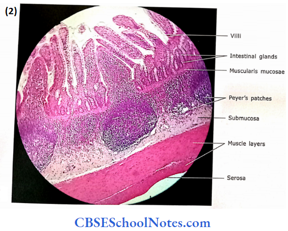
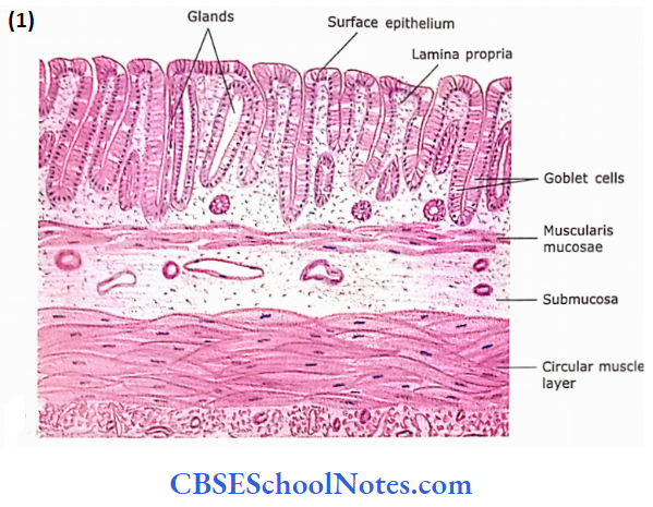
- The surface area of the mucosa is further increased because of the presence of mucosal villi. The plicae are covered with villi. Villi are fingerlike mucosal projections about 0.5 mm in length.
- The core of each villus is formed by loose connective tissue of lamina propria containing fenestrated capillaries, smooth muscle fibres and lymphatics (blind-end lacteals).
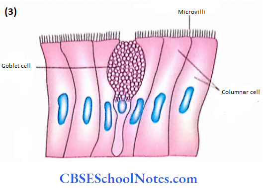
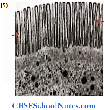
- The surface of the villi is covered by epithelium consisting predominantly of columnar cells with striated borders (microvilli). In between the columnar cells, a few goblet cells are also present.
Mucosa Remember:
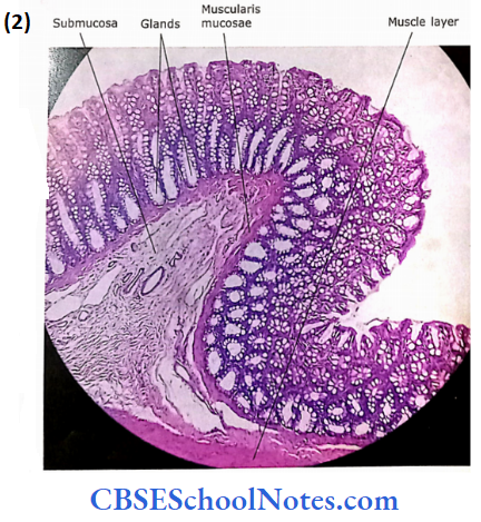
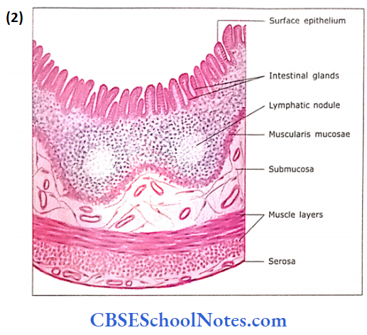
- The mucosa of the intestine consists of villi, intestinal glands along with lamina propria, lymphocytic collections and muscularis mucosae.
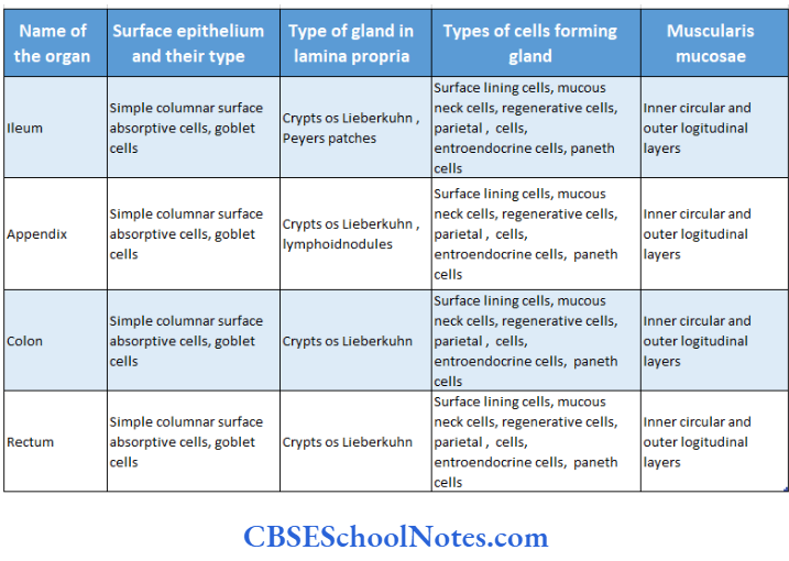
- In between the bases of two intestinal villi, the epithelium is invaginated in lamina propria to form intestinal glands (crypts of Lieberkuhn). These glands are short tubular glands that open into the lumen of the intestine at the base of the intervillous space. These glands are lined by columnar cells, goblet cells, Paneth cells and enteroendocrine cells.
- The lamina propria, in between tubular intestinal glands, consists of loose connective tissue, reticular cells and diffuse lymphoid tissue. Occasionally, lamina propria may also contain submucosal Brunner’s glands. The muscularis mucosae sends smooth muscle fibres towards the core of the villi.
2. Submucosa:
The submucosa is almost completely occupied by highly branched, tubuloacinar duodenal glands. Brunner’s glands. The ducts of these glands pass through muscularis mucosae and open into the lumen of the duodenum. The acini of duodenal glands secrete mucus, hence taking light stain.
- These acini are lined with cuboidal or low columnar cells.
- Duodenal glands secrete alkaline mucus with high concentrations of bicarbonates that protect the duodenal mucosa from acid secreted by the stomach.
- Brunner’s gland also secretes “human epidermal growth factor,” which increases the mitotic activity of epidermal cells.
- This factor also inhibits the secretion of HCl.
3. Muscular Layer: It is made up of outer longitudinal and inner circular layers of smooth muscle.
4. Serosa/Adventitia: As most of the duodenum is retroperitoneal, some parts of its surface may show serosa. Otherwise, it is covered by adventitia.
Brunner’s gland Remember:
Brunner’s gland secretes “human epidermal growth factor” (which increases the mitotic activity of epidermal cells) besides producing mucous and alkaline fluid to neutralize the acidic chyme.
2. Jejunum
It also consists of four layers.
1. Mucosa:
Similar to the duodenum, the mucosa and submucosa of the jejunum are also thrown into permanent circular folds (plicae circularis), which bear tongue-shaped villi of different heights.
- Each villus is covered with tall columnar cells having a striated border. The core of each villus is formed by lamina propria containing diffuse lymphatic tissue, blood vessels, smooth muscle fibres and central lacteal.
- The crypts of Lieberkuhn (intestinal glands) are present in lamina propria below the bases of villi.
- These glands contain four different types of cells, i.e., absorptive columnar cells, goblet cells, enteroendocrine cells and Paneth cells
- Paneth cells play an important role in controlling the bacterial flora of the small intestine by producing antibacterial agents lysozyme and defensin.
Following are the main hormones secreted by gastric and intestinal enteroendocrine cells glucagon, serotonin, endorphin, histamine, gastrin, somatostatin, secretin, cholecystokinin (CCK), gastric inhibitor.’ peptide (GIP), noradrenaline and vasoactive intestinal peptide (VIP). The muscularis mucosae are in the usual two layers.
2. Submucosa
It is made up of connective tissue, blood vessels, nerve plexus (Meissner’s plexus) and lymphatics. Although no Peyer’s patches are seen, occasionally a lymph nodule may occur. No Brunner’s glands are seen.
3. Muscular Layer
It is the same as in the duodenum.
4. Serosa
It covers the jejunum to form an outermost layer.
Jejunum Remember:
Crypts of Liberkuhn or intestinal glands: These are simple tubular or branched tubular glands present in lamina pro-pria below the bases of villi. They increase the surface area of the intestinal lining. These glands consist of surface absorptive cells, goblet cells, enteroendocrine and Paneth cells.
Enterocytes are tall columnar and have elongated nuclei situated at the base. They are absorptive cells and transport water and substances from the lumen of the Intestine to the blood capillaries. They are also secretory cells, producing enzymes, which help In digestion and absorption.
3. Ileum
The ileum also has the usual four layers in its wall, i.e., mucosa, submucosa, muscle layer and serosa.
1. Mucosa
The epithelial lining of villi consists of the same absorptive columnar cells as present in the jejunum. The villi arc is few, short and finger-like. Lamina propria contains intestinal glands and aggregation of lymphatic nodules called Peyer’s patches. The number of lymphatic nodules increases in the distal ileum where they are covered with rudimentary villi.
In the ileum where Peyer’s patches are much larger they extend into the submucosa after disrupting the museularis mucosae. The muscular mucosae is thin or absent whereas Peyer’s patches are large.
Submucosa, Muscle Layer and Serosa
These layers are the same as in the jejunum.
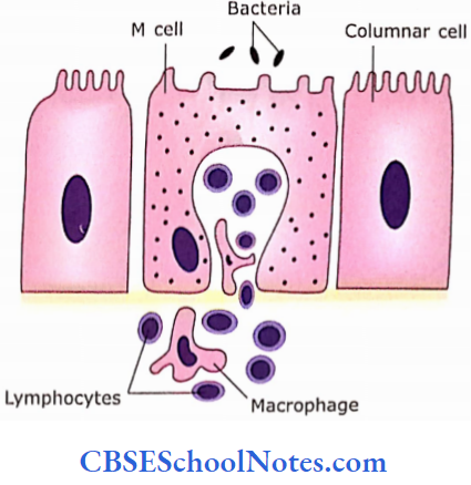
Peyer’s Patches and “M” Cells
The mucosa intestine is exposed to toxins and microorganisms. To protect the gut from this invasion, there is the presence of lymphocytes, plasma cells and macrophages in the connective tissue of lamina propria. In the terminal portion of the ileum, there are aggregations of lymphatic nodules (Peyer’s patches).
- Overlying these lymphatic nodules, there are specialized “M” cells in between the columnar cells of the epithelium.
- These cells are broad and have few micro folds on their apical surface.
- The base of these cells is invaginated and occupied by lymphocytes and macrophages of underlying lymphoid tissue.
- The formation of a deep recess in the M cell facilitates the lymphocytes to come in close contact with the bacteria and macro-molecules present on the apical surface of M cells.
- The M cells are capable of attracting bacteria and transporting their antigen across the cell to macrophages. The macrophages present the antigen to the surrounding lymphocytes.
- These lymphocytes then migrate to the lym¬phatic nodules in lamina propria. Mere, these lymphocytes then differentiate into plasma cells and produce the specific antibody.
- The antibodies (IgA) then pass from lamina propria to the surface of the epithelium through epithelial cells.
- Where (hey sil in the glycocalyx to neutralize toxins and microorganisms. Antibodies produced in lamina propria arc also re-circulated through the liver and gall bladder to the lumen of the intestine.
Ileum Remember:
M cells transport bacteria and other macromolecules from the lumen to cells of the immune system in Peyer’s patches and other large lymphocytic nodules. Thus, the M cell is considered an antigen-transporting cell
Large Intestine
The large intestine consists of the colon, rectum and anal canal.
Similar to the small intestine, the large intestine also has a mucosa, submucosa, muscle layer and serosa.
1. Colon
Mucosa:
The mucosal folds (plicae circularis) and villi are absent. Hence, the mucosal surface is smooth. The surface epithe¬lium principally consists of columnar cells and a few goblet cells. The columnar cells are absorptive and bear microvilli (striated border). These cells are involved in the absorption of lipids, water and salts.
- The lamina propria is occupied with tubular glands, which extend from surface to mus-cularis mucosae. The glands are straight and much longer than the glands of the small intestine.
- Glands are lined with few absorptive columnar cells and widely scattered enteroendocrine cells, but goblet cells now become Iho principal cells.
- Paneth cells may occur occasionally. In the lower third of the gland, the epithelium consists almost entirely of goblet cells. The lamina propria contains rinse lymphatic tissue and sometimes lymph nodules. The muscularis mucosae is typical.
Submucosa:
Its histological structure is typical.
Muscle Layer:
The outer longitudinal muscle layer is very thin. It is arranged in thick bands (taenia coli) only at three places. The inner circular layer of muscle is also thin compared to the small intestine.
Serosa:
It covers the transverse colon but parts of ascending and descending colon are covered by adventitia.
Colon Remember:
The mucosal surface of the colon is smooth as it has no villi or mucosal folds. The lining cells of mucosa consist predominantly of surface absorptive cells, which are involved in the absorption of lipids, water and salts. The number of goblet cells increases from the ascending colon to the sigmoid colon.
The lamina propria is occupied with tubular glands (crypts of Luberkhun), which extend from the surface to muscularis mucosae.
2. Rectum
Mucosa:
- The rectum is structurally similar to the colon. The intesti¬nal glands are straight like test tubes. The surface epithe¬lium and glands are lined predominantly with goblet cells.
- Entero-endocrine cells may be found, but Paneth cells are absent. Toward the termination of the rectum, the long intestinal glands decrease in length.
- The temporary longitudinal folds may be present in the upper rectum that has a core of submucosa.
- The permanent longitudinal folds of the rectum (rectal columns) appear in the lowermost rectum. The muscularis mucosae, in the lowermost rectum, appears as a thin layer.
Submucosa: It has a typical structure.
Muscle Layer:
In the rectum, the taenia coli are absent and a complete longi¬tudinal external muscle coat is present. The inner muscle coat is circular.
Serosa/Adventitia:
As the rectum is retroperitoneal, the serosa is present only
Functions of Colon and Rectum
- Absorption or remaining lipids, which are not absorbed by the small intestine.
- Absorption of water and salts that results in concentra¬tion of faeces.
- Secretion of mucus that facilitates the movement of intestinal contents.
- The mucus also protects the epithelium against abrasions by the concentrated faeces.
3. Anal Canal
Mucosa:
The mucosa of the anal canal has different histological structures in its upper, middle and lower parts.
- The upper part of the anal canal (the longitudinal anal columns) is covered by simple columnar epithelium similar to the rectum. There are few short intestinal glands in lamina propria, which gradually disappear as traced distally.
- The mucosa in the middle part is covered by stratified squamous non-keratinized epithelium.
- The uppermost portion of the middle part sometimes may show stratified cuboidal or columnar epithelium.
- The mucosa in the lowest part of the anal canal is covered with true skin and contains associated sebaceous and large apocrine glands. The muscularis mucosae disappear at the level of the middle part of the anal canal.
- The submucosa contains large thin-walled veins. These vessels are prime targets of varicosities or dilatations and are known as haemorrhoids.
- A thin layer of muscularis mucosa is present that disappears at the junction of the upper ar middle parts of the anal canal
Muscle Layer:
The circular muscle coat gets thickened to form an internal anal sphincter. The longitudinal muscle coat extends up to the middle part of the anal canal and then disappears.
Adventitia: It is the outermost layer.
Anal Canal Function
- Retention and elimination of wastes.
Diarrhoea Clinical Applications
Diarrhoea is the production of stools that are more watery, more frequent or greater in volume than normal. Sometimes, diarrhoea is accompanied by abdominal pain, loss of appetite and vomiting.
- Severe diarrhoea can lead to dehydration and may be life-threatening, especially in infants.
- Diarrhoea, which starts all of a sudden is mostly caused by food poisoning. Diarrhoea may also be caused by viral or bacterial infections.
- Persistent diarrhoea may be due to chronic inflammation of the intestine due to disorders such as ulcerative colitis (inflammation of the large intestine) or malabsorption.
- Lactose intolerance (in this condition person can not digest the milk, i.e., lactose of milk) can also cause diarrhoea.
- If dehydration is treated properly by taking plenty of fluids then diarrhoea clears up within a day or two.
- If it lasts longer than 3-4 days, specific treatment is needed.
4. Appendix
The vermiform appendix is the appendage at the base of the caecum. It is the narrowest part of the large intestine. Its wall contains all four layers typical of the intestinal tract

Mucosa:
The epithelial lining of the mucosa is made up of absorptive 5 columnar cells, goblet cells and M cells.
- The M cells are antigen-transporting cells, which are present among the columnar cells of the epithelium overlying the lymphatic nodules.
- The lamina propria contains a few short intestinal glands. Enteroendocrine cells may be present in the intestinal glands, particularly those that secrete serotonin.
- The lamina propria is occupied with many large and small lymphatic nodules that may also extend into the submucosa.
- Lymphatic nodules with germinal centres can increase in size to such a degree that the narrow lumen of the appendix may be occluded.
- The muscularis mucosae are disrupted at places where lymphatic nodules extend into the submucosa.
Submucosa:
The submucosa is typical and may show the extension of lymphatic nodules.
Muscle Layer and Serosa:
The muscle layer consists of usual circular and longitudinal smooth muscle coats. The serosa forms the outermost layer covering the appendix completely.
- Innervation of Alimentary Canal The alimentary canal (from the oesophagus to the anus) is supplied by the enteric nervous system.
- This system consists of ganglia known as Meissner’s submucosal plexus and Auerbach’s myenteric plexus.
- These ganglia contain a large number of neurons (about 100 million). The enteric nervous system controls the mobility and secretion of the alimentary canal.
- Although the enteric nervous system is self-sufficient, its functions are modified by sympathetic and parasympathetic components of the autonomic nervous system.
Cancers of Intestine Clinical Application
Cancers of the intestine may arise from either surface epi- j thelium or glandular epithelium. Malignant tumours of the stomach and small intestine usually arise from surface 1 epithelial cells. However, in the case of the large intestine, malig¬nant tumours usually arise from the epithelium of intestinal glands. These tumours are called adenocarcinomas. Adenocarcinoma of the colon is the second most common cancer in humans.
Microscopic Structure of, mucosa of the gastrointestinal tract: