
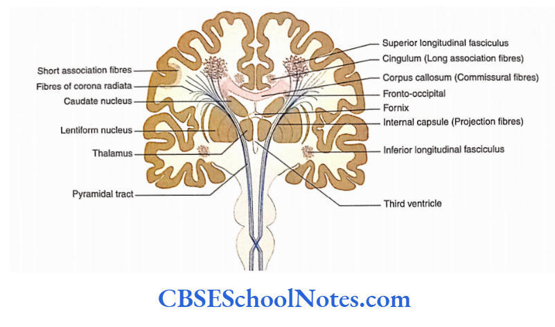
White Matter Of Cerebrum
- Each cerebral hemisphere consists of an enormous mass of white matter lying deep in the gyri and sulci of the cerebral cortex.
- White matter consists of a large number of fibres (axons) which connect various parts of the cortex. These fibres also connect various parts of the cortex with other parts of the central nervous system (CNS).
Classification of Fibres
Based on the nature of their connections, the fibres can be divided into three different groups:
1. Association fibres: These fibres connect various parts of the cerebral cortex of the same hemisphere. They do not cross the midline and are therefore confined to a single cerebral hemisphere.
2. Commissural fibres: These fibres connect the cortical areas of the two cerebral hemispheres and in the process cross the midline. Therefore, these fibres are also referred to as cerebral commissure.
Read and Learn More Neuroanatomy
3. Projection fibres: These fibres establish connections between the cortex and other subcortical regions of the CNS (thalamus, corpus striatum, brainstem and spinal cord). These fibres may or may not cross the midline at various levels of their descent.
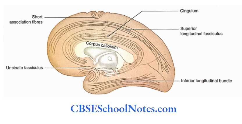
Association Fibres
Association fibres are the most numerous types of white fibres. They are usually grouped into short and long-association fibres.
The short association fibres pass from one part of a gyrus to another part of the same gyrus, or they may connect the adjacent gyri by looping around the intervening sulcus.
The long association fibres run for long distances, for example between two lobes of the same cerebral hemisphere. These fibres connect the functionally related regions of the cerebral cortex.
The long-association fibres are named as follows.
Cingulum: The cingulum is present deep to the cingulate gyrus on the medial surface of the cerebral hemisphere.
These fibres interconnect the cingulate gyrus, parahippocampal gyrus and septal area below the genu of the corpus callosum.
- Superior longitudinal bundle: The superior longitudinal bundle runs on the superolateral surface of the cerebral hemisphere above the level of the insula. The fibres of this bundle connect various areas of the frontal, parietal occipital and temporal lobes.
- Inferior longitudinal bundle: The inferior longitudinal bundle connects the visual association area (areas 18 and 19) of the occipital lobe with the temporal lobe.
- Uncinate fasciculus: Uncinate fasciculus connects Broca’s area and gyri on the orbital surface of the frontal lobe with the cortex of the temporal lobe. This bundle hooks around the stem of the lateral sulcus.
- Fronto-occipital fasciculus: Fronto-occipital fasciculus is located deep in the cerebral hemisphere. It connects the frontal lobe with the occipital and temporal lobes.
Commissural Fibres
Examples of the commissural fibres are corpus callosum, anterior commissure, posterior commissure, and habenular Examples of the commissural fibres are corpus callosum, anterior commissure, posterior commissure, and habenular.
Corpus Callosum
- The highest number of commissural fibres is present in the corpus callosum.
- The corpus callosum interconnects the cortical areas of one cerebral hemisphere with the corresponding cortical areas of the opposite cerebral hemisphere.
- The axons (fibres) of the corpus callosum run in both directions transversely and form a thick, wide Corpus callosum
- The highest number of commissural fibres is present in the corpus callosum.
- The corpus callosum interconnects the cortical areas of one cerebral hemisphere with the corresponding cortical areas of the opposite cerebral hemisphere.
- The axons (fibres) of the corpus callosum run in both directions transversely and form a thick, wide sheet that connects the medial surfaces of the two cerebral hemispheres.
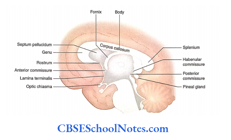
Corpus Callosum Location: The corpus callosum lies on the floor of the longitudinal fissure. It is about 10 cm long in the anteroposterior direction.
Corpus Callosum Shape: In the sagittal section of the cerebrum, the corpus callosum is seen as an arched or a ‘C ’-‘-shaped mass of white matter.
Corpus Callosum Surfaces: The superior surface of the corpus callosum is related to the indusium griseum and is concerned with the cingulate gyrus. The inferior surface is concave and is attached to the fornix through the septum pellucidum.
Parts of the corpus callosum: In the anteroposterior direction, it consists of four parts:
- Rostrum,
- Genu,
- Trunk and
- Splenium.
Rostrum: It connects the genu to the upper end of the lamina terminalis. In the midline, its superior surface gives attachment to the septum peculium.
The fibres of the rostrum interconnect the orbital surfaces of the right and left frontal lobes.
Genu: The genu is the fold of the corpus callosum located at its anterior end. The genu continues as the trunk of the corpus callosum posteriorly. The posterior surface of the genu gives attachment to the septum pellucid, in the midline.
Its fibres pass transversely to the anterior part of the right and left frontal lobes and appear like limbs of forceps. Therefore, these fibres are also called forceps minor.
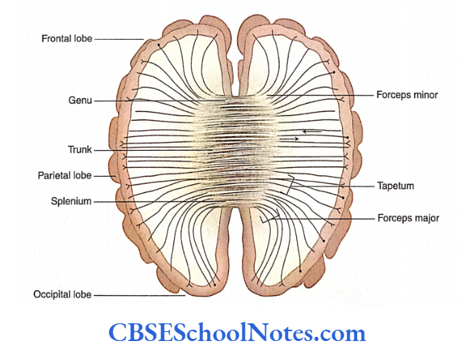
Trunk: It extends between the genu anteriorly and the splenium posteriorly. In the midline, the inferior surface of the trunk gives attachment to the septum pellucidum anteriorly and to the fornix posteriorly.
The fibres of the trunk interconnect most of the corresponding areas of the frontal and parietal lobes.
The fibres of the posteriormost part of the trunk and a few fibres of the anterior part of the splenium form the roof of the posterior horn. These fibres are known as tapetum.
The fibres of the tapetum interconnect the corresponding part of the temporal and occipital lobes.
Splenium: It is the posterior enlarged part of the corpus callosum. It ends behind as a free border overhanging the pulvinars, pineal gland and tectum of the midbrain.
The fibres of splenium interconnect the corresponding part of the right and left parietal, occipital and temporal lobes.
These fibres of splenium which connect two occipital lobes curve backwards as forceps major. The fibres of forceps major bulge in the posterior horn and produce an elevation on its medial wall that is known as bulbs of the posterior horn.
Blood supply of corpus callosum: Most of the corpus callosum is supplied by the anterior cerebral artery. The posteriormost part of the corpus callosum is also supplied by the posterior cerebral artery.
Functions of the corpus callosum: The fibres of the corpus callosum and other commissures provide connections between two cerebral hemispheres.
Thus, any sensory information or knowledge is collected and shared by both the cerebral hemispheres. For example, if a person learns a task with one hand, then information is transferable to another hand due to commissural connections.
The congenitally absent or surgically transected corpus callosum results in an isolated cerebral hemisphere (two separate brains). This condition is referred to as split-brain.
2. Anterior commissure: The anterior commissure is a bundle of axons that crosses the midline in the lamina terminalis, anterior to the column of the fornix.
It connects the anterior perforated substance, olfactory tract, and middle and inferior temporal gyri of two sides. Thus, most of the right and left temporal lobes
are connected through the anterior commissure rather than the corpus callosum.
Posterior commissure: The posterior commissure crosses the midline in the lower stalk of the pineal gland. This commissure connects the interstitial nucleus of Cajal, superior colliculi and pretectal nuclei of two sides.
4. Habenular commissure: This bundle is present in the upper stalk of the pineal gland and connects the habenular nuclei of the two sides.
5. Hippocampal commissure: The hippocampal commissure is also known as the commissure of fornix. It interconnects two crura of fornix, i.e. two hippocampi with each other.
6. Optic chiasma: The fibres from the right and left nasal halves of the retina cross in the optic chiasma.
Projection Fibres
- The projection fibres establish connections between the cerebral cortex and the subcortical regions of the CNS. The projection fibres run in both directions.
- In the cerebrum, the projection fibres are present in the forms of corona radiata and internal capsule.
Corona radiata: The fibres of corona radiata are continuous below the internal capsule which is present around the periphery of the lentiform nucleus.
Above the level of the internal capsule, the fibres spread out in a fan-like fashion into the cerebral cortex. Corona radiata consists of both afferent and efferent fibres.
The fibres of corona radiata are intersected by the fibres of corpus callosum (commissural fibres) and are intermingled with the association fibres.
Internal capsule: The projection fibres of the corona radiata are concentrated in the form of an internal capsule.
The internal capsule is present between large masses of grey matter (nuclei) of the cerebrum. The internal capsule is discussed in detail as a separate topic in the subsequent text.
External capsule: The external capsule is a thin layer of white matter between putamen and claustrum. It consists of projection fibres connecting the cerebral cortex with the putamen and reticular nuclei.
Fornix: The fornix.
Internal Capsule
The internal capsule is an aggregation of projection fibres which are arranged compactly in the cerebrum.
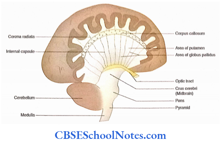
Internal Capsule Extent: The fibres of the internal capsule are continuous above as corona radiata and below as the crus cerebri of the midbrain
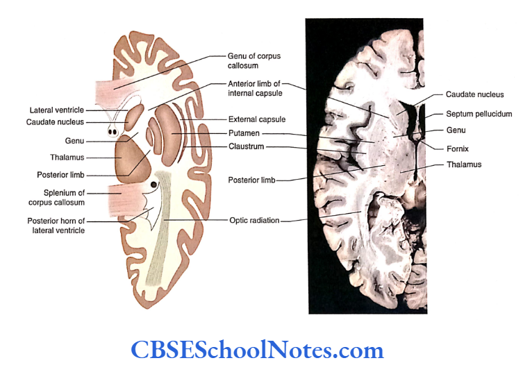
Internal Capsule Relations: ‘Hie lentiform nucleus is present on the lateral aspect of the internal capsule while the thalamus and caudate nucleus are present on its medial aspect.
Internal Capsule Shape: In a horizontal section of the cerebrum, the internal capsule appears like a ‘V’ with its concavity directed laterally.
The internal capsule consists of both ascending and descending (afferents and efferents) fibres which interconnect the cerebral hemisphere with the brainstem and spinal cord.
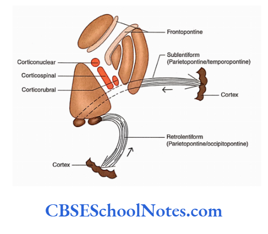
Internal Capsule Parts
- The internal capsule consists of an anterior limb, a genu, a posterior limb, a retrolentiform part and a sublentiform part.
- The anterior limb is bounded laterally by the lentiform nucleus and medially by the head of the caudate nucleus.
- The genu is located medial to the apex of the lentiform nucleus. It is a bent between the anterior and posterior limbs of the internal capsule.
- The posterior limb is situated between the lentiform nucleus laterally and the thalamus medially.
- The retrolentiform part is situated behind the lentiform nucleus.
- The sub-lentiform part consists of fibres that pass beneath the posterior part of the lentiform nucleus.
Fibres in the Internal Capsule
The various parts of the internal capsule consist of both efferent (corticofugal/motor) and afferent (corticospinal/ sensory) fibres. These are presented.
Sensory Fibres
The sensory fibres or the thalamic radiation establish a reciprocal connection between the thalamus and the cerebral cortex.
Anterior thalamic radiation: These fibres are situated in the anterior limb of the internal capsule.
They establish connections between the mediodorsal thalamic nucleus and the prefrontal cortex.
Superior thalamic radiation: This is situated in the genu and posterior limb of the internal capsule.
Fibres provide a connection between the ventral nuclei of the thalamus and motor, premotor and supplementary motor areas of the frontal lobe and sensory areas of the parietal lobe.
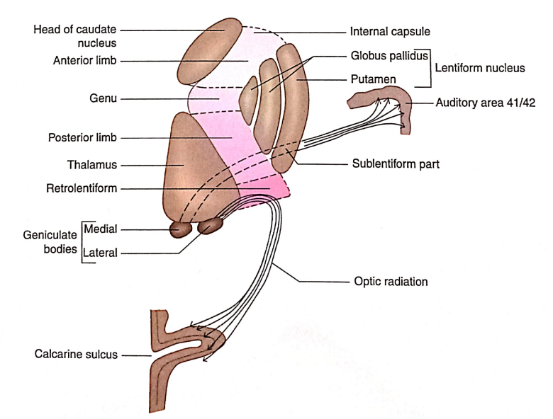
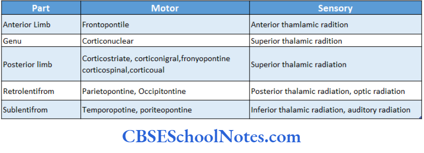
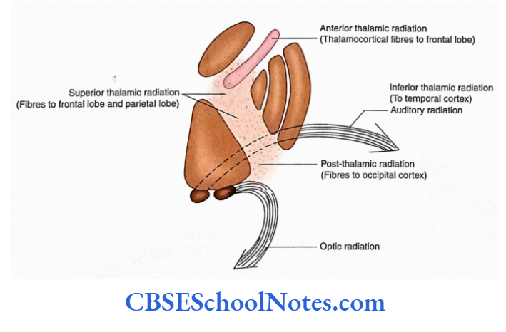
Posterior thalamic radiation: These fibres connect the thalamus with the visual cortex in the occipital lobe.
These fibres are present in the retrolentiform part of the internal capsule and connect the lateral geniculate body with the visual cortex (optic radiation).
Inferior thalamic radiation: These fibres run in the sublentiform part of the internal capsule that connects the medial geniculate body with the superior temporal gyrus (auditory area).
Motor Fibres
- The motor fibres are the projection fibres, also called centrifugal fibres.
- The efferent fibres present in the internal capsule are corticopontine, pyramidal and extrapyramidal
Corticopontine fibres: They originate from all four lobes of the cerebral cortex:
- Frontal, parietal, occipital and temporal. These fibres terminate in the nuclei points located in the basal part of the pons. Accordingly, these are called frontopontine, parietopontine, occipitopontine and temporopontine fibres.
- Fibres of the pyramidal motor system: The corticonuclear and corticospinal fibres originate in the motor, premotor, supplementary motor and cingulate areas of the frontal lobe.
- The genu of the internal capsule is occupied by the corticonuclear fibres while the posterior limb has the corticospinal fibres.
- Corticonuclear fibres terminate on the motor nuclei of cranial nerves of the opposite side while corticospinal fibres terminate on the anterior horn cells of the opposite side of the spinal cord.
Extrapyramidal fibres: These are corticorubral, corticostriate, corticoreticular and corticonigral fibres. Most of these arise from the motor areas of the frontal lobe and terminate in red nucleus, caudate nucleus, putamen, reticular formation and olivary nuclei.
The extrapyramidal fibres are situated in the posterior limb of the internal capsule and accompany the fibres of the pyramidal system.
Arterial Supply
- The internal capsule consists of a compact bundle of motor and sensory fibres.
- Any occlusion of blood vessels supplying the internal capsule can potentially lead to a serious clinical outcome (paralysis and sensory loss on the opposite side of the body).
Arteries supplying various parts of the internal capsule are described as follows:
Anterior limb: The upper part of the anterior limb is supplied by the striate branches of the middle cerebral artery.
The lower part of the anterior limb is supplied by the medial striate artery, a branch of the Anterior cerebral artery (also called the recurrent artery of Huebner).
Genu: The genu of the internal capsule is supplied by a striate branch of the middle cerebral artery, medial striate artery, a striate branch of the posterior communicating artery and direct branches from the internal carotid artery.
Posterior limb: The posterior limb of the internal capsule is supplied by the striate branches of the middle cerebral artery including a large Charcot artery and striate branches of the anterior choroidal artery.
Retrolentiform part: This is supplied by the striate branches of the posterior cerebral artery.
Sublentiform part: This part of the internal capsule is supplied by the striate branches ofthe posterior cerebral artery, striate branches of the anterior choroid artery and branches from the internal carotid artery.
- The ascending sensory and descending motor axons course through the Internal capsule. Occlusion of the vascular supply of the internal capsule can result in paralysis and loss of general sensation on the opposite side of the body (hemiplegia).
- The most common lesion affecting the Internal capsule Is haemorrhage or thrombosis.
- This occurs most commonly due to rupture of the lateral striate branch of the middle cerebral artery which supplies the posterior limb of the internal capsule.
- This artery is also known as the artery of cerebral haemorrhage or Charcot’s artery of cerebral haemorrhage. This is the most common artery to rupture because it remains under constant high pressure and has no collateral vessels.
- The rupture of this artery leads to the upper motor neuron type of paralysis, on the opposite side of the body (hemiplegia).
White Matter Of Cerebrum Summary
The white matter of the cerebrum is divided into three groups:
- Association, Commissural and Projection.
- The association fibres contain axons that conduct nerve impulses between gyri in the same hemisphere.
- The commissural fibres conduct nerve impulses between the corresponding areas of the right and left cerebral hemispheres.
- The projection fibres connect the cerebral hemisphere with the lower parts of the CNS.
- The corpus callosum has the highest number of commissural fibres. It has four parts:
- Rostrum, Genu, Trunk and Splenium.
- A congenitally absent or transected corpus callosum results in an isolated cerebral hemisphere (split brain).
- The projection of corona radiata is continuous below the internal capsule. Parts of the internal capsule are the anterior limb, genu, posterior limb, retrolentiform and sublentiform.
- The internal capsule consists of both ascending (sensory) and descending (motor) fibres.
- As the internal capsule consists of a compact bundle of motor and sensory fibres, occlusion of blood supply can result in paralysis and loss of sensation on the opposite half of the body (hemiplegia).
White Matter Of Cerebrum Multiple Choice Questions
Question 1. Which of the following statement(s) is/are true?
- White matter consists of a large number of fibres (axons)
- White matter connects various parts of the cortex
- White matter connects the cerebral cortex with other parts of the CNS
- Many of these fibres pass through the internal capsule
- All of the above
Answer: 2. White matter connects various parts of the cortex
Question 2. The white matter of the cerebrum is divided into the following groups except
- Association fibres
- Commissural fibres
- Projection fibres
- Decussating fibres
Answer: 4. Decussating fibres
Question 3. The following statements about association fibre(s) are/are true except
- Association fibres are the most numerous white fibres
- Short association fibres run between two cerebral lobes
- The cingulum is an example of long-association fibres
- Long association fibres are arranged in distinct bundles
Answer: 2. Short association fibres run between two cerebral lobes
Question 4. The following are examples of commissural fibres except
- Cingulum
- Corpus callosum
- Anterior commissure
- Optic chiasma
- Habenular commissure
Answer: 1. Cingulum
Question 5. Which of the following statements about corpus callosum is false?
- Axons of the corpus callosum run in both directions
- Corpus callosum is located about 6 cm away from the occipital pole
- Much of the cortices of the temporal lobe of two sides connect through the corpus callosum
- Corpus callosum consists of four parts—rostrum, genu, trunk and splenium
Answer: 3. Much of the cortices of the temporal lobe of two sides connect through the corpus callosum
Question 6. The following facts about corpus callosum are true except
- The fibres of the rostrum connect the orbital surface of the right and left frontal lobes
- The fibres of genu are arranged like the limbs of forceps and are called forceps minor
- The fibres of the posteriormost part of the trunk form ‘tapetum’
- The fibres of splenium curve backwards to form forceps major
- The fibres of forceps major connect two parietal lobes
Answer: 5. The fibres of forceps major connect two parietal lobes
Question 7. The blood supply of the corpus callosum is due to the following arteries:
- Anterior cerebral artery
- Middle cerebral artery
- Posterior cerebral artery
- Central branches from cerebral arteries
Answer: 1. Anterior cerebral artery
Question 8. Which of the following facts about projection fibres are true?
- Projection fibres establish connections between the cerebral cortex and the subcortical regions of the CNS
- Projection fibres run in both directions
- In cerebrum, projection fibres are present in corona radiata and internal capsule
- Projection fibres below the corona are concentrated in the internal capsule
- All of the above
Answer: 5. All of the above
Question 9. The internal capsule consists of the following parts except
- Anterior limb
- Posterior limb
- Inferior limb
- Genu
- Retrolentiform part
Answer: 3. Inferior limb
Question 10. Which of the following are extrapyramidal fibres of the internal capsule?
- Corticorubral
- Corticostriatal
- Corticoreticular
- Cortico-olivary
- All of the above
Answer: 5. All of the above