
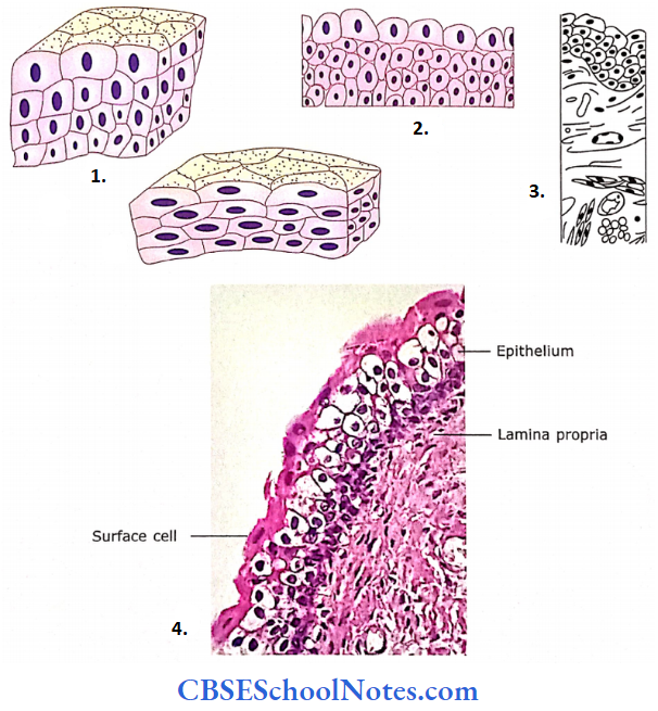
Epithelium Tissue
What is Histology?
The subject of histology deals with the microscopic and ultramicroscopic structure of cells, tissues and organs of the body.
- An aggregation of similar types of cells (and material surrounding them) is defined as tissue (For Example., epithelial tissue, connective tissue, muscular tissue and nervous tissue).
- All the cells of a tissue work in a collective manner to perform a particular function. Various tissues combine to form an organ of the body (For Example., the intestine, kidney, liver, etc).
- The intestine is formed by the combination of various types of tissues like epithelial (epithelium lines the mucous membrane), connective (forms lamina propria, submucosa and serosa), muscular (forms muscularis mucosae and muscle coats) and nervous (forms Meissner’s and myenteric nerve plexus).
- Similarly, various organs of our body aggregate to form a system of the body (For Example., the urinary system is formed by the kidney, ureter, bladder and urethra).
A system of the body consists of related organs that have a common function. All the systems of the body function synchronously to form one living person.
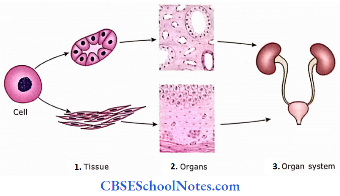
- Cells aggregate to form various kinds of tissues (epithelial and
muscular tissue) - Various tissues aggregate to form organs (kidney and ureter)
- Various organs form an organ system (urinary system).
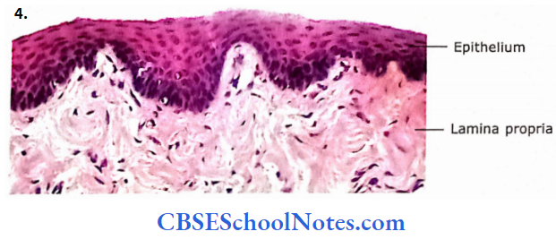
Epithelium
The epithelium is the basic tissue of the body. It consists of cells arranged as continuous sheets, in either single or multiple layers. Cells forming epithelium, on their lateral surfaces, are in close apposition with one another and are held tightly together by various kinds of cell junctions.
One of the surfaces of epithelial cells is a free surface that is exposed to the body cavity, lumen of an internal organ or external surface of the body. The basal surface adheres to a thin, continuous, supporting layer called basal lamina or basement membrane.
Epithelium Remember
The epithelium is defined as a tightly bound continuous sheet of cells, covering the free surface (inner or outer surface) of the body.
What are epithelioid tissues?
At certain places in the body, cells are found aggregated in close apposition with one another like epithelial cells but do not present a free surface. These kinds of issues are called epithelioid tissues.
For Example., parenchyma of the adrenal gland, Leydig cells of the testis, islets of the Langerhans, pituitary gland, parathyroid gland and luteal cells in the ovary.
epithelioid tissues Remember
Epithelial cells when showing the absence of a free surface are called epithelioid tissue.
- Epithelial tissue is avascular but derives its nutrition from capillaries present in the connective tissue layer beneath the basement membrane. Although epithelial tissue is devoid of blood supply, it is supplied by nerves.
- Epithelial tissue is subjected to constant wear and tear and, hence has a high capability to regenerate. The secretory portion of glands and cells lining the ducts are epithelial as the glands are epithelial in origin.
Functions Of Epithelium
- Protection: As epithelial tissue forms a boundary layer between the body and external environment (epithelium of skin), and lines the body cavities, it is protective.
- Absorption: The epithelia of organs like kidneys and intestines are absorptive.
- Acts as barrier: Epithelium may also act as a barrier, as in the skin and urinary bladder, which resists the absorption of water and toxic substances.
- Excretion: Epithelium of a certain portion of the nephron of the kidney is involved in the excretion of harmful metabolites.
- Secretion: Epithelium may also act as secretory as in intestine, For Example., goblet cells, and gastric and intestinal glands.
- Detection of sensations: Epithelial tissue may combine with nervous tissue to form special organs of sense, For Example., organs of hearing, smell and vision.
Classification Of Epithelia
Epithelia are classified according to the number of cell layers, the shape of cells, the arrangement and the specialization of their free surface.
- Epithelium with a single layer of cells is called as simple. While epithelium consisting of multiple layers of cells is called stratified.
- A third type of epithelium that has the appearance of being stratified is classified as pseudo-stratified.
- The fourth class of epithelium is called transitional because it can change its shape when stretched.
Epithelia Remember
Classification of epithelia is based on the shape and arrangement of the epithelial cells.
Classification Of Epithelia
- Simple (single layer of cells)
- Squamous
- Cuboidal
- Columnar
- stratified (multiple layers of cells)
- St. squamous
- St. cuboidal
- St. columnar
- Pseudostratified (epithelium appears as if stratified)
- Transitional (shape of epithelium is not fixed)
Based on the shape of the cells, simple epithelium is further classified as simple squamous, simple cuboidal and simple columnar.
Similarly, the stratified epithelium is further classified as stratified squamous, stratified cuboidal and stratified columnar depending on the shape of the cells on its uppermost layer (free surface).
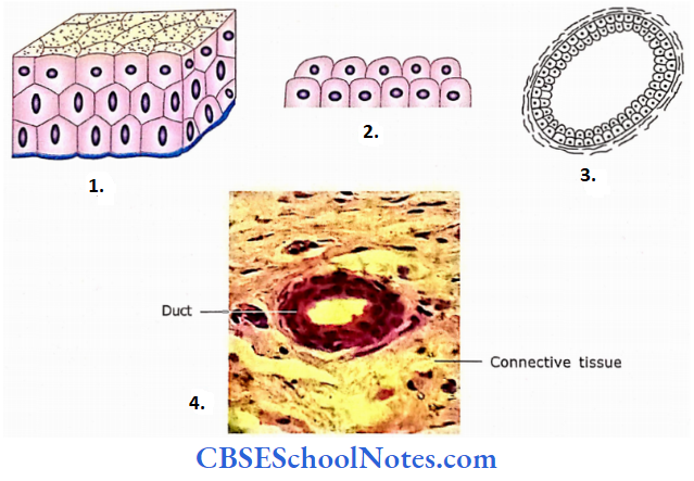
Simple Epithelium
Simple Epithelium Squamous
Squamous Description
- Consists of a single layer of fat cells.
- The nucleus is oval or flat, situated in the centre of the cell.
- On the surface view, cells appear to be arranged like floor tiles.
Squamous Location
- It lines the heart, blood vessels and lymphatics. Here, it is called the endothelium.
- It also lines the serous membranes of body cavities and the surface of viscera. Here, it is called mesothelioma.
- It is present in lung alveoli, a parietal layer of Bowman’s capsule, certain tubules of the kidney and at certain places on the inner aspect of the tympanic membrane.
Squamous Functions
It helps in rapid transport of substances, filtration of fluids, diffusion of gases and osmosis.
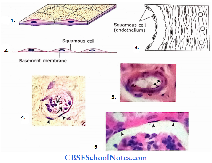
- Three-dimensional arrangement.
- Sectional view.
- Squamous epithelium lining the lumen of blood vessels.
- Section of a venule lined with simple squamous epithelium called endothelium (arrowheads). Few blood cells are seen in the lumen of the vessel (arrows).
- Endothelium of arteriole.
- Squamous epithelium lining the parietal layer of Bowman’s capsule.
Simple Epithelium Cuboidal
Cuboidal Description
- In sectional view, cells appear cuboidal in shape.
- Nuclei are round and centrally placed. All nuclei are arranged at the same level.
- When viewed from the top, cells are hexagonal
Cuboidal Location
- Epithelium lining the follicles of the thyroid gland
- Ducts of exocrine glands
- Pigmented epithelium of the retina
- Lens capsule
- Surface of ovary
- Certain ducts and tubules of the kidney
Cuboidal Function
Secretion and absorption.
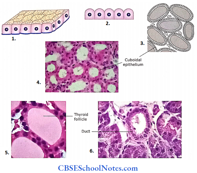
- Three-dimensional view.
- As seen in sectional view.
- This epithelium is present in the thyroid gland.
- Collecting ducts of the kidney lined by simple cuboidal epithelium.
- Lining the thyroid follicles.
- Lining the duct of a glands
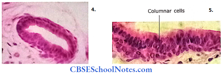
Simple Epithelium Columnar
Columnar Description
- Cells of the epithelium are much taller compared to their width.
- Nuclei are elongated and located in the lower half of the cells. All nuclei are placed at the same level in neighbouring cells).
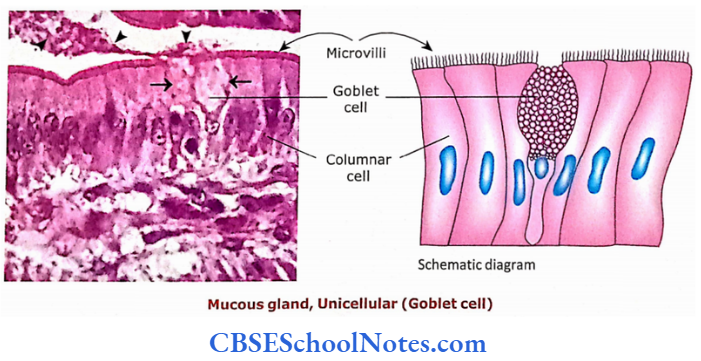
- On their free surface, modifications like microvilli or cilia may be seen. When viewed from the top, cells look hexagonal.
- It may also contain goblet cells.
Columnar Location
Epithelium lining gall bladder, ducts of glands, gastrointestinal tract (from the stomach to anus), uterine tube, uterine cavity, cervical canal and central canal of the spinal cord.
Columnar Function
- Secretion and absorption.
- Ciliary action moves mucus in the respiratory tract and ovum in the uterine tube.
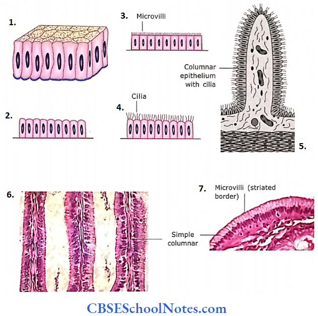
- Three-dimensional view.
- Simple columnar (without any modification on their apical surface, Example., stomach).
- With microvilli, Example., small intestine.
- With cilia.
- The simple columnar epithelium with cilia is seen in the uterine tube.
- Simple columnar epithelium from the intestine.
- Photomicrograph showing simple columnar epithelium with microvilli taken from the lining of the gall bladder.
Simple Epithelium Remember
In cross-section, the cells of the simple cuboidal and simple columnar epithelia are shaped like hexagonal solids.
Stratified Epithelium
Stratified epithelium is named based on the shape of the cells lining the topmost layer (free surface).
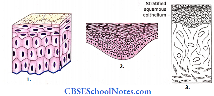
Stratified Squamous (non-keratinized) Description
- Cells are arranged in many layers.
- The basal layer is attached to the basement membrane and is usually columnar, cuboidal or rounded in shape.
- Intermediate cells are irregularly polyhedral in shape and become increasingly flattened as they move towards the superficial layer.
- The superficial layer consists of thin squamous cells.
- Basal cells replace surface cells as they are shed off.
StratifiedSquamous (non-keratinized) Location
- Epithelium lining the oral cavity, tongue, part of epiglottis, oesophagus and vagina.
- StratifiedSquamous (non-keratinized) Functions
- Protection of deeper tissue.
- Three-dimensional view.
- Sectional view.
- As seen in the mucosal lining of the vagina.
- Stratified squamous non-keratinized epithelium taken from the lining of the oesophagus.
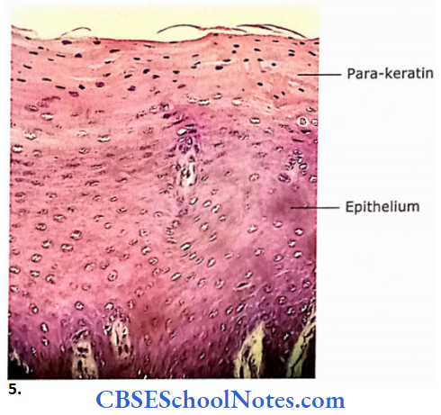
- Para-keratinized epithelium from the tongue. Parakeratin is an intermediate stage between non-keratinized to keratinized epithelium.
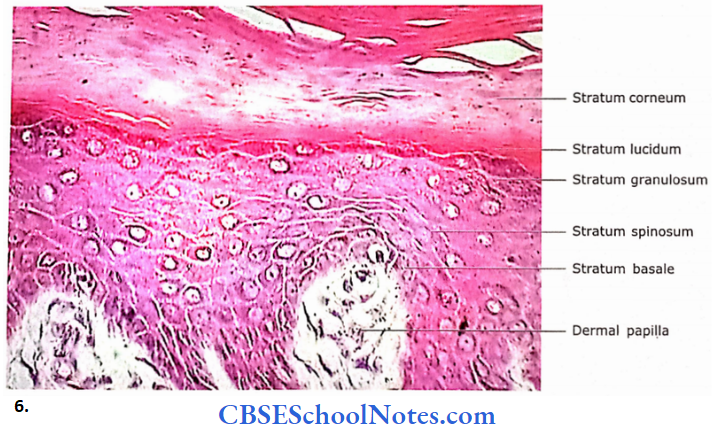
- The photomicrograph shows the stratified squamous keratinized epithelium from the epidermis of the skin.
Stratified Squamous (keratinized) Description
- In this type of epithelium, superficial cells become dead, dehydrated andnon-nucleated like scales.
- These dead cells become hard (comified) as they are filled with keratin.
- Stratified Squamous (keratinized) Location
- Epithelium of the skin
Stratified Squamous (keratinized) Functions
- Protection of deeper structure.
- It prevents the absorption of water.
- Keratin prevents dehydration of underlying tissue.
Stratified Squamous (keratinized) Remember
In keratinized epithelium, cells lining the free surface are dead and filled with keratin. Nuclei are also absent in these superficial cells.
Stratified Cuboidal Description
- The epithelium consists of two or more layers of cells.
- Cells of the superficial layer are cuboidal in shape
Stratified Cuboidal Location
- Ducts of sweat glands.
Stratified Cuboidal Functions
- Provides passage to the secretion and acts as a barrier.
- Three-dimensional view.
- Sectional view.
- As seen in the duct of the sweat gland.
- The stratified cuboidal epithelium is present in the excretory duct of an exocrine gland.
Stratified Columnar Description
- Two or more layers of cells.
- Cells of the superficial layer are columnar
Stratified Columnar Location
Epithelium lining large ducts of some glands, fornix of the conjunctiva and cavernous urethra.
Stratified Columnar Functions
Provides passage to the secretion and acts as a barrier.
Stratified Columnar Remember
The stratified epithelium is composed of more than one layer. It is classified based on the shape of cells forming the uppermost layer (surface layer).
- Three-dimensional view.
- Sectional view.
- As seen in the duct of the salivary gland.
- image
- The Duct of the pancreas,
- Epithelium lining the duct of the serous salivary gland.
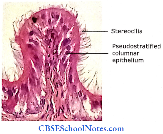
Pseudostratified Epithelium
Columnar Description
- It is not a true stratified epithelium but appears to be stratified.
- All cells are attached to the basement membrane but are of different heights. Hence, not all reach the apical surface. Because of this, the nuclei of cells are at different levels.
- The epithelium may be ciliated or non-ciliated and may contain goblet cells.
Columnar Location
- The non-ciliated epithelium is found in the large excretory ducts, auditory tube and male urethra.
- The ciliated epithelium is found in the upper respiratory tract.
Columnar Function
- Protection of underlying tissue.
- Ciliary movements remove mucus, while goblet cells secrete mucus.
- Pseudostratified Epithelium Remember
- Although pseudostratified epithelium looks as if cells are arranged in many layers all the cells of this epithelium are attached to basal lamina.
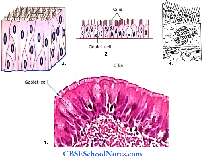
- Three-dimensional view
- Sectional view
- Trachea
- The Pseudostratified Ciliated Columnar epithelium is taken from the lining of an intrapulmonary bronchus.
Transitional Epithelium (urothelium) Description
- The appearance of epithelium varies during stretched and relaxed conditions. When this epithelium is stretched then it looks like stratified squamous epithelium.
- But when the epithelium is in a relaxed condition, it appears stratified cuboidal. Due to this apparent change in the shape this epithelium is called transitional epithelium.
- This stratified epithelium is made up of 2-3 layers of cells as seen in a distended urinary bladder. However, when the bladder is relaxed many layers of cells are seen due to the folding of epithelial cells.
- These changes in shape are confined to the urinary bladder only and not observed at any other place in the urinary tract. The deeper cells are cuboidal or polyhedral, while superficial cells are large and have rounded free surfaces (domeshaped or umbrella-shaped cells).
- The apical surface of dome-shaped cells is thickened and more eosinophilic. This is due to the presence of plaques of intramembranous glycoprotein particles. Some cells of superficial cells may show two nuclei.
Transitional Epithelium (urothelium) Location
Epithelium lining the urinary tract.
Transitional Epithelium (urothelium) Functions
- The presence of occluding junctions and intramembranous plaques forms an effective barrier, i.e., prevents the absorption of toxic substances in urine.
- Distention
Transitional Epithelium (urothelium) Remember
Transitional and pseudostratified epithelia are a special class of epithelium.
- Transitional epithelium
- Sectional view.
- Urinary bladder.
- The transitional epithelium is taken from the urinary bladder.
The Basement Membrane
The basement membrane is the thin supporting layer placed between the basal surface of the epithelium and underlying connective tissue.
- This membrane is not seen very clearly in most of the organs in H and E (haematoxylin and eosin) preparations. However, when stained with the PAS (periodic acid Schiff) technique.
- It appears as a well-defined magenta(pink) layer. This colour reaction is due to the presence of carbohydrates (sugar)in the basement membrane.
The Basement Membrane Structure
When seen under an electron microscope, the basement membrane appears to be made up of two layers, i.e., basal lamina and reticular lamina.
The basal lamina is associated closely with the basal layer of the cell surface and the reticular lamina is near the connective tissue layer. The reticular lamina consists of reticular fibres.
Basal Lamina
The basal lamina is further divided into lamina lucida and lamina densa. The lamina lucida is a layer of very low density and lies immediately beneath the epithelium.
- The lamina densa is the outer layer of greater density, facing the underlying reticular lamina. Lamina lucida is almost featureless while lamina densa contains a network of extremely fine (3-4 nm) filaments (type 4 collagen) in an amorphous matrix.
- Anchoring fibrils are type 7 collagen, which links basal lamina to reticular lamina. Chemically the basal lamina consists of proteoglycans, laminin and typeIV collagen.
- Lamina lucida consists of molecules of glycoproteins and aminin while a network of type 4 collagen is Present in lamina densa. The type 4 collagen of basal lamina is produced by epithelial cells.
- The basal lamina and basement membrane are sometimes used as interchangeable terms. However, the term basement membrane is mostly used in light microscopy and basal lamina in electron microscopy. Basal lamina in non-epithelial cells is known as external lamina.
Reticular Lamina
It is placed between the basal lamina and underlying connective tissue. It is manufactured by fibroblasts and is composed of type 1 and type 3 collagen.
These collagen fibres are bound to anchoring fibrils (type 7 collagen) of the lamina reticularis.
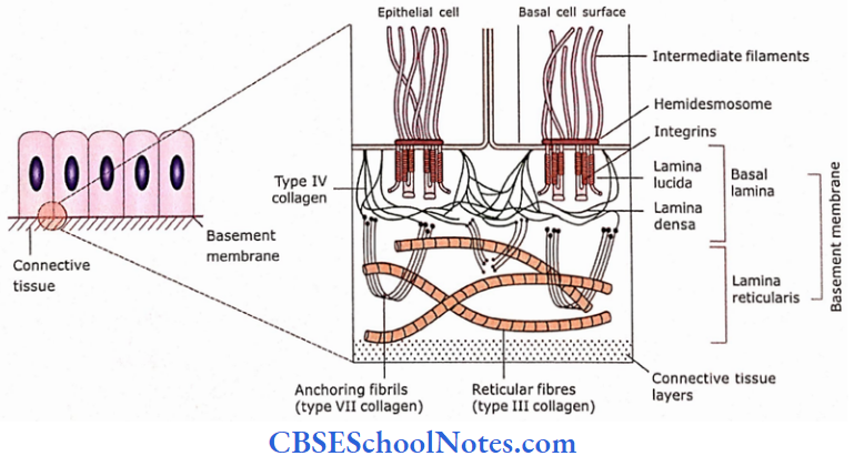
The Basement Membrane Remember
Basement membrane, as seen by a light microscope, is made up of two layers, i.e., basal lamina and lamina reticularis.
The Basement Membrane Functions
- It provides support to the epithelium.
- The epithelium is attached to the underlying connective tissue with the help of basal lamina.
- It provides support to the epithelium.
- The epithelium is attached to the underlying connective tissue with the help of basal lamina.
- It acts as a mechanical barrier preventing malignant cells from invading deeper tissues.
- It provides a selective filtration barrier. Molecules of certain shapes, sizes and electrostatic charges only are allowed to pass through the basal lamina.
- In some diseases of the kidney, the basal lamina of glomerular capillaries is thickened.
Intercellular Contacts
The cells of epithelia on their lateral surfaces are in contact with each other. These contacts provide adhesion and communication between adjoining cells.
- The plasma membranes of two adjacent cells are usually separated from each other by a 15-20 nm gap. This gap is occupied by cell adhesion molecules (CAM), which are glycoproteins in nature.
- Besides these adhesion molecules, opposing membranes also show some specialization for intercellular contacts that are called junctional complexes (cell junction). The following four junctional complexes are described.
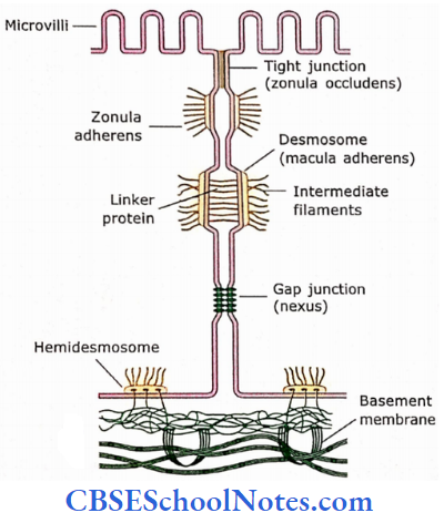
Zonula Occludens (Tight Junction)
This junction is located near the apical part of cells of tissues that line the body cavities. It is in the form of a circumferential (belt-like) band or ring that encircles the entire circumference of the cell.
In this junction, surrounding each cell near its apex, the outer surfaces of adjacent plasma membranes arc fused by a web-like strip of proteins. These transmembrane junctional proteins are called claudins and occludins.
Zonula Occludens (Tight Junction) Remember
The zonula occludens is present near the apical end of epithelial cells. This intercellular cell junction consists of localized sealing of the plasma membrane of adjacent epithelial cells.
Zonula Occludens (Tight Junction) Functions
It is a barrier device. As the adjacent plasma membranes are fused, it prevent the passage of large and small water-soluble molecules between cells.
- It also prevents the leak of contents of organs into the blood or surrounding tissues, For Example., urine from the urinary bladder.
- This kind of junctional complex is common in kidney tubules, intestines and urinary bladder.
Zonula Adherens
This junctional complex is present immediately below the zonula occludens. This complex also completely encircles the cell. There is a gap of 15-20 nm between opposing cell membranes.
- In this complex, the electron-dense material is found along the cytoplasmic side of the membrane of each cell. The microfilaments (actin) are seen embedded in this electron-dense area.
- These actin microfilaments are continuous with the filaments of the terminal web situated near the apex of the cell.
- A specific glycoprotein i.e., the cell adhesion molecule (CAM) is present in the gap between two opposing membranes. This helps membranes to maintain their adherence to one another.
Zonula adherens Functions
It provides strong adhesion between adjacent cells. It gives stability to the terminal web of the cell. Terminal webs of adjacent cells are interconnected by zonula adherens.
Zonula Adherens Remember
Zonula adherens is present below occludentes and provides encircling band-like lateral adhesion between epithelial cells.
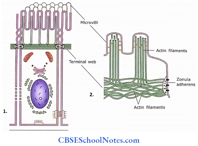
Terminal Web
The terminal web is a network of actin filaments present horizontally in the apical part of the cytoplasm just beneath the microvilli. The actin filaments of the terminal web are cross-linked and contractile.
- On the periphery, this network is attached to the intracellular density of zonula adherens. The terminal web also gives attachment to the actin filaments of microvilli.
- The contraction of the terminal web causes the microvilli to spread apart, thus increasing the space between microvilli thereby exposing more surface for absorption.
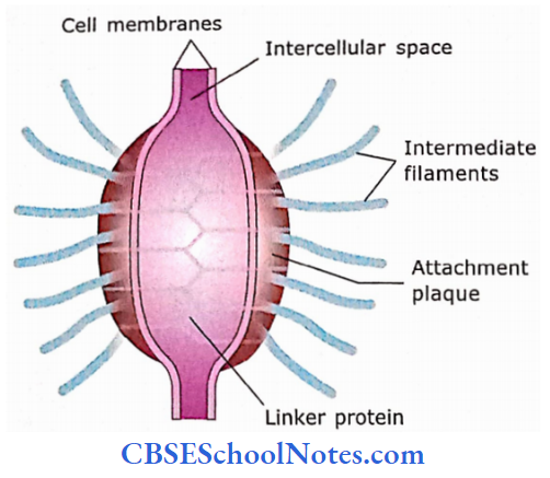
Desmosomes (Macula Adherens)
This type of junctional complex is not in the form of an entire cling band but is in the form of discs scattered over the lateral surface of cells. The opposing membranes are separated from each other by 30 nm distance.
- This junction shows the presence of a dense disc (attachment plaque) on the cytoplasmic side of opposing membranes. The intermediate filaments of the cytoplasm converge and terminate on this dense disc.
- The transmembrane linker proteins extend between opposing dense discs across the intercellular space. These linkerproteins are cadherins, For Example., desmogleins, desmocollins, etc.
- Desmosomes are predominantly present in epithelia that are subjected to abrasion and physical stress (For Example., stratified squamous epithelium of epidermis).
Desmosomes (Macula Adherens) Functions
- It provides stability to epithelium as a whole by linking cytoskeletons of adjacent cells.
- It provides strong adhesion between cells.
Desmosomes (Macula Adherens) Remember
The desmosomes are spot-like (disc-like) junctions between epithelial cells, while hemidesmosomes are cell junctions between the basal cell membrane and underlying basal lamina.
Hemidesmosomes
It looks like half a desmosome and connects cells to the basal lamina. Thus, they are found at the basal surface of cells. The basal lamina that faces the hemidesmosome is usually thick and connecting strands (integrins) extend between it and the plasma membrane.
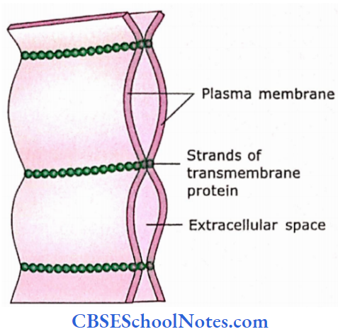
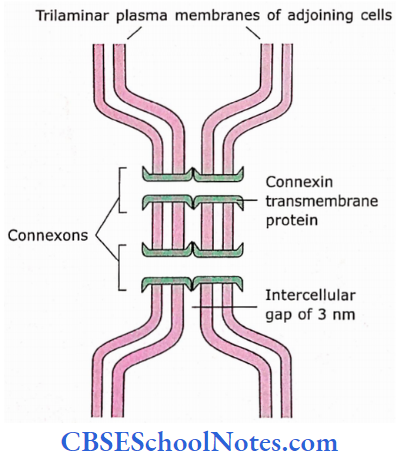
Gap Junction (nexus)
Gap junctions allow ions and small molecules to pass from the cytosol of one cell to another. The two adjacent plasma membranes come close to each other and a gap of 3 nm is observed between the two.
- These intercellular gaps are bridged by transmembrane protein channels called connexons. These six rod-like protein subunits (connexins) are so arranged around a central pore that they form a minute fluid-filled tunnel.
- The diameter of this tunnel (central pore) is about 1.5-2 nm. The connexons of opposite membranes face each other and project 1.5 nm into the intercellular gap where they are linked end to end. Channels thus formed connect the cytoplasm of neighbouring cells.
- The opening and closing of gap junction is regulated. An increase in calcium concentration or decrease in cytoplasmic pH closes gap junctions. While channels are open when there is a decrease in Ca+ concentration or an increase in pH in the cytoplasm.
Gap Junction Functions
- It coordinates the activities of cells in the epithelium, heart and smooth muscles.
- Gap junctions enable nerve or muscle impulses to spread rapidly between cells.
- It coordinates the activities in embryonic cells by distributing signalling molecules throughout the cell mass.
Gap Junction Remember
The nexus or gap junctions allow communication between adjacent epithelial cells by allowing the passage of small molecules, ions, hormones, amino acids and vitamins
Gap Junction Clinical Applications
Tumours of Epithelial Cells
- A tumour is a swelling that results due to excessive proliferation of cells. Epithelial cells can give origin to both benign (harmless, non-cancerous) and malignant (cancerous) tumours.
- Epithelial cells are a common site of origin of malignant tumours (cancers) and are called carcinomas. Cancers derived from glandular epithelial tissues are called adenocarcinomas.
Specialization Of The Free Surface Of Cell
Following modifications may occur on the free surface of epithelial cells to perform duties related to tissue function:
- Microvilli
- Stereocilia
- Cilia
Microvilli
Under the light microscope, the epithelia lining the tubules of the kidney and intestine show fine vertical striations near their free surface.
- This surface modification in intestinal absorptive cells is called a striated border and brush border for kidney tubule cells.
- Under an electron microscope, this border is found to consist of cytoplasmic finger-like protrusions from the apical cell surface.
- These cell processes arc closely packed and measure about 1-2 pm in length and about 80-90 ran in diameter. These processes are called microvilli. Microvilli are covered by a fuzzy coat called glycocalyx.
- Each microvillus contains a bundle of 25-35 actin filaments. These actin filaments at one end arc attached to the terminal web in the apical cytoplasm and at the other end to the membrane at the tip of the microvillus.
- In epithelia not involved in absorption, microvilli are few and lack actin filament.
Specialization Of The Free Surface Of Cell Functions
The formation of microvilli is a special modification of the cell surface to increase the surface area of the membrane. It achieves a 15-30-fold increase in the area.
- Increased surface area is an adaptation to increase the capability for absorption. Microvilli are non-motile processes.
- However, in some microvilli oscillatory, contractile movements may be seen due to the presence of actin filaments. This helps in the process of absorption.
- Microvilli are small finger-like cytoplasmic projections from the free cell surface. It contains the core of actin filaments.
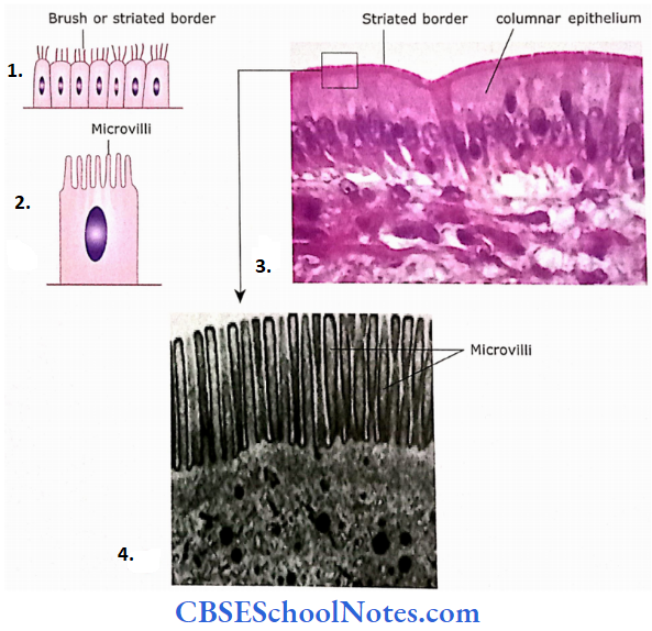
- Seen as a brush or striated border under a light microscope.
- Seen as small projections in an enlarged view.
- Simple columnar epithelium showing striated border (microvilli).
- Electron micrograph of microvi li from the apical surface of cell lining intestinal villi. The microvilli are finger-like protrusions measuring about 1 to 2 pm.
Stereocilia
Stereocilia are found on the apical surface of the epithelium lining epididymis, ductus deferens and on the sensory hair cells of the cochlea of the inner ear.
- Stereocilia are long, non-motile cytoplasmic processes measuring about 100-120 pm in length. Their structure resembles that of microvilli and is therefore called a large microvilli.
- Their function is not known exactly. It is believed that they increase cell surface area and serve as an absorptive device. In the hair cells of the ear, they probably function in signal generation.
Stereocilia Remember
Stereocilia are very long non-motile microvilli. Their core consists of actin filaments.
Cilia
Cilia are structural modifications of the cell surface that are capable of rapid, regular and synchronous to-and-fro movements. Cilia covering the epithelium move as waves in a grass field.
- Cilia are short (5-10 pm in length, 0.3 pm in diameter), fine, hair-like structures arranged in rows on the apical cell surface. The shaft of each cilium is covered by the cell membrane.
- The core of it consists of a central pair of microtubules (which are separated from each other) with nine pairs of evenly spaced microtubules around them, The outer pairs of tubules are connected to the central pair.
- At the base of cilia are basal bodies that are believed to have originated from the centriole. Each basal body is similar in structure to a centriole and is made up of nine triplet microtubules around the periphery.
- The 9+2 microtubules of cilia extend between the tip of the cilium to its base where the basal body is situated. Each of the paired microtubules of the cilium is continuous with the two inner microtubules of the triplet of the basal body.

- Under the light microscope, cilia are seen as hair-like structures.
- In an enlarged view, they are seen as finger-like projections.
- Photomicrograph shows the ciliated cells of the respiratory epithelium (trachea). A single cell may have up to 300 cilia.
Cilia Remember
Cilia are hair-like motile structures arranged on the api¬ cal surface of the cell. The core of cilium contains longitudinal microtubules arranged in a 9+2 organization called the axoneme.
Cilia Functions
- The wave-like (rapid back-and-forth) movements of cilia on the surface of bronchial and tracheal epithelium help to move the mucus in one direction (towards the pharynx).
- Cilia are responsible for the movements of ova in the oviduct.
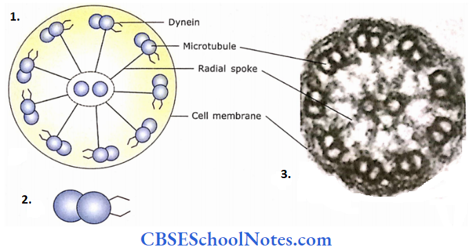
- Schematic diagram showing cross-section of a cilium.
- To one of the peripheral microtubules, protein dynein is attached.
- Electron micrograph showing a cross-section of the cilium.
Glandular Epithelia
A gland is an organ that consists of specialized secretory cells. The material secreted by the gland is usually a liquid (enzyme, hormone, mucus or fat).
Glands are epithelial in origin. They may be unicellular and multicellular. The unicellular gland consists of a single cell distributed among non-secretory cells, For Example., the goblet cells.
The multicellular glands are formed by the invagination of surface epithelium in deeper tissue. These are classified into the following two types depending on how their secretion is released.
Exocrine glands – These types of glands remain in contact with the surface epithelium by the ducts and pour their secretions on its surface.
Endocrine glands – On the other hand, some glands lose their epithelial contact because of the disappearance of ducts. These types of glands pour their secretion directly into the blood.
They secrete hormones that act on target cells, which are usually situated some distance away from the gland.
Glandular Epithelia Remember
Exocrine glands secrete their product through a duct. Endocrine glands are ductless and secrete directly into the blood bloodstream or lymphatic system. Paracrine gland secretion remains locally and affects the surrounding cells.
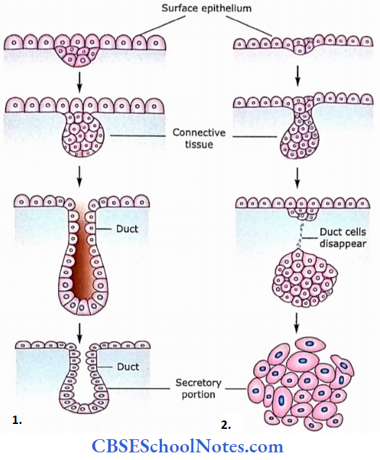
- Formation of exocrine gland.
- Formation of the endocrine gland.
Exocrine Glands
Classification of Exocrine Glands
1. Classification based on the shape and branching pattern of the duct
The classification of exocrine glands is based on the shape of secretory units (tubular or alveolar) and the branching pattern of their ducts. If the ductofa gland is unbranched, it is called as simple while glands with branched ducts are called compound.
- Simple Glands: Simple tubular (For Example., crypt of Lieberkuhn); simple coiled tubular(For Example., sweat glands); simple branched tubular (where only the secretory part is branched,
- For Example., fundic glands stomach); simple alveolar (mucous glands of the urethra); simple branched alveolar (where only secretory units are branched, For Example., meibomian gland).
- Compound Glands: Compound tubular (Brunner glands); compound alveolar (For Example., mammary gland); compound tubulo-alveolar (submandibular gland).
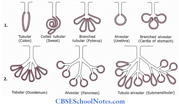
- Various types of simple glands were duct unbranched.
- Various types of compound glands
2. Classification based on the mode of release of their product
Exocrine glands secrete their products by three different methods.
- Merocrine: Secretion is released by the exocytosis of secretory granules (For Example., pancreas and parotid gland. Here, neither cell membrane nor cytoplasm becomes part of the secretion).
- Apocrine: In this process of secretion, the apical portion of the cell along with the secretory product is pinched off (For Example., the lipid component of milk from the mammary gland is secreted by this method. However, the protein component of milk is secreted by merocrine method).
- Holocrine: In this process of secretion, the whole cell is shed along with secretory products (For Example., the sebaceous gland). As the secretory cell matures, it dies and becomes the secretory product.
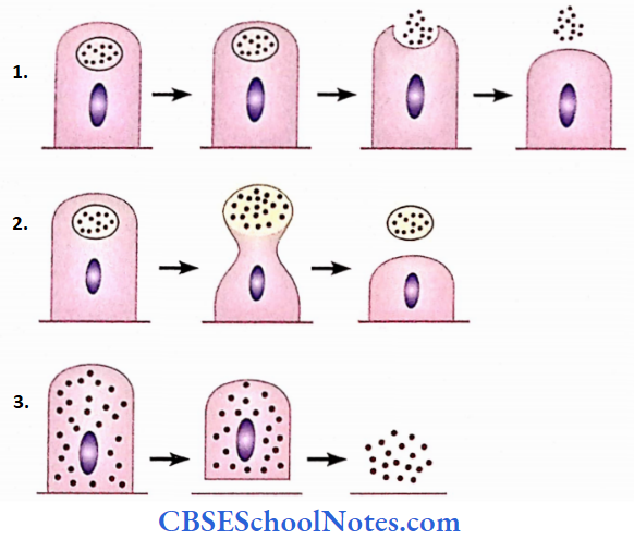
- Merocrine (secretion through exocytosis).
- Apocrine (an apical portion of the cell is pinched off).
- Holocrine (cell dies and becomes secretory product)
3. Classification based on the nature of their secretion
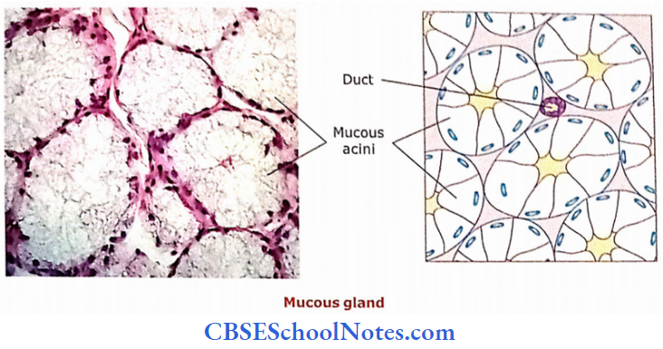
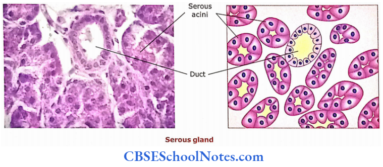
- Mucous Glands: Mucous glands secrete mucus.
- Serous Glands: They secrete thin watery secretion rich in enzymes.
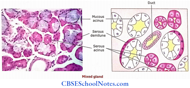
- Mixed Glands: They contain both mucous and serous secretory units. Sometimes, most of the secretory units are mucous acini and serous cells form crescentic caps on the acini. These crescentic caps are called serous demilunes.
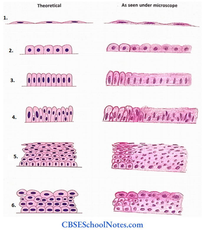
- Simple squamous
- Simple cuboidal
- Simple columnar
- Pseudostratified columnar
- Stratified squamous and
- Transitional.