Connective Tissue Proper
Classification Of Connective Tissue
- Embryonic connective tissue
- Mesenchyme
- Mucous connective tissue(areolar tissue)
- Connective tissue proper
- Loose connective tissue(areolar tissue)
- Adipose tissue
- Reticular tissue
- Dense connective tissue
- Irregular dense connective tissue
- Regular dense connective tissue
- Elastic connective tissue
- Specialized connective tissue
- Cartilage tissue
- Bone tissue
- BIood
In the previous chapter, we studied the basic components of general connective tissue, i.e., ground substance, fibers, and cells. All three components are present in all types of connective tissues.
- However, the proportions of various fibrous and amorphous components vary from one connective tissue type to the other.
- For example, dense connective tissue consists predominantly of fibers, but a very small amount of ground substance and few cells.
- On the other hand, loose connective tissue consists predominantly of ground substances and cells but very few fibers. Following is a brief description of various connective tissues.
Embryonic Connective Tissue
Embryonic connective tissue is classified into two subtypes, i.e., mesenchyme and mucous connective tissue.
Mesenchyme
It consists of small spindle-shaped cells with many processes. Processes of neighboring cells are in contact with each other through a gap junction.
Space between cells is occupied by ground substance and reticular fibers. Mesenchy mal cells are found in the embryo.
Mucous Connective Tissue
It consists of spindle-shaped cells with long and thin cytoplasmic processes. Large intercellular spaces are occupied by jelly-like matrix and thin collagen fibers.
The ground substance of the umbilical cord is also called Wharton’s jelly.
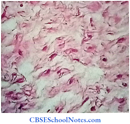
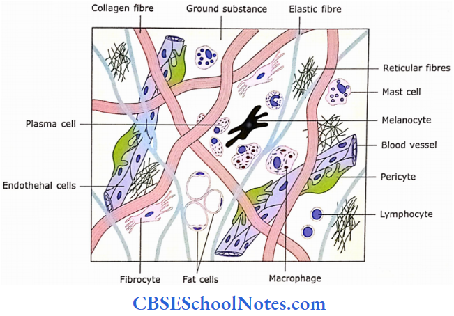
Loose Connective Tissue
Loose Connective Tissue Description
Loose or areolar connective tissue is the most widespread
of all the connective tissues. It consists of a loosely woven network of fibers (all types of fibers are present-collagen, elastic, and reticular).
- Almost all kinds of connective tissue cells(fibroblasts, macrophages, plasma cells, fat cells, white blood cells, and mast cells) are present.
- Fibers and cells are embedded in a semi-fluid ground substance that consists of hyaluronic acid and chondroitin sulfate.
Loose Connective Tissue Location
- It lies beneath the epithelium as lamina propria.
- It is present in superficial fascia along with the adipose tissue.
- It surrounds blood vessels, nerves, viscera muscle, etc.
- It is also present in the mesentery.
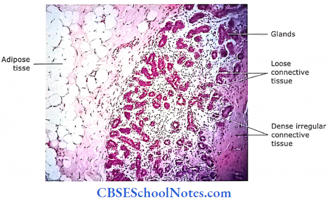
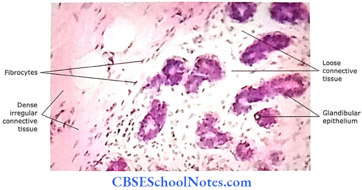
- It surrounds the parenchyma of glands.
- It lies below the mesothelium of body cavities.
Loose Connective Tissue Functions
It provides support to the epithelium. It acts as packing material around various structures and thus provides strength, support, and elasticity.
- There occurs the secretion of histamine by mast cells in inflammatory conditions. Histamine causes increased permeability of blood capillaries.
- This leads to a collection of excessive fluid in loose connective tissue resulting in swelling or edema.
Loose Connective Tissue Remember
Loose connective tissue consists of a loosely arranged network of fibers and almost all kinds of connective tissue cells embedded in a gel-like ground substance.
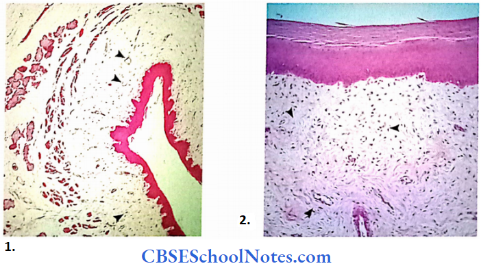
- Low magnification photomicrograph of oesophagous.
- Magnified view of loose connective tissue.
Adipose Tissue
Adipose tissue or fat can be classified into two types, i.c., white adipose tissue(unilocular fat) and brown adipose tissue (multilocular fat).
White Adipose Tissue Description
The color of adipose tissue(fat) is white or yellowish. This tissue is formed by the aggregation of adipocytes(fat cells). Fat cells arc polyhedral or oval.
- Their size is quite large(up to 100 pm), Within a fat cell there is the presence of a single large droplet of lipid (unilocular) that pushes the nucleus to one side and the cytoplasm is present in the form of a thin rim around it.
- Adipocytes are surrounded by the reticular fibers; while groups of adipocytes arc surrounded by connective tissue septa. Adipose tissue is rich in blood vessels. Adipose tissue stores the nutritional calories.
White Adipose Tissue Location
Adipose tissue is present in superficial fascia deep to the skin, bone marrow, omentum, mesentery, and around some viscera(heart, kidney, eyeball, etc).
White Adipose Tissue Functions
It serves as an energy reserve that supports and protects the structures, which surround and prevent heat loss through the skin.
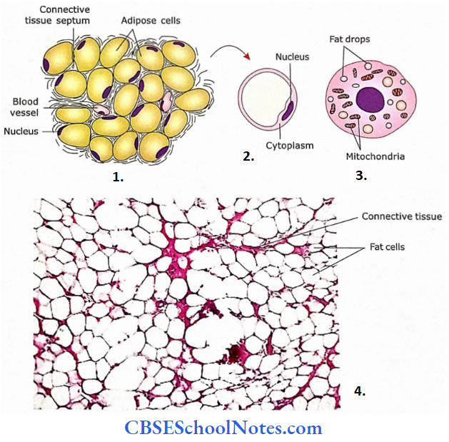
- Adipose cells are placed close to each other. These cells are separated by a small amount of connective tissue in which blood vessels run
- An adipose(fat cell) appears as an empty cell with a flattened nucleus compressed in the peripheral rim of the cytoplasm
- Multilocular adipose cells of brown adipose tissue
- In this photomicrograph of adipose tissue(H and E) lipid contents of fat cells are extracted during tissue processing.
Brown Adipose Tissue
The brown adipose tissue of fat is found only at certain places in the newborn human body, i.e., the posterior triangle abdominal wall. Here, it prevents the excessive heat loss from the body surfacing.
The cells of brown fat differ from the white or yellowish fat in two ways, i.e., the nucleus is not flat but may be pushed to one side, and fat contained in the cytoplasm is in the form of small drops (Multilocular).
Brown Adipose Tissue Remember
Brown fat is not present in mature humans. It is present only in newborns and children and is gradually replaced by white fat (thus changing from multilocular droplets to unilocular droplets of fat within adipose cells).
Brown Adipose Tissue Functions
The function of the brown adipose tissue is to maintain normal body temperature in newborns. This is regulated by the thermogenin protein present in the mitochondria.
Reticular Connective Tissue
Reticular Connective Tissue Description
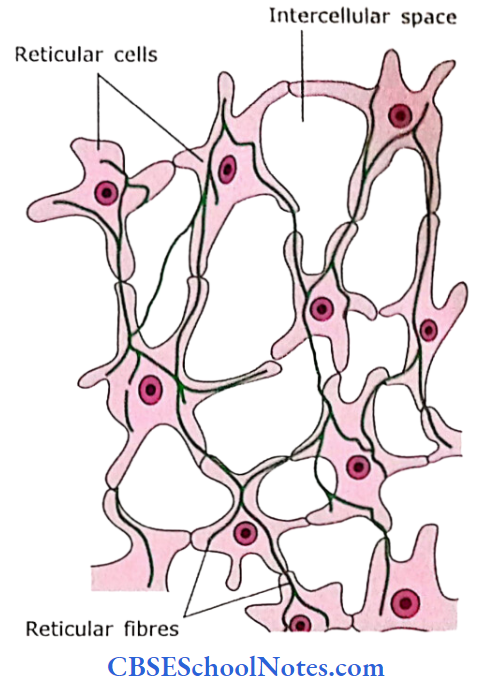
The reticular tissue consists of a mesh-like network of reticular fibers and reticular cells. Reticular fibers are type 3 collagen.
These cells tend to be star-shaped with radiating processes. Some consider that these cells are not distinct types but fibroblasts. Reticular tissue also consists of macrophages.
Reticular Connective Tissue Location
This tissue forms the architectural framework in the spleen, lymph nodes, liver, glands, bone marrow, reticular lamina of the basement membrane, and around smooth muscle cells.
Reticular Connective Tissue Functions
- It provides structural support to various organs by forming stroma or reticular framework.
- Reticular cells may be phagocytic.
- It binds smooth muscle cells.
Reticular Connective Tissue Remember
It is believed by some that reticular cells are not different from fibroblasts.
Dense Connective Tissue
Dense connective tissue contains a large amount of fibers and few cells as compared to loose connective tissue. This connective tissue may be subdivided into three types, i.e., dense irregular, dense regular, and elastic connective tissues.
Dense Irregular Connective Tissue Description
This tissue consists predominantly of collagen fibers. In this tissue, the bundles of collagen fibers are randomly(irregularly) oriented.
Fiber bundles are coarse and form a compact network with little spaces occupied by few connective tissue cells and little ground substance. Fibroblasts are present between bundles.
Dense Irregular Connective Tissue Location
It is present in the dermis of the skin, dura mater, epimysium, epineurium, pericardium, periosteum, tunica albuginea of testis, sclera, capsule of various organs, and submucosa of the intestinal tract.
Dense Irregular Connective Tissue Functions
It provides strength to the tissue.
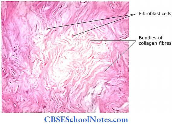
Dense Regular Connective Tissue Description
This tissue also consists predominantly of collagen fibers (type 1). Bundles of collagen fibers are arranged regularly and parallel to the direction of stress, i.e., as in the case of tendons and aponeuroses.
Fibroblasts are present in rows between bundles of collagen fibers. Bundles of collagen fibers are so closely packed that very little space is left for ground substance and loose connective tissue through which run small blood vessels.
Dense Regular Connective Tissue Location
This tissue is present in tendons and aponeuroses of muscles and ligaments.
Dense Regular Connective Tissue Functions
Tendons, aponeuroses, and ligaments provide great resistance to pulling force but at the same time are flexible.
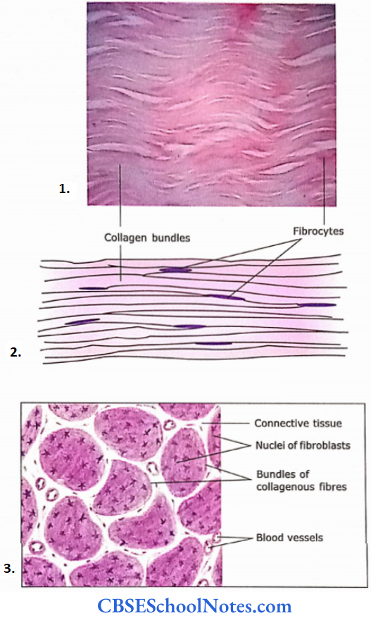
- Longitudinal section of tendon showing bundles of collagen fibers arranged parallel to each other.
- Transverse section of a tendon. Between the collagen bundles note the presence of loose connective tissue and blood vessels.
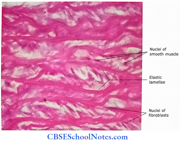
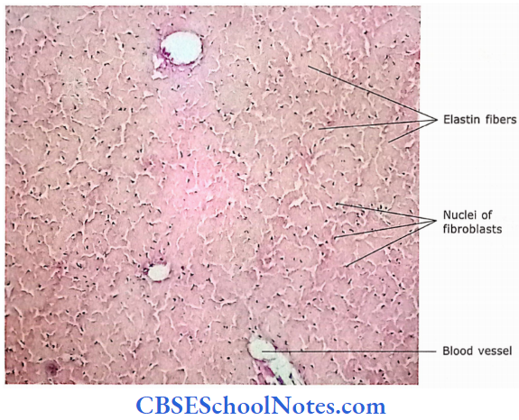
Elastic Tissue Description
Few elastic fibers are found in almost all types of connective tissues. However, in certain situations, elastic fibers are predominantly present.
- In these situations, connective tissue is called clastic connective tissue. This type of connective tissue consists of layers of freely branching elastic fibers.
- Fibroblasts and ground substances are present in small spaces intervening between fibers.
Elastic Tissue Location
Elastic tissue is found in the fascia of the anterior abdominal wall; a wall of the aorta and large arteries; trachea and bronchi; vocal cords; ligamentum nuchae; ligamentum flavum and in the suspensory ligament of the penis.
Elastic Tissue Functions
K provides elasticity to tissue.
Elastic Tissue Clinical Applications
- Obesity
- Obesity in adults may result due to the accumulation of excessive fat in white adipose cells so that cells become larger than usual. Sometimes, an increase in the number of adipose cells may also occur.
- Lipoma
- The most common adipose tissue tumor is called lipoma. They are usually found in the subcutaneous tissue of middle-aged persons. These are benign tumors (not cancerous).
- Liposarcoma
- It is a malignant tumor of adipose tissue, which is quite rare.