
Thalamus, Metathalamus And Epithalamus
The diencephalon forms the central core of grey matter surrounded by cerebral hemispheres. Each half of the diencephalon is located between the midbrain and the cerebrum.
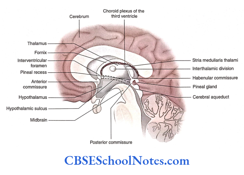
It extends from the region of the interventricular foramen to the region of the posterior commissure. Laterally, it is bounded by the posterior limb of the internal capsule.
The third ventricle may be regarded as the cavity of the diencephalon that divides it into two symmetrical parts.
Read and Learn More Neuroanatomy
Components Of Diencephalon
The hypothalamic sulcus divides the diencephalon into dorsal and ventral parts. The dorsal diencephalon lies above the hypothalamic sulcus while the ventral diencephalon below the sulcus.
Dorsal Diencephalon
The dorsal diencephalon consists of:
- Thalamus
- Metathalamus (medial and lateral geniculate bodies)
- Epithalamus (habenular nucleus and commissure, pineal gland and posterior commissure)
Ventral Diencephalon
The ventral diencephalon consists of:
- Hypothalamus: It lies below the hypothalamic sulcus
- Subthalamus: It lies below the posterior part of the thalamus and consists of the subthalamic nucleus
Thalamus
The thalamus is the largest part of the diencephalon. It consists of paired egg-shaped oval masses of grey matter, situated one on each side ofthe third ventricle. It is about 4 cm long and placed obliquely.
External Features
Each thalamus has two ends (anterior and posterior) and four surfaces (superior, inferior, medial and lateral;
Ends
Anterior end: The anterior end (pole) is narrow, placed close to the midline and lies just behind the interventricular foramen.

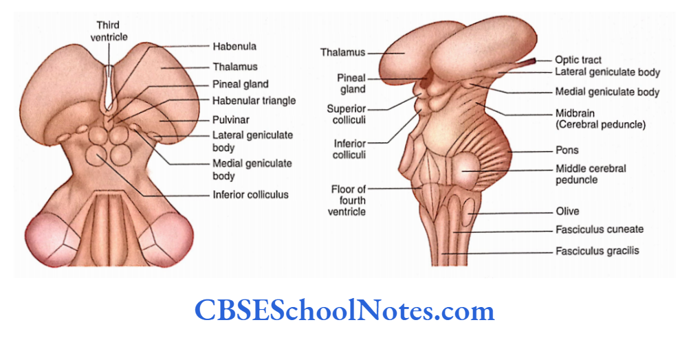
Posterior end: The posterior end (pole) is wide and directed dorsolaterally.
It is situated above and lateral to the superior colliculus. This expanded end of the thalamus is called the pulvinar. The medial and lateral geniculate bodies are present on the inferior aspect of the pulvinar
Surfaces
Medial surface: The medial surface forms the upper part of the third ventricle, i.e. above the hypothalamic sulcus.
The hypothalamic sulcus extends from the interventricular foramen to the upper end of the cerebral aqueduct.
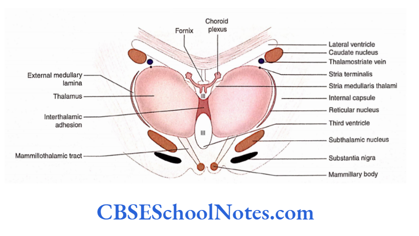
This surface is lined by ependyma. The medial surfaces of the right and left thalami are usually interconnected by a band of grey matter called interthalamic adhesion.

Superior (dorsal) surface: The superior (dorsal) surface is a slightly convex and curved surface. It extends between the stria medullaris thalami (medially) and the caudate nucleus (laterally).
The superior surface is separated from the caudate nucleus by a thalamostriate vein and a bundle of fibres called stria terminalis The superior surface of the thalamus forms the floor of the central part of the lateral ventricle.
3. Lateral surface: The lateral surface of the thalamus concerns the external medullary lamina, reticular nucleus and the posterior limb of the internal capsule
Inferior surface: The inferior surface of the thalamus concerns the superior part of the hypothalamus (anteriorly) and subthalamus (posteriorly).

Internal Structure
The internal structure of the thalamus consists of
- White matter and
- Grey matter
White Matter
The white matter covering the lateral surface of the thalamus is called the external medullary lamina
Grey Matter
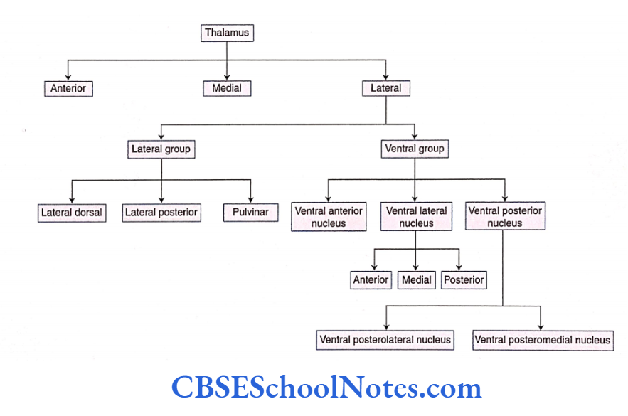
Each thalamus is mainly composed of grey matter which is subdivided into three main parts by a Y-shaped vertical sheet of white matter known as the internal medullary lamina
Anterior part
- Medial part
- Lateral part
Thalamic Nuclei
The three parts of the thalamus (anterior, medial and lateral parts) are further subdivided into various nuclei,
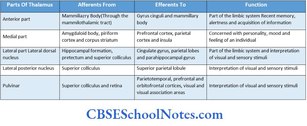
Anterior Part
The nuclei in the anterior part of the thalamus lie between the two limbs of ‘Y’.
Medial Part
The medial part of the thalamus lies medial to the internal medullary lamina. This part consists of a large medial dorsal nucleus and a smaller medial ventral nucleus or midline nucleus.
Lateral Part
The lateral part is subdivided into many nuclei. These nuclei in simple terms may be classified into ventral and lateral groups of nuclei
White Matter
The white matter covering the lateral surface of the thalamus is called the external medullary lamina.
Grey Matter
Each thalamus is mainly composed of grey matter which is subdivided into three main parts by a Y-shaped vertical sheet of white matter known as the internal medullary lamina.
- Anterior part
- Medial part
- Lateral part
Thalamic Nuclei
The three parts of the thalamus (anterior, medial and lateral parts) are further subdivided into various nuclei
Anterior Part
The nuclei in the anterior part of the thalamus lie between the two limbs of ‘Y.
Medial Part
The medial part of the thalamus lies medial to the internal medullary lamina. This part consists of a large medial dorsal nucleus and a smaller medial ventral nucleus or midline nucleus.
Lateral Part
The lateral part is subdivided into many nuclei. These nuclei in simple terms may be classified into ventral and lateral groups of nuclei.
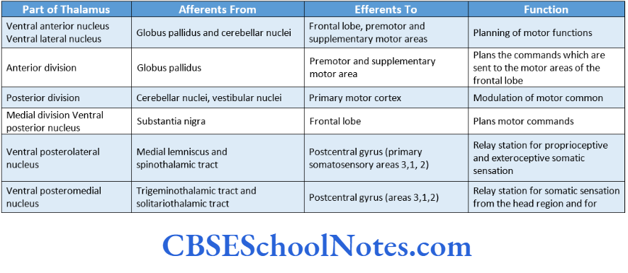
Ventral group of nuclei: In the anteroposterior direction, this group consists of three nuclei:
Ventral anterior nucleus, Ventral lateral nucleus and Ventral posterior nucleus. The ventral posterior nucleus is further divided into the ventral posterolateral nucleus and the ventral posteromedial nucleus.
Lateral group of nuclei: From the anterior to the posterior side, this group consists of three nuclei lateral dorsal nucleus, lateral
posterior nucleus and pulvinar.
Other Nuclei
In addition to the anterior, medial and lateral parts, the thalamus also contains the following nuclei.
Intralaminar Nuclei
Intralaminar nuclei are a collection of grey matter-forming nuclei within the internal medullary lamina of thalamus The important nucleus of this group is known as the centromedian nucleus.
Medial and Lateral Geniculate Bodies
These were previously considered a part of the metathalamus but are now considered an integral part of the thalamus.
Connections of Thalamus: An Overview
The thalamus has extensive afferent and efferent connections.
Afferents
The following afferents bring various kinds of impulses to the thalamus.
- Visual impulses: These are brought to the lateral geniculate body through the optic tract.
- Auditory impulses: These are brought to the medial geniculate body through the lateral lemniscus.
- Olfactory impulses: These are brought to the thalamus indirectly through the amygdaloid complex.
- Taste impulses: These are brought through the solitariothalamic tract.
- Exteroceptive impulses: The impulses carrying pain, touch and temperature sensations are brought through the spinothalamic and trigeminothalamic tracts.
- Proprioceptive impulses: The sensory impulses from muscles, joints and tendons are brought to the thalamus through the medial lemniscus and trigeminothalamic tracts.
- Visceral information: This information is brought through the hypothalamus and reticular formation.
- In addition to the above afferents, the thalamus also receives afferents from all parts of the cerebrum, cerebellum and corpus striatum.
Efferents
The efferents from the thalamus project to the following structures:
Cerebral cortex: The thalamocortical fibres project to all parts of the cortex.
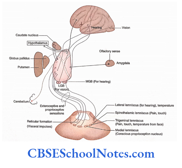
- Hypothalamus, cerebellum, corpus striatum and reticular formation With its extensive afferent and efferent connections, the thalamus is regarded as a great integrating centre and is believed to perceive the sensations of crude pain and temperature.
The connections and functions of important nuclei of the thalamus are summarized
Functions of Thalamus
Thalamic nuclei act as primary relay nuclei. All sensory impulses (except smell) terminate in various nuclei of the thalamus.
From here, the sensory impulses project to different specific cortical areas through thalamocortical radiations.
Ultimately, the cerebral cortex is responsible for the interpretation of various kinds of sensory stimuli.
However, in case of the destruction of the cerebral cortex, the thalamus can appreciate the sense of crude pain and temperature.
The thalamus serves as an integrative centre for motor functions.
Through the ascending reticular activating system (ARAS), the intralaminar nuclei of the thalamus regulate the state of consciousness, alertness and attention.
Thalamic Syndrome
The thalamic syndrome occurs due to a vascular lesion of the artery supplying to the thalamus, i.e. thalamogeniculate branch of the posterior cerebral artery.
In this condition, emotional instability (spontaneous laughing or crying) and disturbances of sensation, i.e. hypersensitivity, are seen. A spontaneous pain on the opposite side of the body is also noticed. This syndrome may be associated with hemiparesis if the lesion also involves the internal capsule.
Metathalamus
The metathalamus consists of medial and lateral geniculate bodies.
Medial Geniculate Body
The medial geniculate body is a small ovoid mass of grey matter on the inferior aspect of the pulvinar.
It consists of medial, ventral and dorsal nuclei. It receives auditory information from the inferior colliculus through the inferior brachium. Its connections are summarised.
Lateral Geniculate Body
The lateral geniculate body is a small ovoid mass of grey matter on the inferior aspect of the pulvinar.
It is connected with the superior colliculus through the superior brachium. The lateral geniculate body consists of an inverted U-shaped lateral geniculate nucleus.
It is composed of six layers of nerve cells in the coronal section. These laminae are numbered 1-6 from the ventral to the dorsal side. Layers 1 and 2 consist of large cells (magnocellular layers) and layers 4-6 have smaller neurons (parvocellular laminae).
Afferents
The lateral geniculate body receives fibres from the retina. The nasal fibres from the opposite retina (crossed fibres) Terminate in layers 1, 4 and 6.
The ipsilateral fibres from the temporal half of the retina (uncrossed fibres) terminate in layers 2, 3 and 5. Thus, each lateral geniculate body receives visual information from the opposite field of vision.
Efferents
The efferents from the lateral geniculate body terminate in the visual areas of the occipital lobe (areas 17, 18 and 19) through optic radiation (geniculocalcarine tract)
Epithalamus
The epithalamus consists of the pineal gland, habenular nucleus, stria medullaris and posterior commissure.
Pineal Gland
The pineal gland is a small conical structure present in the posterior wall of the third ventricle. It lies below the splenium of the corpus callosum in a depression between two superior colliculi.
It is attached to the posterior wall of the third ventricle by a stalk (pineal stalk). The pineal stalk divides anteriorly into superior and inferior laminae to enclose the pineal recess of the third ventricle.
The superior lamina of the pineal stalk contains a habenular commissure and the inferior lamina contains the posterior commissure.
The pineal gland was previously considered a rudimentary gland but now it is a well-established endocrine gland which secretes various hormones.
Functions
The pinealocyte secretes serotonin and melatonin, which influence the activities of other endocrine glands.
The secretion of the hormone melatonin is associated with circadian rhythm and is influenced by light. Thus, the pineal gland acts as a biological clock.

Habenular Nucleus, Stria Medullaris Thalami and Posterior Commissure
The habenular nuclei are situated in the habenular triangle. These nuclei are regarded as cell stations in olfactory and visual pathways. These nuclei are also involved in the limbic system.
The stria medullaris thalami are a bundle of white fibres. These fibres are afferent to the habenular nuclei.
The posterior commissure is the band of white fibres which crosses the midline by passing through the inferior lamina of the stalk of the pineal gland. Many nuclei are present concerning the fibres of the posterior commissure.
Thalamus, Metathalamus And Epithalamus Summary
- The diencephalon is located between the cerebrum (above) and the midbrain (below).
- The third ventricle is the cavity of the diencephalon that divides it into two symmetrical parts.
- The hypothalamic sulcus divides the diencephalon into ventral and dorsal parts.
- The dorsal part of the diencephalon lies above the sulcus and consists of the thalamus, metathalamus and epithalamus.
- The ventral part of the diencephalon lies below the hypothalamic sulcus and consists of the subthalamus and hypothalamus.
Thalamus
- The thalamus of each side is a large, egg-shaped mass of grey matter, situated on either side of the third ventricle.
- Each thalamus is subdivided into three main parts by a Y-shaped vertical sheet of white matter known as the internal medullary lamina.
- The three main subdivisions of the thalamus are the Anterior, Medial and lateral parts.
Anterior part
- The medial part consists of the medial dorsal and midline nucleus.
- The lateral part is further divided into ventral and lateral groups of nuclei. The lateral group consists of lateral dorsal, lateral posterior and pulvinar.
- The ventral group consists of the ventral anterior, ventral lateral and ventral posterior.
- Intralaminar nuclei are present within the internal medullary lamina.
- The thalamus has extensive afferent and efferent connections. It receives visual impulses, auditory impulses, olfactory impulses, taste impulses, exteroceptive impulses, proprioceptive impulses and visceral information.
- The efferents of the thalamus go to cerebral cortex, hypothalamus, cerebellum, corpus striatum and reticular formation.
- Thalamic nuclei are the principal relay station for sensory impulses that reach the cerebral cortex from various parts of the CNS.
Medial and lateral geniculate bodies
- These bodies are present on the inferior aspect of the pulvinar.
- The medial geniculate body is connected with the inferior colliculus through the inferior brachium.
- The efferents of the medial geniculate body project to the auditory area of the cortex. The medial geniculate body is the relay centre in the auditory pathway.
- The lateral geniculate body is connected with the superior colliculus through the superior brachium. It receives fibres from the retina and sends efferents to the visual cortex in the occipital lobe.
- The lateral geniculate body acts as a final relay station in the visual path.
Epithalamus
- The epithalamus consists of pineal gland, habenular nucleus, stria medullaris and posterior commissure.
- The pineal gland is a small conical structure present in the posterior wall of the third ventricle. The gland secretes serotonin and melatonin which influence the activities of other endocrine glands
Thalamus, Metathalamus And Epithalamus Multiple Choice Questions
Question 1. Which of the following statements about diencephalon are true?
- Each half of the diencephalon is located between the cerebrum (above) and the midbrain (below)
- It extends from the region of the interventricular foramen to the region of the posterior commissure.
- Laterally, it is bounded by the posterior limb of the internal capsule
- The third ventricle is regarded as the cavity of the diencephalon
- All of the above
Answer: 5. All of the above
Question 2. The diencephalon consists of the following subdivisions except
- Dorsal thalamus
- Metathalamus
- Epithalamus
- Lateral thalamus
- Subthalamus
- Hypothalamus
Answer: 4. Lateral thalamus
Question 3. The following features are seen on the medial surface of the thalamus except
- Hypothalamic sulcus
- Stria medullaris thalami
- Taenia thalami
- Stria terminalis
Answer: 4. Stria terminalis
Question 4. The grey matter of the thalamus is subdivided into the following parts except
- Anterior part
- Posterior part
- Medial part
- Lateral part
Answer: 2. Posterior part
Question 5. Which of the following are the nuclei of the ventral group of the lateral part of the thalamus?
- Ventral anterior
- Ventral lateral
- Ventral posterolateral
- Ventral posteromedial
- All of the above
Answer: 3. Ventral posterolateral
Question 6. Which of the following statements is true?
- The nuclei of the anterior group of the thalamus are concerned with memory and emotion as they are part of the limbic system
- The nuclei of the medial group of the thalamus are connected with the prefrontal cortex; thus, the person becomes aware of emotions
- The lateral group of nuclei projects to the postcentral gyrus. Thus, this group of nuclei is a relay station for exteroceptive sensations
- All of the above
Answer: 4. All of the above
Question 6. The following facts about the medial geniculate body are true except
- It is a relay centre in the auditory pathway
- It receives fibres from the inferior colliculus of the same side through the inferior brachium
- It does not receive fibres from the inferior colliculus of the opposite side
- It consists of three nuclei—medial, ventral and dorsal nuclei
Answer: 4. It consists of three nuclei—medial, ventral and dorsal nuclei
Question 7. The following facts about the lateral geniculate body are true except
- It is connected with the superior colliculus through the superior brachium
- It is a relay station in the visual pathway
- It receives fibres from the retina on the same side
- The nucleus of the lateral geniculate body consists of six layers of nerve cells.
Answer: 3. It receives fibres from the retina on the same side
Question 8. The following facts about the lateral geniculate body are true except
- It is connected with the superior colliculus through the superior brachium
- It is a relay station in the visual pathway
- It receives fibres from the retina on the same side
- The nucleus of the lateral geniculate body consists of six layers of nerve cells.
Answer: 3. It receives fibres from the retina of the same side.