
Diencephalon-2 Subthalamus And Hypothalamus
The ventral part of the diencephalon lies below the hypothalamic sulcus and consists of the subthalamus and hypothalamus.
Ventral Thalamus (Subthalamus)
The ventral thalamus was previously described as the subthalamus.
However, many nuclei of the subthalamus, for functional reasons, are now considered a part of basal nuclei. The ventral thalamus appears to be the upward continuation of the tegmentum of the midbrain.
It is bounded medially and ventrally by the hypothalamus and laterally by the lowest part of the internal capsule. The ventral thalamus contains two main nuclei:
Read and Learn More Neuroanatomy
- Zona incerta
- Reticular nucleus
Zona Incerta
Zona incerta is a thin layer of grey matter that is interposed between the thalamus and the subthalamic nucleus. It is continuous with the reticular nucleus Above.
Reticular Nucleus
- The reticular nucleus is a thin layer of grey matter situated lateral to the dorsal thalamus.
- It is separated from the thalamus by a thin layer of white matter known as the external medullary lamina. Inferiorly, it is continuous with zona incerta.
- Laterally, the reticular nucleus lies concerning the posterior limb of the internal capsule.
- This nucleus has nothing to do with the reticular formation of the brainstem. It is believed that the reticular nucleus may play a role in the regulation of thalamic activities.
Hypothalamus
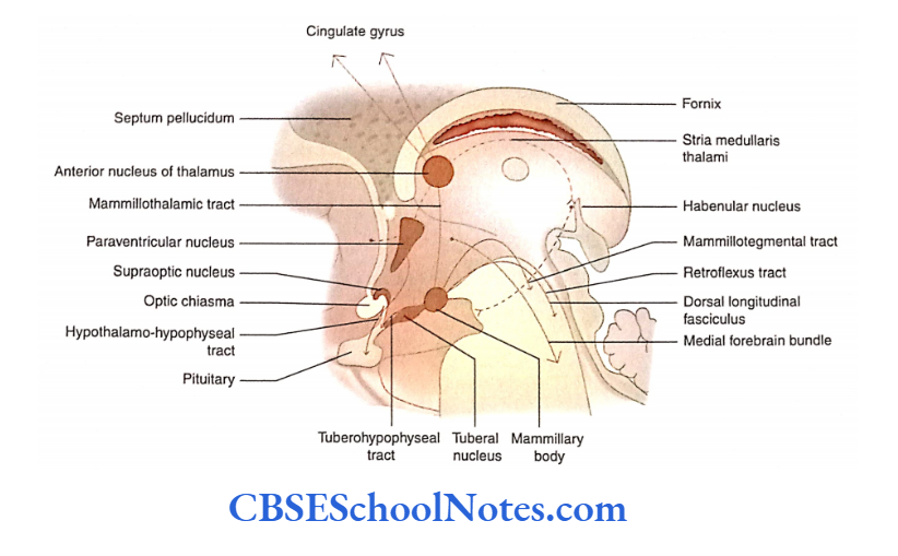
- The hypothalamus, on the right and left sides, lies below the thalamus and is separated from the latter by the hypothalamic sulcus. It is present in the lateral wall and the floor ofthe third ventricle.
- The hypothalamus is concerned with many important functions which include autonomic, visceral, and endocrine functions. Most of the hypothalamus is hidden (not visible) to the naked eye when a specimen of the brain is examined.
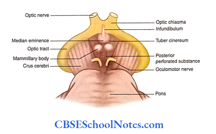
- However, some parts of the hypothalamus can be seen on the external (ventral) surface of the brain.
- These visible parts of the hypothalamus are located in the interpeduncular fossa.
- These include optic chiasma, the median eminence of the tuber cinereum, infundibulum, and mammillary bodies. All these structures also form the floor of the third ventricle.
Boundaries of Hypothalamus
In the midsagittal section of the brain; also, the extent of the hypothalamus can be visualized as follows:
Anterior boundary: Anteriorly, the hypothalamus is bounded by lamina terminalis. The lamina terminalis extends from the anterior commissure to the optic chiasma.
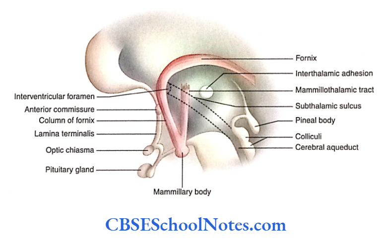
Posterior boundary: Posteriorly, the hypothalamus extends up to a vertical plane posterior to mammillary bodies.
Superior boundary: Superiorly, the hypothalamus is separated from the thalamus by the subthalamic sulcus.
Inferior boundary: Inferiorly, the hypothalamus is bounded by structures that form the floor of the third ventricle and is visible externally in the interpeduncular fossa.
Lateral Boundary: Lterally, The hypothalamus concerning the internal capsule
Medial boundary: Medically, the hypothwamulues form the ventrolateral wall of the cavity of the third ventricle below the hypothalamic sulcus.
Subdivisions of Hypothalamus
The hypothalamus (on both the right and the left sides) consists of grey and white matter.
There are many distinct nerve cell areas known as nuclei. To locate the position of these nuclei, the hypothalamus is divided into two different planes:
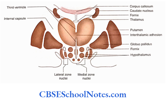
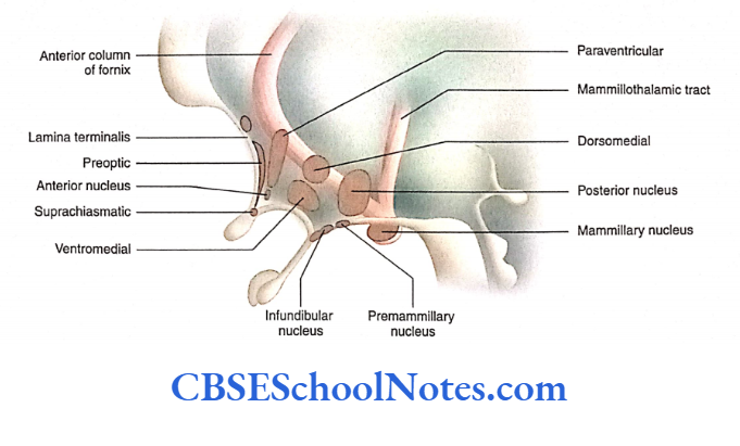
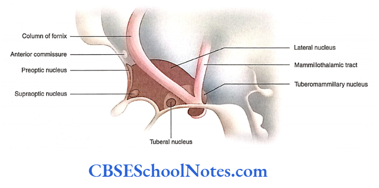
Mediolateral subdivision: Based on the presence of prominent myelinated fibers of the column of the fornix and mamillothalamic tract, the hypothalamus is divided into medial and lateral zones. The medial zone contains many small nuclei while the lateral zone contains only a few.
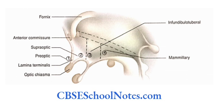
Anteroposterior subdivision: The hypothalamus can also be divided anteroposteriorly into four regions:
- Preoptic, Supraoptic, Infundibulotuberal and Mammillary regions
- Nuclei in Various Regions of Hypothalamus
The hypothalamus is divided into four regions:
Prcoptic region: The preoptic region lies adjacent to the lamina terminalis. It has a preoptic nucleus, which extends in both medial and lateral zones.
Supraoptic region: The supraoptic region of the hypothalamus lies above the optic chiasma.
In the medial zone, it has three nuclei:
- Paraventricular,
- Anterior and
- Suprachiasmatic
In the lateral zone, the supraoptic nucleus is present The lateral zone also contains a large lateral nucleus extending through the supraoptic, tuberal, and mammillary regions.
Infundibulotuberal region: The infundibulotuberal region consists of infundibulum, tuber cinereum and the region above it.
The medial zone of this region contains a dorsomedial nucleus, ventromedial nucleus, infundibular nucleus, and a small premammillary nucleus. The lateral zone of this region consists of a small tube nucleus.
Mammillary region: The mammillary region consists of the posterior nucleus and mammillary nucleus in the medial zone and the tuberomammillary nucleus in the lateral Zone. The Lateral Zone Extending Through The Supraptic, Infundibular, and Mammilary regions contains a large lateral nucleus.
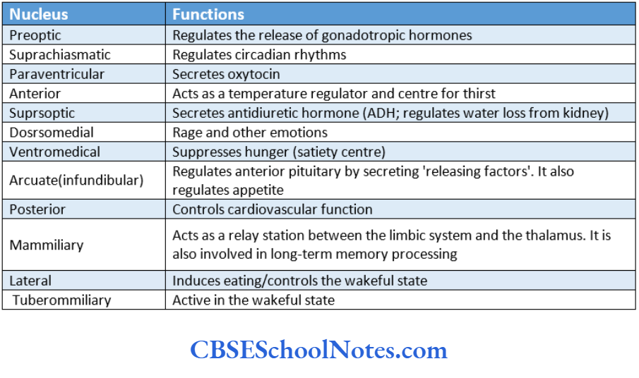
The physiological significance of the nuclei in the hypothalamus is summarised.
Nervous Connection of Hypothalamus
The afferent and efferent connections of the hypothalamus are described in the following text.
Afferent Connections
The hypothalamus receives the afferent connections from the following structure
Afferent from the spinal cord: Reticular formation and collaterals of sensory lemnisci terminate in the lateral hypothalamus.
Afferent from the brainstem: From visceral (autonomic) nuclei and nuclei of the solitary tract to various hypothalamic nuclei.
Afferent from the limbic system: From the hippocampus through the fornix to the mammillary body.
Afferent from the retina: To suprachiasmatic nuclei.
Afferent from the thalamus: To various nuclei of the hypothalamus.
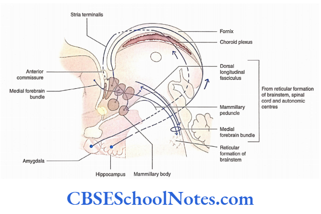
Efferent Connections
The efferent fibers from the hypothalamus go to the same structures from where it has received afferent fibers:
Efferents to hippocampal formation: Through the medial forebrain bundle
Efferents to the amygdaloid nuclear complex: Through stria terminalis.
Efferents to the thalamus and tegmentum: Through the mamillothalamic tract and mamillotegmental tract.
Efferents to the autonomic motor neurons of the brainstem and spinal cord: Through the medial forebrain bundle to the autonomic nuclei of the brainstem and spinal cord (sympathetic T1 to L2 and parasympathetic S2 to S4).
Efferent connections to the pituitary gland: This is described in detail separately.
Hypothalamic Control Of The Pituitary Gland
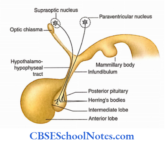
- The endocrine secretions from the pituitary gland are under the direct control of the hypothalamus.
- This control is because the median eminence and part of the infundibulum (both are parts of the hypothalamus) secrete certain substances that control the release or inhibition of hormones from pars distalis.
- These substances are called releasing and release-inhibiting factors or hormones.
- The releasing and release-inhibiting factors (hormones) are produced by a group of neurons (nuclei) situated in the median eminence and upper part of the infundibulum.
- The axons of these neurons end concerning capillaries present in these areas of the hypothalamus and pour their hormones into blood circulation see in the following text).
- The production of releasing and release-inhibiting hormones is under the control of signals from the nervous system and/or chemical changes in the blood (feedback mechanism).
Hypothalama-Hypophyseal Portal System
- The releasing or release-inhibiting factors from the hypothalamus reach the pars distalis (anterior pituitary) through the portal system of blood capillaries.
- This system is called the hypothalami-hypophyseal portal System the system is called the hypothalamic hypophyseal portal system.
- The release of release-inhibiting factors is taken into the primary capillary plexus of the portal system
- From the primary plexus, these factors reach the secondary plexus situated in the anterior pituitary where they act on the cells of the anterior pituitary gland,
- The hormones secreted by the anterior pituitary gland pass into the secondary plexus of the portal system.
- These hormones then pass into the anterior hypophyseal veins for distribution to target tissues throughout the body.
Connection of Hypothalamus with Neurohypophysis (Posterior Pituitary)
- Neurohypophysis consists of structures such as median eminence, infundibular stalk, and a posterior (neural) lobe of the pituitary gland.
- The supraoptic and paraventricular nuclei of the hypothalamus synthesize vasopressin and oxytocin hormones, respectively.
- The axons from these nuclei project to the posterior lobe of the pituitary through the infundibular stalk.
- The arrival of an action potential results in the release of hormones from Herring bodies which diffuse through capillaries to enter the general circulation.
- The hormone ‘vasopressin’ (ADH) helps in the restriction of water loss from the kidney while oxytocin causes the contraction of smooth muscles during childbirth.
Functions Of Hypothalamus
The hypothalamus, in general, serves to marry functions. These are as follows:
- Controls the autonomic nervous system (both sympathetic and parasympathetic) and acts as an integrating center.
- The limbic system sets emotional states and controls sexual desire and behavior.
- Creates new memory
- Regulates body temperature (the anterior hypothalamus facilitates heat loss by vasodilatation and sweating while the posterior hypothalamus conserves the body heat by vasoconstriction and shivering)
- Serves as a link between nervous and endocrine systems to regulate growth, metabolism, reproductive and stress responses
- Secretes hormones such as ADH and oxytocin
- Controls appetitive drives such as thirst and hunger (control food and water intake)
- Controls circadian rhythms
- Regulates sleep and wakefulness states.
Diabetes Insipidus
- Diabetes insipidus is a condition in which there is increased secretion of dilute urine (polyuria).
- This condition may result due to destruction of the supraoptic nucleus which is specifically concerned with the maintenance of body water balance.
- This nucleus produces ADH which helps in the reabsorption of water from the distal convoluted and collecting tubules of the kidney. Although ADH is produced in the hypothalamus, it is stored and secreted by the posterior pituitary.
- Therefore, it is usually considered a pituitary hormone. In the absence or reduced secretion of this hormone, water is not absorbed, and polyuria results.
- This also results in increased water intake (polydipsia). This condition is known as hypothalamic syndrome.
- The reduced secretion of ADH may be due to the destruction of the supraoptic nucleus, hypothalami-hypophyseal tract, or posterior pituitary (due to a tumor near the hypothalamus, pituitary tumor, surgery, and radiation therapy, or head injury).
- Diabetes insipidus may also occur due to the disease of the kidney in which it fails to respond to ADH.
Subthalamus And Hypothalamus Summary
- The hypothalamus is situated below the hypothalamic sulcus in the lateral wall and across the floor of the third ventricle.
- Some parts of the hypothalamus are visible on the external (ventral) surface of the brain, which are located in the interpeduncular fossa.
- The hypothalamus consists of many nuclei. To locate the position of these nuclei, the hypothalamus is divided into two different planes:
- Mediolaterally and Anteroposteriorly.
- From the medial to the lateral side, it is divided into medial and lateral zones because of the presence of columns of the fornix and mamillothalamic tract between the two zones.
Anteroposteriorly, it is divided into four regions:
- Pre-optic,
- Supraoptic,
- Infundibulotuberal and
- Mammillary regions.
The following nuclei are present in each region of the hypothalamus:
- Preoptic region—
- preoptic nucleus
- Supraoptic region—paraventricular, anterior, and suprachiasmatic nuclei in the medial zone and lateral nucleus in the lateral zone.
- Infundibulotuberal region—dorsomedial nucleus, ventromedial nucleus, infundibular nucleus, pre mammillary nucleus in the medial zone, and lateral tuberal nucleus in the lateral zone
- Mammillary region—posterior nucleus and mammillary nucleus in the medial zone; tuberomammillary nucleus and large lateral nucleus in the lateral zone
- The afferent connections of the hypothalamus are from various sources, i.e. limbic system, cerebral cortex, visual and olfactory systems, thalamus, ascending visceral and somatic sensory systems from the brainstem and spinal cord.
- The efferents of the hypothalamus go to most of the above-mentioned sources.
- The hypothalamus is concerned with many important autonomic, visceral, and endocrine functions.
Multiple Choice Questions
Question 1. Which of the following facts about habenular nuclei is are false?
- They are situated in the habenular triangle medial to the pulvinar above the superior colliculus
- They are regarded as cell stations in olfactory and visceral pathways
- Afferents of habenular nuclei run in stria medullary thalami
- Nuclei of two sides are connected with habenular commissure
- None of the above
Answer: 5. None of the above
Question 2. The following parts of the hypothalamus are visible on the the external surface of the brain (in the interpeduncular fossa) except
- Lamina terminalis
- Optic chiasma
- Median eminence of tuber cinereum
- Infundibulum
- Mamillary body
Answer: 1. Lamina terminalis
Question 3. In the midsagittal section of the brain, the following structures indicate the extent of hypothalamus except
- Anteriorly—lamina terminalis
- Posteriorly—vertical plane posterior to mammillary bodies
- Superiorly—subthalamic sulcus
- Inferiorly—structures visible in the interpeduncular fossa
- Medially—floor of the third ventricle
Answer: 3. Inferiorly—structures visible in the interpeduncular fossa
Question 4. The hypothalamus is divided anteroposteriorly into the following regions except
- Preoptic
- Supraoptic Infraoptic
- Infundibulotuberal
- MammiUary regions
Answer: 3. Infundibulotuberal
Question 5. Which of the following nuclei is/are present in the hypothalamus?
- Preoptic
- Paraventricular
- Supraoptic
- Dorsomedial
- Ventromedial
- All ofthe above
Answer: 6. All of the above
Question 6. The following are the connections of the mammillary body except
- Afferents from the hippocampus through the fornix to the mammillary body
- Mammillary peduncle from tegmental nuclei to the mammillary body
- Mammillothalamic tract
- Mammillotegmental tract
- Mammillospinal tract
Answer: 3. Mammillothalamic tract
Question 7. The hormone vasopressin (antidiuretic) is synthesized by
- Paraventricular nucleus
- Supraoptic nucleus
- Suprachiasmatic nucleus
- Preoptic nucleus
Answer: 2. Supraoptic nucleus
Question 8. The releasing and release-inhibiting hormones produced by
- Nuclei of median eminence and upper part of infundibulum
- Nuclei located in neurohypophysis
- Adenohypophysis
- Herring bodies
Answer: 1. Nuclei of the median eminence and upper part of the infundibulum
Question 9. The following statement(s) about the hypothalamohypophyseal portal system is/are false:
- The release or release-inhibiting factors from the hypothalamus reach the anterior pituitary by the portal system of blood capillaries
- The portal system consists of two sets of capillary networks
- The first capillary network is present in median eminence and infundibulum
- The second capillary plexus is present in the anterior pituitary
- None of the above
Answer: 5. None of the above
Question 10. The following functions may be attributed to the hypothalamus except
- Control of circadian rhythms
- Control of food and water intake
- Control of coordinated muscular movement during walking
- Secretion of hormones
- Control of autonomic functions
Answer: Control of coordinated muscular movement during walking