
Spinal Cord
The spinal cord is a long, cylindrical structure present in the upper two-thirds of the vertebral canal.
Within the vertebral canal, the spinal cord is well protected by bony vertebrae, meninges, and cerebrospinal fluid (CSF).
External Structure Of The Spinal Cord
The spinal cord extends from the level of the upper border of the first cervical vertebra up to the lower border of the first lumbar vertebra.
The length of the cord varies from 42 to 45 cm and its weight is about 30 grains. The diameter of the spinal cord is minimum in the thoracic region, about 2 cm.
Read and Learn More Neuroanatomy
The gross anatomical features that are examined externally are as follows:
- Enlargements
- Conus medullaris
- Filum terminal
- Fissure and sulci
- Spinal nerves
- Spinal cord segments
Enlargements
The spinal cord shows the fusiform enlargement in the cervical and lumbosacral regions. These enlargements are due to the collection of a large number of spinal neurons for the nerve supply of the upper and lower limbs.
Therefore, these enlargements are also sometimes called limb enlargements, i.e. cervical and lumbosacral enlargements.
The cervical enlargement extends from C4 to T2 spinal cord segments.
The ventral rami of C5 to T1 spinal nerves form the brachial plexuses, which supply the upper limb.
The lumbosacral enlargement extends front LI to S3 spinal cord segments.
The ventral rami of the 1.2 to S3 spinal cord segment forms the lumbosacral plexus to supply the lower limb.
Conus Medullaris
Just below the lumbosacral enlargement, the lower end of the spinal cord is tapering and is known as conus medullaris.
Filum Terminale
The Filum terminal is a slender filament, which extends from the tip of the conus medullaris to the dorsum of the first coccygeal vertebra.
Filum terminale lies in the middle of cauda equina. The cauda equine is a bunch of spinal nerves around the filum terminale. Cauda equine consists of lumbar, sacral, and coccygeal spinal nerves.
Fissure and Sulci
On the anterior aspect of the spinal cord, there is an anterior median fissure, which extends throughout its length. On either side of this fissure, there is the presence of shallow anterolateral sulci.
Similarly, on the posterior aspect of the cord, a single median posteromedian sulcus and two posterolateral sulcus, one lying on each side of the posteromedian sulcus, are present. This fissure and sulci can be better appreciated in the transverse section of the spinal cord.

Spinal Nerves
Thirty-one pairs of spinal nerves are attached to the spinal cord. These are grouped into 8 cervical, 12 thoracic, 5 lumbar, 5 sacral and 1 coccygeal.
Spinal Cord Segments
Based on the origin of the 31 pairs of spinal nerves, the spinal cord is also divided into 31 spinal segments.
The spinal segment is the portion of the spinal cord to which a pair of dorsal and ventral spinal roots is attached.
The spinal cord is divided into the following segments; 8 cervical, 12 thoracic, 5 lumbar, 5 sacral, and 1 coccygeal.
Spinal Cord Segments and Vertebral Levels
Students should note that the spinal cord segments do not
correspond to the vertebral column segments in adults.
However, in early fetal age, there is a perfect correlation between spinal cord segments and vertebral segments. However, after the early fetal age, the vertebral column grows at a much faster rate as compared to the growth of the spinal cord.
Therefore, the lengthening of spinal nerve roots between the spinal cord and the intervertebral foramen occurs. This lengthening of spinal nerves is more marked in lumbar and sacral spinal roots.
The spinal nerves at the lower level (lower thoracic and lumbar) have to pass obliquely downwards for a considerable distance to get out of their respective foramina.
Thus, the nerve roots from the lumbosacral region (conus medullaris) emerge in the form of a bunch.
These nerve roots surround the filum terminale and are known as cauda equina (resembling the tail of the horse).

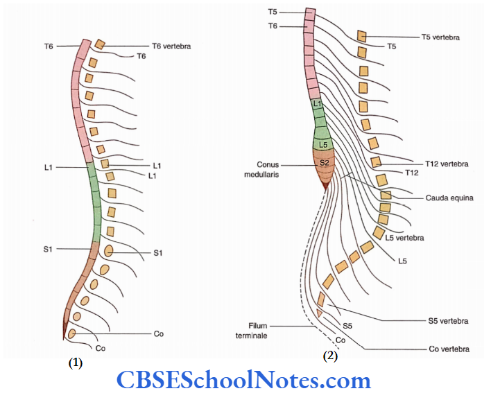
As there are seven cervical vertebrae and eight pairs of cervical spinal nerves, spinal nerves from Cl to C7 come out above the corresponding vertebrae while spinal nerve C8 comes out between C7 and T1 vertebrae.
The T1 spinal nerve and all others below this level come out below their corresponding vertebrae.
Internal Structure Of The Spinal Cord
By studying the transverse section of the spinal cord, one can learn the basics of its internal structure. The transverse section of the spinal cord shows that it is composed of grey and white matter.
Grey matter is placed centrally and white matter is on the periphery. Grey matter is composed of cell bodies (neurons) and their processes while white matter is composed of myelinated and nonmyelinated nerve fibers.
Grey Matter
The grey matter of the spinal cord, on each side, is subdivided into anterior (ventral), posterior (dorsal), and lateral grey columns (horns).
The anterior, posterior, and lateral grey columns (horns) lie anterior, posterior, and lateral to the transverse band of grey commissure.

Anterior (Ventral) Grey Columns (Horns)
- The anterior grey columns contain cell bodies of motor neurons (alpha and gamma) and Renshaw cells. The Renshaw cell is a type of interneuron.
- The cells of this column give origin to axons, which form the ventral root of the spinal nerve. These fibers carry nerve impulses for the contraction of skeletal muscles.
Spinal Cord Function And Structure
Posterior (Dorsal) Grey Columns
The posterior grey columns are directed backward and laterally toward the surface. This column contains somatic and autonomic sensory neurons and receives the central processes of pseudo-unipolar cells of the dorsal root ganglion.
The posterior grey column gives origin to the axons, which form ascending (sensory) tracts of the white matter. Conventionally, the dorsal horn is divided dorsoventrally into the apex, body, and base.
Lateral Grey Columns
The lateral grey columns are present between the anterior and the posterior grey columns on each side.
These columns extend from Tl to L2 and S2 to S4 spinal segments.
They contain the preganglionic autonomic motor neurons that carry nerve impulses for the regulation of the activities of the smooth muscle, cardiac muscle, and glands (see in the following text).
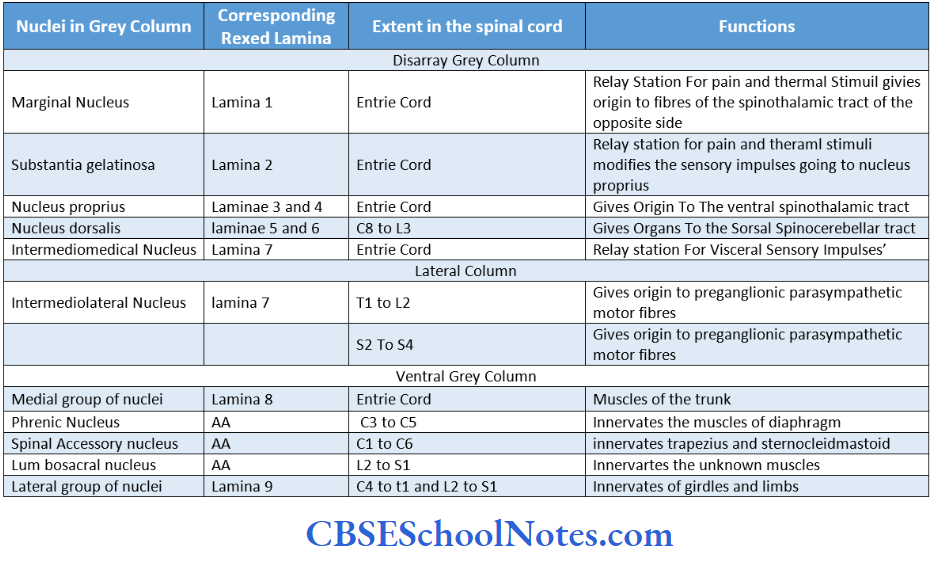
Nuclei in the Grey Matter
The H-shaped grey matter of the spinal cord consists of neurons of various sizes and shapes. These nerve cells are organized in different groups and each group extends longitudinally within the grey matter of the spinal cord.
It is due to this reason that each nerve cell group is called a column or nucleus.
Nuclei in the anterior grey column (ventral horn): The nerve cell groups in the anterior column or ventral horn are motor nuclei.
These are arranged in three groups:
- Medial,
- Lateral and
- Central.
Medial group: This group is present throughout the length of the spinal cord.
Spinal Cord Function And Structure
It is subdivided into two groups:
Ventromedial and Dorsomedial. The ventromedial group supplies the muscles of the back while the dorsomedial group supplies anterolaterally situated trunk muscles.
Central group: This group is present in the cervical and lumbosacral segments of the spinal cord.
It consists of three nuclei:
- Phrenic (between C3 and C5 spinal segments),
- Spinal accessory (between Cl and C6 segments) and
- Lumbosacral nuclei (between L2 and SI spinal segments).
Lateral group: This group is present only in cervical and lumbar enlargements of the cord as it supplies the muscles of the upper and lower limbs.
It is subdivided into three subnuclei:
- Ventrolateral,
- Dorsolateral and
- Retrodorsolateral
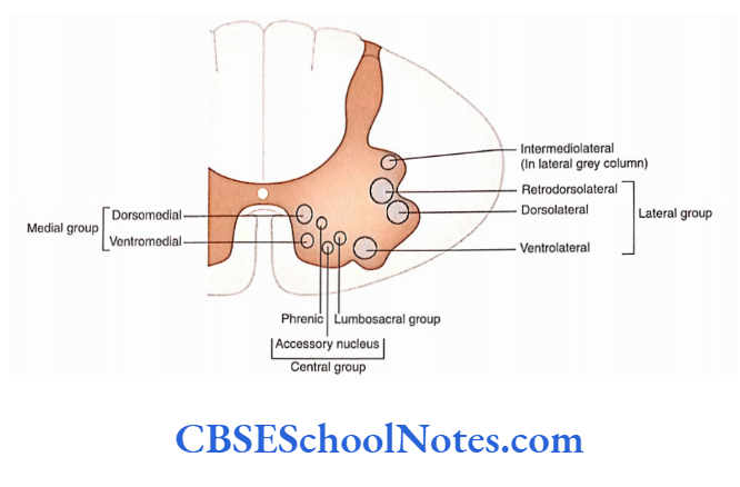
Nuclei in the lateral grey column: The lateral grey 3. column, or the intermediate nucleus, is present between the anterior and posterior grey columns on either side of the central canal.
- A distinct group of nerve cells is present in the lateral grey column between the T1 and L2 spinal segments.
- The axons of these cells give rise to preganglionic sympathetic motor fibers, which travel in the ventral spinal root.
- A similar less distinct group is also seen between S2 and S4 spinal segments. The axons of these cells give origin to preganglionic parasympathetic motor fibers.
Spinal Cord Function And Structure
Nuclei in the posterior grey column (dorsal horn): This group consists of sensory nerve cells.
The sensory neurons of the dorsal grey column are much smaller in size as compared to the motor neurons of the anterior grey column.
A brief description of these groups is arranged in the dorsoventral direction.
Marginal nucleus: It is a thin layer of small neurons situated dorsal to substantia gelatinosa at the apex of the dorsal grey column.
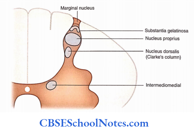
Substantia gelatinosa: Substantia gelatinosa cells are present near the apex of the posterior grey column.
This group extends throughout the spinal cord. Substantia gelatinosa is continuous above (in the medulla and lower pons) with the nucleus of the spinal tract of the trigeminal nerve.
Nucleus proprius: It (magnocellular nucleus) is a large nucleus of the posterior grey column. This nucleus extends throughout the spinal cord.
Dorsal nucleus (Clarke’s column): It (thoracic nucleus or Clarke’s column) extends from C8 to L4 spinal segments. It is present at the base of the posterior grey column.
Intermediomedial nucleus: It (visceral afferent) is situated ventral to the dorsal nucleus at the base of the dorsal grey column lateral to the central canal.
It extends throughout the entire spinal cord. Probably, it receives sensory impulses from the viscera.
Laminar Architecture of the Spinal Grey Matter
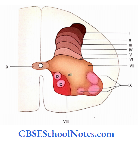
- The grey matter of the spinal cord may be divided into 10 laminae (layers) based on the cytoarchitectonic organization (i.e. shape, size, and packing density of neurons).
- Though Rexed studied these laminae in the spinal cord of cats, however, it is believed that a similar lamination exists in all mammals including man.
- These laminae are designated by Roman numerals I to X. Laminae I to IX are designated starting from the tip of the posterior horn and moving towards the ventral horn.
- Laminae I to 4 occupy the dorsal grey horn, lamina VII occupies the intermediate grey matter (between dorsal and ventral grey horns) while laminae 8 and 9 are present in the ventral grey horn. Lamina X is the grey matter surrounding the central canal.
- The nuclei of the ventral, dorsal, and lateral grey horns are summarised.
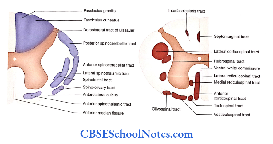
White Matter
The white matter of the spinal cord is organized into three broad areas called funiculi (or columns).
These are anterior (ventral) funiculus, lateral funiculus and dorsal (posterior) funiculus.
Each funiculus consists of distinct tracts (a tract is defined as bundles of nerve axons having a common origin, termination, and carrying similar impulses).
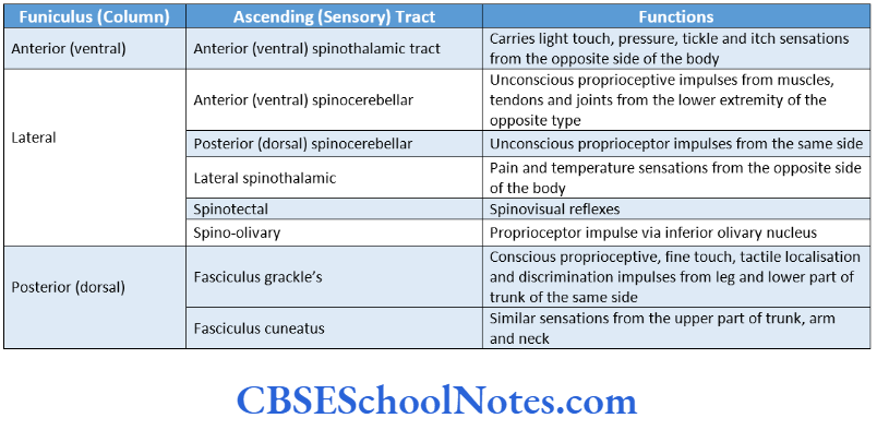
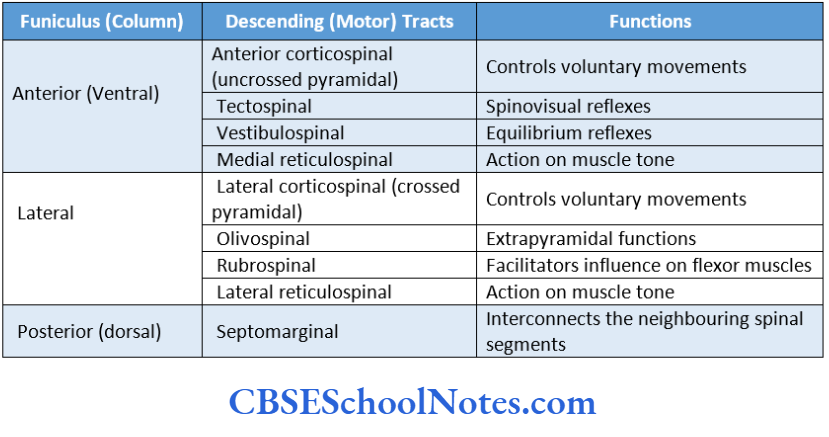
- In the white matter of the spinal cord, tracts are mainly of three types. These are sensory or ascending tracts, motor or descending tracts, and intersegmental tracts.
- Sensory (ascending) tracts carry sensory impulses from the spinal cord to the brain, for example spinothalamic tract.
- The descending (motor) tracts carry motor impulses from the brain to the spinal cord, for example, pyramidal and extrapyramidal tracts.
The intersegmental tracts run for short distances; that is, they remain confined within a few segments of the spinal cord.
They connect the neurons within segments or of different segmental levels.
Dorsal Funiculus
- The dorsal (posterior) funiculus is bounded medially by the posteromedian septum and laterally by the dorsal grey column. It consists of medially placed gracile and laterally placed cuneate fasciculi.
- The gracile fasciculus extends throughout the length of the spinal cord but the cuneate fasciculus is present only above the level of the midthoracic region. Both the gracile and the cuneate asciculi are sensory.
Lateral Funiculus
The lateral funiculus is present between dorsal and ventral grey columns (between ventral spinal roots and posterolateral sulcus). It consists of both motor (descending) and sensory (ascending) tracts.
Ventral Funiculus
The ventral (anterior) funiculus is present between the anterior median fissure and the lateral-most fibers of the ventral roots.
Axons decussate in the ventral white commissure. It also consists of both motor (descending) and sensory (ascending) tracts. Enumerate the ascending and descending tracts in various funiculi of the spinal cord.
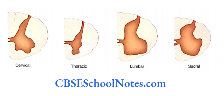
Regional Differences In The Arrangement Of White And Grey Matter
- Some regional differences are observed when transverse sections of the spinal cord are examined in different regions.
- The amount of white matter gradually increases from the sacral to the cervical region (below upwards) of the spinal cord. The white matter is the maximum in the cervical part of the spinal cord.
- The amount of grey matter is much higher in the cervical and lumbosacral regions (enlargements). The neurons of these enlargements are responsible for the innervation of limbs.
- The grey matter is much less in the thoracic region than at each segmental level. It mainly innervates the intercostal space only.
Spinal Cord Function And Structure
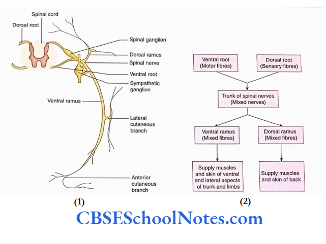
Formation Of A Typical Spinal Nerve
A typical spinal nerve arises from the spinal cord in the form of two roots: Ventral and Dorsal.
Ventral Root
The ventral root (anterior root) contains bundles of efferent (motor) fibers, which arise from the motor neurons situated in the anterior grey column. These fibers supply the skeletal muscles.
Dorsal Root
- The dorsal root (posterior root) contains bundles of afferent (sensory) fibers.
- These fibers arise from the nerve cells present in the dorsal root ganglion. The dorsal root ganglion cells are pseudo-unipolar.
- ‘The cell body of these neurons gives a single process, which divides into central and peripheral processes.
- Hie peripheral process goes peripherally through the spinal nerve to supply the tissues while the central process passes through the dorsal root to the dorsal grey column of the spinal cord.
The trunk of the Spinal Nerve
The dorsal and ventral nerve roots unite to form the trunk of the spinal nerve. Within the trunk of the spinal nerve, both sensory and motor fibers are present.
Branches
The trunk of the spinal nerve immediately divides into two rami: dorsal and ventral. These rami soon come out of the intervertebral foramen. Both the rami contain mixed fibers (motor and sensory).
Spinal Cord Function And Structure
Ventral Ramus
- The ventral ramus and its branches supply motor and sensory nerve fibers to the anterior and lateral regions of the trunk and the upper and lower limbs. The ventral rami is larger than dorsal rami.
- They supply the limbs and anterolateral aspect of the trunk (thorax and abdomen). In the thoracic region, ventral rami are known as intercostal nerves.
- They run independently and retain their segmental innervations. However, the ventral rami of the cervical, lumbar, and sacral regions form nerve plexuses by uniting with each other. In the cervical region, they form cervical and brachial plexuses.
- The brachial plexus supplies the muscles of the upper limb. The lumbar and sacral ventral rami form lumbosacral plexuses which supply the muscles of the lower limb.
Dorsal Ramus
The dorsal ramus and its branches carry motor fibers to the deep muscles of the back and sensory fibers to the overlying skin. The dorsal rami of spinal nerves supply the dorsal aspect of the body.
They are usually smaller than the ventral rami. Similar to thoracic ventral rami (intercostal nerves), dorsal rami also have segmental distribution.
Spinal Cord Function And Structure
The dorsal rami do not form nerve plexuses. A dorsal ramus usually divides into medial and lateral branches and supplies muscles and skin of the posterior region of the neck and trunk.
Spinal Cord Summary
- The spinal cord is a long cylindrical structure present in the upper two-thirds of the vertebral canal. The vertebral canal is well protected by bony vertebrae, meninges (dura, arachnoid, and pia), and a cushion of cerebrospinal fluid.
- The upper end of the spinal cord lies at the level of the upper border of the first cervical vertebra while the lower end lies at the lower border of the LI vertebra. The length of the cord varies from 42 to 45 cm.
- On the external surface of the spinal cord, we get the following fissures and sulci: one anterior median fissure, two anterolateral sulci, one posteromedian sulcus, and two posterolateral sulci.
- The spinal cord shows the fusiform enlargement in the cervical and lumbosacral regions. Just below the lumbosacral enlargement, the lower end of the spinal cord is tapering and is known as conus medullaris.
- Filum terminate is a slender filament, which extends between the tip of the conus medullaris and the dorsum of the first coccygeal vertebra.
- Thirty-one pairs of spinal nerves are attached to the spinal cord, i.e. 8 cervical, 1 2 thoracics, 5 lumbar, 5 sacral, and 1 coccygeal.
- The transverse section of the spinal cord shows that it is composed of grey and white matter. The grey matter is placed centrally and the white matter is on the periphery.
- The grey matter of the spinal cord, on each side, is subdivided into anterior (ventral), posterior (dorsal), and lateral grey columns (horns).
- Various nerve cell nuclei in the spinal grey columns (horns) are presented,
- The white matter of the spinal cord is organized into three broad areas called funiculi (or columns). These are anterior (ventral) funiculus, lateral funiculus and dorsal (posterior) funiculus.
- Each funiculus consists of distinct tracts, i.e. sensory or ascending tract, motor or descending tract, and intersegmental tract.
Spinal Cord Multiple Choice Questions
Question 1. The spinal cord extends from the upper border of the atlas vertebra to
- The lower border of S2
- Lower border of
- Lower border ofL3
- Lower border ofL2
Answer: 2. Lower border of
Question 2. At birth, the spinal cord usually lies at the level of
- The lower border of S2
- The lower border of SI
- The lower border of LI
- The lower border of L3
Answer: 4. Lower border of L3
Question 3. Which ofthe following statements about the spinal cord is false?
- The anteromedian sulcus is present in the midline anteriorly
- Anterolateral sulci are present on either side of the midline
- Posterolateral sulci are present on either side of the posteromedian sulcus
- Posteromedian sulcus is present in the midline posteriorly
Answer: 1. Anteromedian sulcus is present in the midline anteriorly
Question 4. Which ofthe following statements about filum terminale is false?
- It extends from the tip of the conus medullaris to the first coccygeal vertebra
- It consists of pia mater surrounding the neuroglial element
- It is a vestigial structure
- It lies above Cauda equine
Answer: 4. It lies above cauda equine
Question 5. Which ofthe following statements about spinal nerves is false?
- There are 33 pairs of spinal nerves
- There are 7 pairs of cervical spinal nerves
- There are 3 pairs of coccygeal nerves
- All of the above
Answer: 4. All of the above
Question 6. All of the following statements are correct regarding the grey matter of the spinal cord except
- The anterior grey horn contains cell bodies of motor neurons
- The posterior grey horn contains cell bodies of sensory neurons
- The lateral grey horn contains postganglionic autonomic motor neurons
- The grey matter of the spinal cord contains interneuron
Answer: 3. Lateral grey horn contains postganglionic autonomic motor neurons
Question 7. Which nerve cell group is not present in a large part of the posterior grey column?
- Marginal nucleus
- Substantia gelatinosa
- Nucleus properties
- Clarke’s column
- Interomediolateral nucleus
Answer: 5. Interomediolateral nucleus
Question 8. Which is not the major ascending tract of the spinal cord?
- Ventral spinothalamic tract
- Pyramidal tract
- Fasciculus gracilis
- Lateral spinothalamic
Answer: 2. Pyramidal tract
Question 9. Sensation of touch is carried by
- Lateral spinothalamic tract
- Anterior spinothalamic tract
- Posterior column tracts
- Anterior spinothalamic and posterior column tract
Answer: 4. Anterior spinothalamic and posterior column tract.