Respiratory System
The respiratory system consists of the nose, pharynx larynx, trachea and lungs.
Respiratory System is concerned with the following functions:
- Perception of smell: The olfactory mucosa of the nasal cavity contains receptors for the sense of smell
- Filtration of inhaled air: The upper respiratory mucosa of the nasal cavity contains receptors for the sense of smell.
- Phonation: The Larynx helps in the production of sounds. The larynx also prevents the entry of solid food, liquid and foreign bodies into the trachea and lungs
- Respiration: Exchange of gases, i.e., intake of O and elimination of ofCOr This process of exchange of gases is called respiration.
- Maintenance of blood pH: It participates in the regulation of blood pH.
Functionally, the respiratory system consists of two factors:
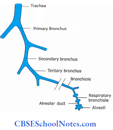
- Conducting part: It conducts the air from nose to lung and consists of the nasal cavity, pharynx, larynx, trachea, bronchi, bronchioles and terminal bronchioles. The trachea bronchi and bronchioles are branching systems of tubes The conducting part also filters, warms and moistens the while passing through it
- Respiratory part: It is located strictly within the lungs and consists of spongy respiratory tissue of the lungs across which blood and air exchange their gases.It consists of respiratory bronchioles, alveolar ducts, alveolar sacs and alveoli
The Conducting Part Of The Respiratory System
The conducting part of the respiratory system consists of the
- Nasal cavity,
- Nasopharynx
- Larynx
- Trachea and Primary bronchi
- Intrapulmonary bronchi and
- Bronchioles.
Histology of Nasal Cavity, Nasopharynx and Larynx:
1. Nasal Cavities
The nasal cavities repaired chambers separated by septum. Each nasal cavity is a hollow organ composed of bone, cartilage and connective tissue covered by mucous membrane. The nasal cavity chamber is divided into three regions, i.e., nasal vestibule, respiratory region and olfactory region.
Nasal vestibule:
This is the most anterior dilated part. the nasal cavity. It is situated inside the nostril and lined by hair skin These hair filter large particles from in Spired air
Respirators- region:
It is the largest part of the nasal cavity. It is occupied by the lower 2/3 part of the nasal cavity. The respiratory portion of the nasal cavity is lined with pseudostratified ciliated columnar epithelium with goblet cells.
- Underlying this epithelium, aminapropria contains ser°us mucus-secreting glands, large venous plexus, lymphatic tissue collection, plasma cells and macrophages
- Cilla on tall colour cells act as filter and their beatings carry foreign matter and mucus towards the oropharynx where they are swallowed’ The goblet cells Produce mucus to trap foreign particles and keep the membrane moist
- The cells in lamina propria (lymphocytes, plasma cells macrophages, etc) serve aprotective function.
- The large venous plexus warms the air, and seromucous glands provide the moisture and mucus.
- Thus, while passing through the nasal cavity, the air is conditioned, i.e., it is wanned, moistened and freed of dust particles.
- The upper respiratory tract is exposed to a wide variety of pathogens in the process of warming, humidifying and filtering the inspired air.
- These organisms may lodge and grow in the host This may lead to an upper respiratory infection (sore throat tonsillitis and infection of paranasal air sinuses)
Respiratory system Remember:
The inhaled air is conditioned before reaching the lung alveoli. This is achieved by warming, humidifying and filtering the inspired air while it is passing through the conducting part of the respiratory system.
Olfactory region:
It is occupied by the upper 1/3 part of the nasal cavity near its apex. It is lined by specialized olfactory mucosa. The total area of olfactory mucosa is only about 1 0 cm² area. The epithelium of olfactory mucosa is pseudostratified.
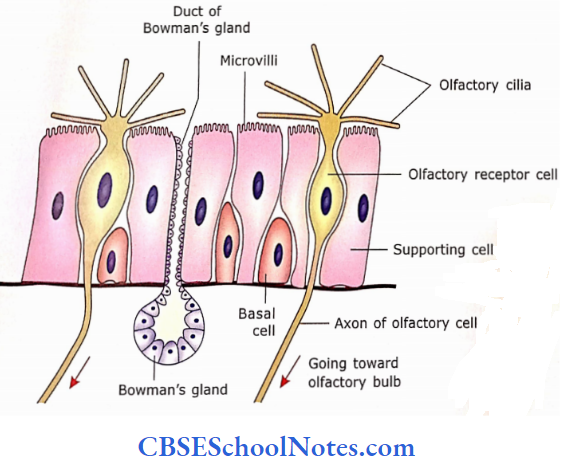
Composed of the following types of cells:
- Olfactory receptor cells: These are modified bipolar neurons that send axons to the olfactory nerve. Their apical portion bears cilia (hair).
- These cilia are involved in olfactory transduction pathways.
- Cilia have numerous odour receptor molecules.
- The molecules of odoriferous substance dissolved in serous fluid bind to the specific receptor molecules.
- This generates the action potential and information is passed to the brain
- Supporting cells: These are long columnar cells which provide mechanical support to olfactory receptor cells
- Basal cells: These are present near the basement membrane. They differentiate into receptor and supporting cells
- Olfactory glands (Bowman’s glands): These are present in the lamina propria. They deliver their proteinaceous secretion via ducts onto the olfactory surface
Olfactory Remember:
The olfactory mucosa is pseudostratified and composed of olfactory receptor cells, supporting cells and basal cells. Olfactory glands (Bowman’s glands) are present in the lamina propria.
2. Nasopharynx
It lies between the nasal cavity and the oropharynx. The nasopharynx is also covered with pseudostratified ciliated columnar epithelium goblet cells.
3. Larynx
It begins with the epiglottis and ends at the trachea. Most ofthe parts ofthe larynx are also covered by pseudostratified ciliated columnar epithelium with goblet cells. Lamina propria at places may contain seromucous glands and lymphatic tissue.
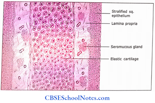
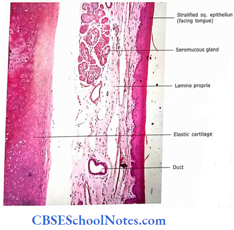
Epiglottis:
The epiglottis consists of a plate of elastic cartilage in the middle. On both the sides of cartilage, there is the presence of lamina propria. Lamina propria contains seromucous glands. The anterior surface (facing tongue) and upper part of the posterior surface of the epiglottis are lined by stratified squamous epithelium.
The remaining pan of the posterior surface is lined by pseudostratified ciliated columnar epithelium with goblet cells.
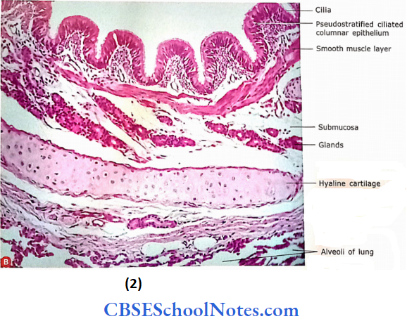
4. Trachea and Primary Bronchi
The histological structures of the trachea and primary bronchi are essentially the same. The trachea is a flexible tube measuring about 11 cm in length and 2 cm in diameter. Its wall contains 1 6-20 C-shaped hyaline cartilages, which keep the lumen of the tube patent. The gap between the free ends of a C is bridged by a muscle called the tracheal
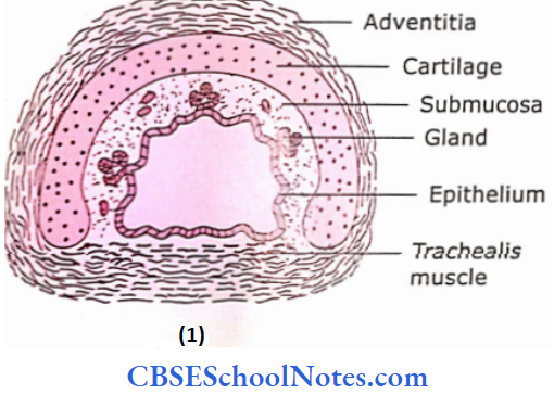
The trachea divides into two principal (or primary) bronchi The trachea and primary bronchi consist of four layers.
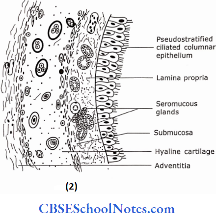
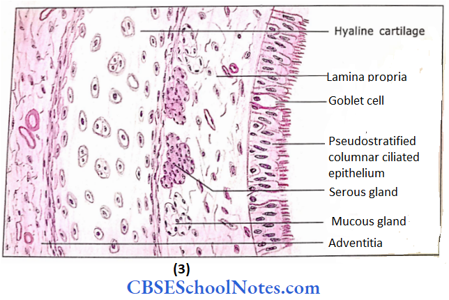
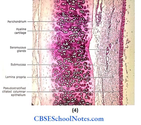
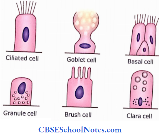
- Mucous membrane (epithelium and lamina propria): The epithelium is a pseudostratified ciliated columnar with goblet cells. The epithelium rests on thick basal lamina. Three different types of common cells of epithelium are ciliated cells (30%), goblet cells (28%) and basal cells (29%).
- The ciliated cells propel the mucus upwards and their percentage increases from the upper to the lower trachea.
- The goblet cells produce mucus. The basal cells are small pyramidal cells situated near the basal lamina.
- They act as stem cells that can divide and differentiate to replace other cell types
- The other two less common cells are granule cells Kulchitsky cells) and brush cells.
- The small granule cells are situated at the base ofthe epithelium and contain dense secretory granules in an intranuclear position.
- These cells discharge their contents in response to hypoxia. The brush cells contain large microvilli with an actin filament core extending down into the cytoplasm.
- The nerve terminals are seen in these cells. These cells contain no secretory granules and may have sensory functions
- The lamina propria of the trachea and principal bronchi is made up of loose connective tissue, which is rich in elastic fibres.
- It may also contain lymphatic tissue, in both diffuse and nodular forms.
- Submucosa: In the trachea, it is difficult to distinguish the boundary between lamina propria and submucosa.
- At the junction of the two, there is a dense layer of elastic fibres, which is seen with a special stain.
- The submucosa contains mixed seromucous glands whose ducts open onto the surface epithelium
- Cartilage and smooth muscle layer: This layer consists of a hyaline cartilaginous ring separated by interspaces bridged by fibroblastic connective tissue.
- The rings of cartilage are incomplete posteriorly.
- The gap is filled by smooth muscle (tracheal) and by fibroblastic tissue.
- Adventitia: External to the cartilaginous ring, there is a layer of connective tissue that is rich in elastic fibres.
Trachea and primary branch Remember:
The trachea and primary bronchi consist of four layers, i.e., mucosa (lined by pseudo-stratified ciliated columnar epithelium with goblet cells resting on lamina propria) submucosa (contains mixed seromucous glands, cartilage and srn°°th muscle layer and adventitia.
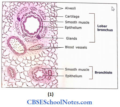
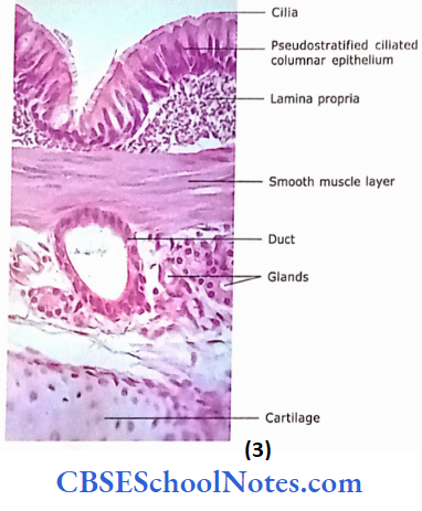
5. Intrapulmonary Bronchi
The primary bronchi enter the lung through the hilum. In the lung, primary bronchi branch into lobar bronchi, which in turn divide into segmental bronchi. The segmental bronchi branch many times as they penetrate further into the lung, resulting in many orders of decreasing diameter.
Following are the layers observed in the intrapulmonary bronchi:
- Mucosa: The epithelium is pscudoslrati tied ciliated columnar with goblet cells. The lamina propria contains many clastic fibres and seromucous glands
- Smooth muscle layer: The smooth muscle layer is present in lamina propria beneath the epithelium Smooth muscle is arranged in bundles, which run spirally. Elastic fibres arc intermixed with muscle
- Submucosa: It consists of the loose connective tissue in which glands are embedded.
- Cartilage layer: In contrast to C-shaped cartilage present in the trachea, the intrapulmonary bronchi contain irregular plates of cartilage. These cartilaginous plates surround the bronchi and keep them open. Therefore, intra-pulmonary bronchi are always cylindrical rather than flattened on one side as arc the trachea and primary bronchi.
- Adventitia: Outside the cartilage layer adventitia is made up of the connective tissue layer
As the bronchi divide repeatedly and become smaller in size each component of the wall becomes thinner and sparser, except the layer of smooth muscle, which remains distinct The submucosal glands gradually decrease in number and end at the level ofthe bronchiole.
Intrapulmonary Bronchi Remember:
In contrast to the trachea, bronchi contain circumferentially arranged smooth muscle layers in lamina propria and irregular plates of cartilage
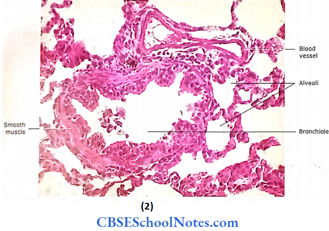
6. Bronchioles
Bronchioles are defined as conducting tubules that are less than 1 mm in diameter and their wall no longer contain The bronchioles further divide to end in the terminal bronchioles, which are the terminal segments ofthe conducting portion of the respiratory tract. Terminal bronchioles subdivide into microscopic branches called respiratory bronchioles.
Following are the layers observed in bronchioles:
- Mucosa: The type of epithelium varies with the size of the bronchiole. Large bronchioles have simple ciliated columnar cells with very few goblet cells.
- The smaller bronchioles have simple cuboidal ciliated cells with no goblet cells. The Clara cells replace the goblet cells.
- These cells are non-ciliated but have many microvilli on their apical surface.
- They first appear in small bronchi but greatly increase in number of terminal bronchioles.
- Clara cells are considered to secrete a lipoprotein, which forms a protective layer of surface active agents throughout the lower respiratory tract.
- Surface active agent reduces the surface tension of the alveolar fluid, which reduces the tendency of alveoli to collapse. It also prevents the adhesion between walls forming a lumen of air passage, especially during expiration.
- Clara cells also produce a 16-kilodalton protein known as Clara cell secretory protein (CC16). Seromucous glands are not present in the lamina propria of bronchioles.
- Smooth muscle layer: It is a prominent layer and a major component of the wall. The smooth muscles are supplied with sympathetic and parasympathetic nerves.
- Normally, the muscle layer regulates the passage of air through it.
- But sometimes the hyperirritability of the respiratory tract may lead to the excessive contraction of smooth muscle
- This results in the constriction of bronchi and bronchioles and may cause difficulty in breathing. This condition may arise in a person suffering from asthma
- The connective tissue layer:
- Outside the muscle coat, the bronchioles are surrounded by lung alveoli.
- The bronchioles and lung alveoli are connected with the connective tissue layer, which is rich in elastic network
Bronchioles Remember:
Bronchioles are less than 1 mm in diameter. Cartilage plates and glands are absent. However, Clara cells are present in their epithelial lining.
Differences between bronchus and bronchiole:
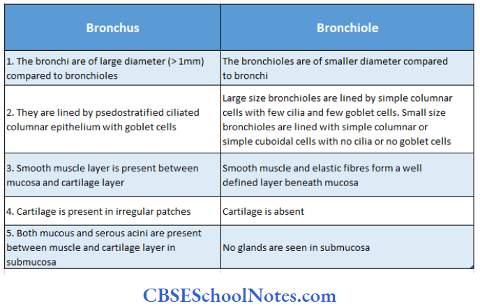
Conducting part of respiratory system Clinical Applications
Asthma
Asthma is an acute, episodic, airway obstruction that results from an allergy to a variety of substances (such as pollen, house dust, mites, moulds or a particular food).
- A person suffering from asthma tends to develop bronchospasm, which leads to obstruction of airways and air trapping.
- The bronchospasm (airway obstruction) may be due to smooth muscle spasm in the wall of smaller bronchi and bronchioles, increased secretion of mucus and swelling of mucosa
- A person suffering from asthma has difficulty in breathing, tightness in the chest, cough and anxiety.
- Asthma is treated with drugs, which relaxes smooth muscles in the bronchioles and opens up the airways.
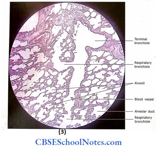
The Respiratory Part Of Respiratory System
Respiratory Bronchioles, Alveolar Ducts and Alveolar Sacs and Alveoli
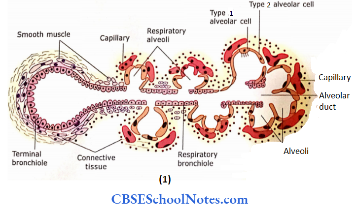
1. Respiratory Bronchioles
- Respiratory bronchioles are the site of transition from the conducting part to the respiratory part.
- It is called respiratory bronchiole because it is partly respiratory in function
- A respiratory bronchiole usually measures less than 0.5 mm in diameter
- The respiratory alveoli are present in its wall as outpouching.
- The epithelium is simple cuboidal with cilia, which are gradu¬ ally lost and are replaced by non-ciliated low cuboidal cells
- The Clara cells are also present in between cuboidal cells
- A smooth muscle layer is still present outside the epithelium
- Outermost covering is ofdelicate connective tissue.
2. AlveolarDucts and Alveolar Sacs
Each respiratory bronchiole ends by dividing into 2 or 3 alveolar ducts. Many alveoli and alveolar sacs open into the alveolar duct. At the end of the alveolar duct, the clusters of alveoli share a common opening to the alveolar duct. This cluster of alveoli is called as alveolar sac.
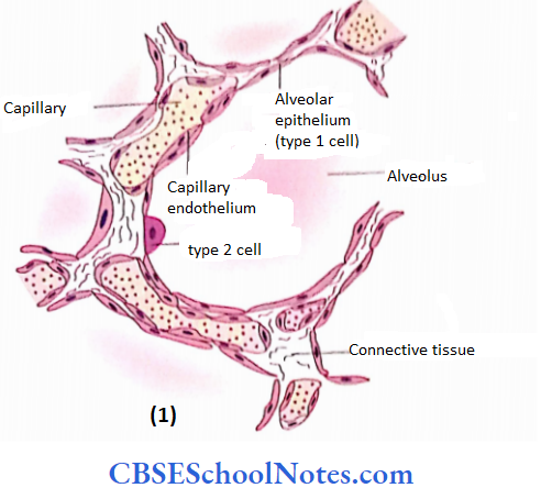
3. Alveoli
These are thin-walled polyhedral sacs, one side of which is always lacking. Alveoli are round or polygonal in shape and about 200 pm in diameter.
- They consist of pure respiratory surfaces across which gaseous exchange occurs.
- Alveoli are closely packed, so the alveolar wall is a partition 2 present in their epithelial lining.
- Remember or septum between two alveoli. The interalveolar septa contain a network of capillaries, supported by reticular and elastic fibres, and occasionally fibroblasts and macrophages between the squamous epithelium of adjacent alveoli.
There may be openings in septa, which are called pores. These pores help in the circulation of air from one alveolus to another, thus equalizing the pressure in the alveoli. The alveolar pores play another important role in case of blockage of air passage.
The alveoli distal to the blockage may get air from the alveoli of neighbouring lobules. Wherever the endothelium of the capillary comes in contact with the alveolar epithelium no connective tissue lies in between the two.
Respiratory bronchioles Remember:
Respiratory bronchioles are partly respiratory in function where the exchange of gases occurs. Alveolar duct, atria and alveoli are supplied by rich capillary network and are the site of gas exchange
The blood-air barrier:
The air in the alveolus is separated from the blood in the capillary by following three structures
- Continuous non-fenestrated endothelium of capillary.
- Squamous epithelium lining the alveolus.
- The fused basal lamina of endothelium and epithelium
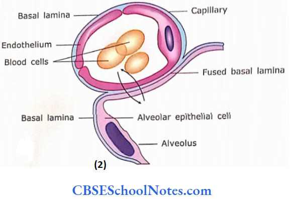
This thin (1.5 – 2μm) blood-air barrier facilitates the diffusion of oxygen and carbon dioxide.
4. Cells of Alveolar Wall (septum)
The alveolar wall (septum) is covered with the following types of epithelial cells
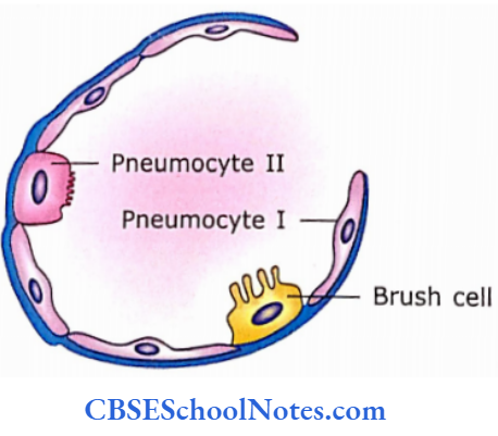
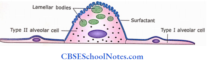
Type 1 alveolar cells (squamous epithelial cells/pneumocyte 1):
These are very thin squamous cells. These cells cover over 95% ofthe alveolar surface
Type 2 alveolar cells (great alveolar cells/ pneumocyte):
These are large rounded cells, which bear microvilli on their apical surface. These cells are placed in between type 1 cells and the remaining 5% surface area of alveoli.
- Type 2 alveolar cells are secretory and secrete the pulmonary surfactant, which lowers surface tension and prevents alveoli from collapsing during expiration.
- When viewed under electron microscope an electron microscope the secretory granules of these cells show a peculiar appearance.
- These granules contain membrane-like layers arranged parallel one upon the other.
- These layers are made up mainly of phospholipids and proteins (precursors of surfactants). The surfactant is secreted continuously by cytosis.
- This kind of secretory granules is called s. Type 2 alveolar cells also give origin to type 1 alveolar cells. After lung injury, they give origin to type 1 and type 2 cells within lung alveoli.
- Besides secreting lipoprotein surfactant, type 2 cells also produce various types of proteins (surfactantprotein A, B, C and D). These proteins modulate alveolar immune responses.
Type 3 alveolar cells (brush cells):
- These cells are found only occasionally in the alveolar epithelium. These cells serve as receptors to monitor the quality of air entering the lung
- All cells forming the alveolar epithelium are joined to each other by zonula occludens.
Alveolar Remember:
95% of the alveolar wall is covered by type 1 pneumocytes, which are involved in the exchange of gases. The remaining 5% of the alveolar wall is covered by type 2 pneumocytes involved in the secretion of surfactant. Brush cells are also found occasionally in the alveolar epithelium. Brush cells serve as receptors to monitor the quality of air entering the lungs.
5. Alveolar Macrophage
These cells are found in the connective tissue of interalveolar septa. Some of these cells enter the connective tissue from blood and pass through the alveolar epithelium to reach into the lumen alveoli. Alveolar macrophages are the first line of defence against pulmonary infection.
They act in the following way:
- They phagocytose dust particles, which may be seen in their cytoplasm.
- Hence, sometimes they are also called dust cells.
- In cigarette smokers, they engulf carbon and tar
- They are also capable of phagocytosing bacteria In patients of heart failure (here pulmonary capillaries are overloaded with blood), erythrocytes may accumulate in alveoli.
- These erythrocytes are phagocytosed by macrophages, which acquire a brick-red colour because of the presence of haemosiderin (a product of haemoglobin degradation and are then called as heart failure cells.
The respiratory part of the respiratory system Clinical Application
Pneumonia
- It is an acute infection of the lung alveoli. Pneumococcal pneumonia is the most common bacterial pneumonia and represents a threat to the aged and chronically.
- When microorganisms enter the lungs, they produce toxins which in turn stimulate inflammation and immune response.
- This leads to a collection of exudates, plasma proteins, RBCs, fibrin and leucocytes in the alveoli.
- This interferes with ventilation and gas exchange.
- The clinical signs include fever, cough, chest pain, blood-streaked sputum and difficulty in breathing. Pneumonia is treated with antibiotics
Cigarette Smoking and Cancer of Lung
The association between cigarette smoking and cancer of the lung is well known.
- Although the bronchial tree is mainly lined by pseudostratified ciliated columnar epithelium, it changes to stratified squamous epithelium in chronic smokers because of the presence of toxic elements in smoke. (The epithelial alteration from one kind to another is known as metaplasia.)
- This transformation is the first step in the development of squamous cell carcinoma, which is the main type of cancer of the lung.
Different Types of Ceils ofRespiratory System and their functions
1. In the epithelium of the trachea, bronchus and bronchiole:
- Ciliated cells: Beating oftheircilia moves the mucus upward
- Goblet cells: These cells produce mucus, which traps the dust particles
- Basal cells: They multiply and give rise to another type of cell
- Granule cells: They are believed to be endocrine cells.
- Brush cells: Sensory
- Clara cells: Produce secretion, which reduces the surface tension in alveoli
2. In the epithelial lining of lung alveoli:
- Type 1 alveolar cells (pneumocytes 1/squamous cells): Exchange of gases occurs through these cells
- Type 2 alveolar cells (pneumocytes 2): These cells produce pulmonary surfactant, which prevents collapse ofthe alveolus during expiration.
- Brush cells: They are considered to be sensory cells.
3. About the lumen of lung alveoli:
Macrophages: They phagocytose dust particles (dust cells) or erythrocytes (heart failure cells).
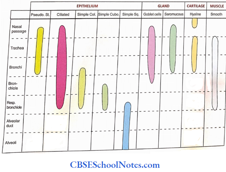
Distribution of epithelium in various parts of respiratory passage: