
Olfactory System
The olfactory system consists of the following structures:
- Olfactory epithelium with olfactory nerves
- Olfactory bulb, tract and striae
- Primary and secondary olfactory cortices
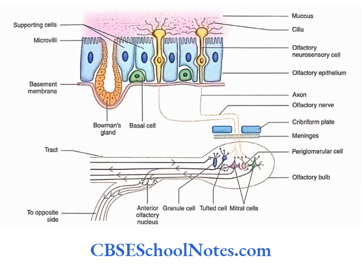
Olfactory Epithelium And Olfactory Nerve
- The olfactory epithelium is present on the roof and back of the nasal cavity.
- The olfactory neurosensory cells of the olfactory epithelium are modified bipolar neurons.
- From the apical pole of each olfactory sensory cell (neuron), a single dendrite runs towards the epithelial surface.
- From each dendrite, about 5-20 thin cilia protrude on the surface while from the basal pole of this sensory neuron, a single axon projects.
- These axons are collected to form about 15-20 olfactory nerves.
- These nerves reach the olfactory bulb through the cribriform plate ofthe ethmoid. The olfactory nerves end in the cells of the olfactory bulb.
Read and Learn More Neuroanatomy
Olfactory Bulb, Tract And Striae
- The olfactory bulb is a small, oval structure that lies above the cribriform plate of ethmoid.
- In the olfactory bulb, the incoming sensory axons synapse with the dendrites of olfactory bulb neurons (mitral cells, tuft cells, and periglomerular cells).
- The mitral cells and tuft cells are the principal cells and their axons form the olfactory tract. The periglomerular and granule cells are interneurons of the olfactory bulb.
- Millions of axons of olfactory sensory receptor cells terminate in the synaptic unit called glomeruli.
- Each glomerulus receives many afferent neurons, which synapse with the dendrites of a few principal cells (mitral and tufted cells).
- The activity of principal cells is modified by inhibitory interneurons of olfactory bulbs (granule cells and periglomerular cells).
- The axons of mitral and tufted cells run in the olfactory tract. They also send collateral branches to the neurons of the anterior olfactory nucleus.
- The fibers that originate in the anterior olfactory nucleus pass through the anterior commissure to the opposite olfactory bulb.
- The fibers that go to the opposite olfactory bulb synapse with the dendrites of interneurons. Sensory information is likely to be extensively processed and refined in the olfactory bulb before it is sent to the olfactory cortex.
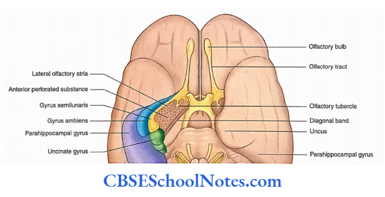
- The olfactory bulb is continuous posteriorly with the olfactory tract and expands into the olfactory triangle at the anterior end of the anterior perforated substance.
The olfactory tract divides into two roots:
- Lateral and Medial olfactory striae. The lateral stria runs posterolaterally on the margin of the anterior perforated substance and carries most of the axons of the tract. These fibers enter the gyrus semilunaris, which lies anterior to the uncinate gyrus.
- The fibers of the medial olfactory stria probably terminate in the anterior perforated substance and the paraterminal gyrus. It is a rudimentary stria.
- The intermediate olfactory stria is not always present. It ends in the olfactory tubercle in the anterior perforated substance.
Primary Olfactory Cortex
The primary olfactory cortex is that region of the cerebral cortex that is responsible for conscious awareness of olfactory stimuli.
The primary olfactory cortex receives direct afferents from the lateral olfactory stria. Olfaction appears to be unique in the sensory system as the sensations reach the primary cortex without relaying in the thalamus.
However, when the primary olfactory cortex projects to the secondary cortex, information reaches through the thalamus. The primary olfactory cortex includes the following
Lateral olfactory stria, when traced backward, ends in gyrus semilunaris, which is present in front of the uncinate gyrus.
The lateral olfactory gyrus covers the lateral olfactory stria It is continuous posteriorly as the gyrus ambiance. Gyrus ambiens lies lateral to gyrus semilunaris. The dorsomedial part of the amygdala
The anterior part of the parahippocampal gyrus includes uncus (uncinate gyrus). It is included in the entorhinal area (Brodmanns area 28).
Secondary Olfactory Cortex
- The lateral part of the orbital surface of the frontal lobe is the olfactory association cortex.
- This part receives direct afferents from the primary olfactory area. The frontal cortex also receives indirect input from the olfactory cortex through the thalamus.
- The frontal and orbitofrontal cortices are known as the olfactory association area. The hypothalamus receives olfactory information through the amygdala.
- The emotional aspects of olfactory sensations are due to their limbic projections involving the hypothalamus and amygdala.
- Olfactory stimuli induce visceral response (salivation following pleasing aromas from food and nausea and vomiting following offensive smell) by modulating the activities of the autonomic nervous system.
Anosmia
Anosmia is defined as a lack of olfactory sensation. It is of two types:
- Specific and
- General.
Specific anosmia: Olfactory acuity varies from person to person. This may be explained as due to the absence of a specific odourant receptor on the olfactory sensory cells.
General anosmia: Complete lack of olfactory sensation Olfactory Hallucination Olfactory hallucination may be due to a lesion involving the parahippocampal gyrus, uncus, and the adjoining areas.
These olfactory hallucinations precede epileptical seizures referred to as ‘uncinate fits’.
Olfactory System Summary
- Humans can perceive thousands of different varieties of odor.
- The olfactory system consists of olfactory epithelium present in the nasal cavity, olfactory bulb, olfactory tract, and olfactory striae.
- The smell is perceived in the primary and secondary olfactory cortices of the cerebrum.
- The olfactory neurosensory cells of the olfactory epithelium can appreciate a large number of smells because there are more than 3000 different receptor proteins in their cilia.
- The axons of olfactory sensory receptor cells terminate on mitral and tuft cells in the olfactory bulb. Their axons form the olfactory tract.
The olfactory tract divides into two roots:
- Lateral and Medial olfactory striae.
- The fibers of the lateral olfactory stria end in the gyrus semilunaris, which is part of the primary olfactory cortex.
- The primary olfactory cortex is responsible for conscious awareness of olfactory stimuli.
- The secondary olfactory cortex (olfactory association cortex) receives direct information from the primary olfactory area and is located in the frontal and orbitofrontal cortices. This area is responsible for the conscious dissemination of odor.
Olfactory System Multiple-Choice Questions
Question 1. The following types of cells are present in the olfactory epithelium except.
- Olfactory neurosensory cells
- Supporting cells
- Pillar cells
- Stem cells
Answer: 3. Pillar cells
Question 2. Which of the following neurons and interneurons are present in the olfactory bulb?
- Granule cells
- Periglomerular cells
- Mitral cells
- Tuft cells
- All of the above
Answer: 5. All of the above
Question 3. Which of the following olfactory striae carry most ofthe axons of the olfactory tract?
- Lateral olfactory stria
- Medial olfactory stria
- Intermediate olfactory stria
Answer: 1. Lateral olfactory stria
Question 4. The primary olfactory cortex includes the following except
- Gyrus semilunaris
- Gyrus ambient
- The dorsomedial part of amygdalae
- Uncus
- The lateral part of the orbital surface of the frontal lobe
Answer: 5. Lateral part of the orbital surface of the frontal lobe
Question 5. The following cells synapse in ‘glomeruli’ of the olfactory bulb except
- Axons of olfactory sensory receptor cells
- Mitral cells
- Tuft cells
- Periglomerular cells
- Cells of amygdalae
Answer: 5. Cells of amygdalae