
Visual System
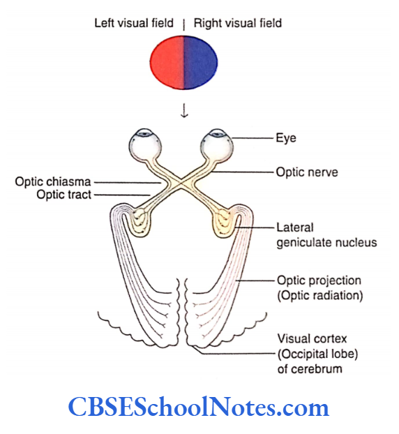
The visual system is concerned with the special sense of vision. The visual system or the optic pathway begins from the retina in the eyeball and ends in the visual cortex of the occipital lobe
Optic Chiasma
The optic nerves of two sides cross and form optic chiasma. In optic chiasma, the fibres of the optic nerve arising from the nasal half cross to the opposite side while fibres from the temporal half do not.
Optic Tract
Each optic tract consists of crossed and uncrossed axons that project from optic chiasma to the lateral geniculate body (LGB) of that side. The optic tracts curve around the midbrain before they terminate in the LGB.
Each optic tract consists of fibres from the temporal half of the retina from the same eye and the nasal half of the retina from the opposite eye.
Read and Learn More Neuroanatomy
Retina
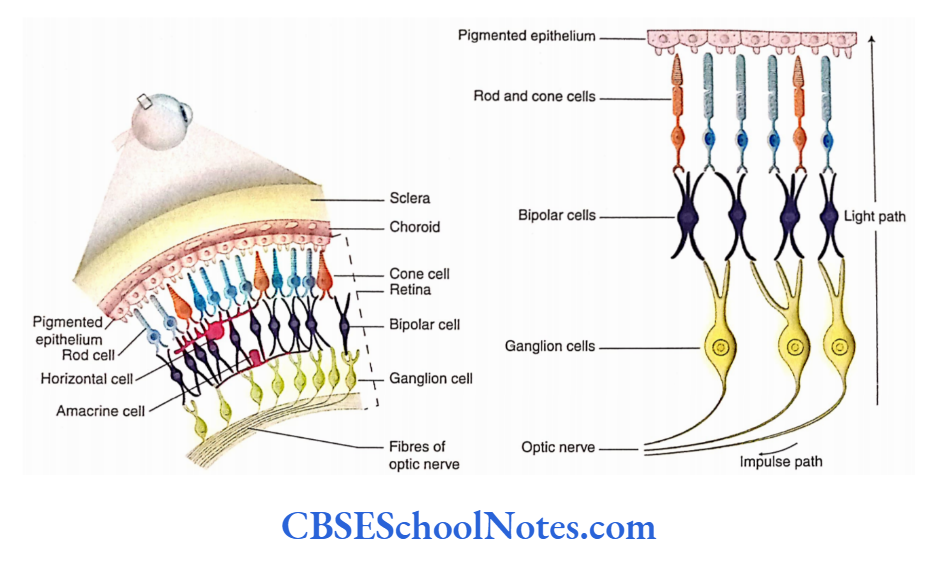
The retina is the innermost coat of the eyeball. The retina consists of a layer of pigmented epithelium, a layer of rods and cones, a layer of bipolar cells and a layer of ganglion cells.
Optic Nerve
The axons of ganglion cells form the optic nerve, and these fibres exit the eyeball at the optic disc’. As soon as optic nerve fibres come out of the sclera, they acquire a myelin sheath.
The optic nerve which has about 1 million fibres is surrounded by the meningeal layers, i.e. pia, arachnoid and dura.
The central artery and central vein of the retina are present in the anterior part of the optic nerve.
Lateral Geniculate Body
- The LGB is the main terminus for input to the primary visual cortex (striate cortex, area 17).
- The LGB is a small projection from the pulvinar part of the thalamus retina of the opposite side (crossed fibres) and terminates in layers 1, 4 and 6 while those from the ipsilateral retina (uncrossed fibres) end in layers 2, 3 and 5.
- The fibres from the temporal half of each retina terminate in the LGB of the same side while those from the nasal half terminate in the LGB of the opposite sides.
Geniculocalcarine Tract (Optic Radiation]
- The fibres arising from the LGB first traverse the sub lentiform and then the retrolentiform parts of the internal capsule.
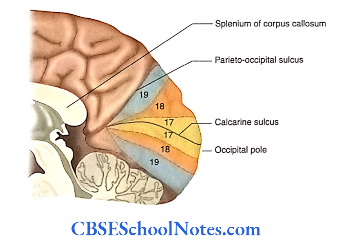
- Thereafter, these fibres terminate in the visual cortex (area 17) ofthe same side. These fibres constitute the geniculocalcarine tract or the optic radiation.
Visual Cortex
The visual cortex consists of a primary area and an association area. The association area or the association cortex is involved in the recognition of objects and perception of colours, depth and motion.
Primary Visual Cortex
- The primary area or the primary visual cortex is an area where optic impulses reach the level of consciousness The visual cortex has a representation of the retina.
- The central part of the retina is represented on the occipital pole while its peripheral part is represented at the anterior part of the visual cortex.
Visual Association Cortex
The association cortex (areas 18 and 19) is involved in the recognition of objects and perception of colours, depth and motion. This is achieved by relating the present to past visual experience.
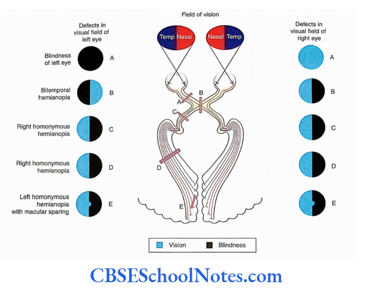
Visual Field
- The area seen by one eye, when one looks ahead with the eyes fixed, is the visual field of that eye. The visual field of each eye is divided into the nasal half and the temporal half.
- The light rays from an object situated in the temporal half of the visual field fall on the nasal half of the retina and the light rays from an object situated in the nasal half of the visual field fall on the temporal half of the retina.
- The upper half of the visual field projects onto the inferior half of the retina while the lower half of the visual field projects to the superior half of the retina.
- Therefore, damage to the upper retina will produce a deficit in the lower visual field. It should be noted that due to the presence of a convex lens in the eye, the visual image that is formed on the retina is inverted.
Visual Field Defects
- Visual field defects are characterised by the loss of a part of the normal area of vision in one or both eyes.
- The visual field defects may range from loss of area at the outer edges of vision (peripheral vision), or from a small blind spot or from a large area to complete blindness.
- Hemianopia means loss of vision in one half of the visual field. The visual defects may be caused by damage to any part of the visual pathway.
Reflexes Associated With Vision
Reflexes associated with vision include pupillary light reflex and accommodation reflex.
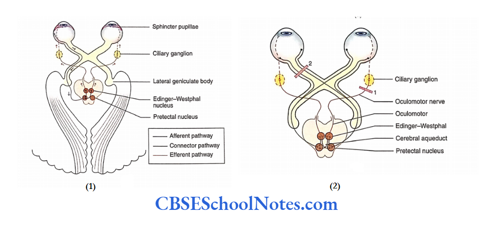
Pupillary Light Reflex
The pupillary light reflex includes both a direct response and a consensual response.
Direct Light Reflex
- When light is thrown on one eye with the help of a torch, it causes constriction of the pupil (iris) in that eye. This is known as direct response or direct light reflex.
- The pupillary constriction or the constriction of the constrictor muscles of the iris occurs due to stimulation of the Edinger-Westphal nucleus.
Consensual Light Reflex
Simultaneously with the direct light reflex, the pupil of the other eye also constricts. This is known as consensual response or consensual light reflex.
This reflex is seen because of the following two facts:
- Each retina sends afferent signals to the optic tracts of both sides. Fibres cross in the optic chiasma; these fibres later terminate in the pretectal nuclei of both sides.
- The pretectal nucleus of each side sends connections to the Edinger-Westphal nuclei of both sides.
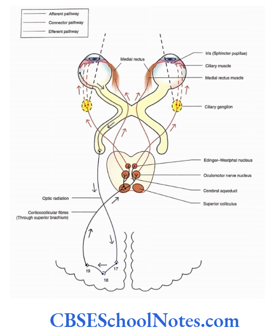
Accommodation Reflex
After looking at a distance for some time and then looking at a near object, the visual responses that are observed are given in the following text.
Ocular Convergence
When we look at a close object, our eyes must rotate medially to focus the light rays on the same corresponding points on both the retinas. This is achieved by the contraction of the medial recti muscles on both sides.
Accommodation
- As the eye focuses on close objects, the lens becomes more curved. This increase in curvature of the lens for the near vision is called accommodation.
- The accommodation is achieved through the contraction of the ciliary muscles.
- As a result of the contraction of ciliary muscles, the tension in the lens and suspensory ligaments is released and the lens therefore becomes more convex.
Constriction of Pupil
- Simultaneously with the accommodation and convergence, the pupil also constricts due to the constriction of circular muscle fibres of the iris.
- This helps to prevent the light rays from entering the eye through the periphery of the lens. This is needed to prevent blurring of vision and to achieve sharpness of image on the retina.
- The pathway of these responses observed during the accommodation reflex is illustrated.
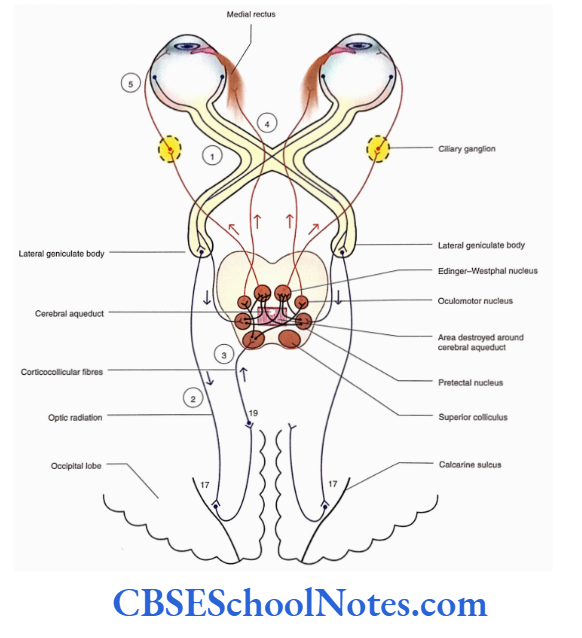
Argyll Robertson Pupil
- In certain central nervous system lesions (e.g. syphilis), the pupillary light reflex is abolished without affecting the accommodation reflex.
- This means that the constriction of the pupil after exposure to torch light gets abolished; however, constriction of the pupil occurs when the eyes are focused on a near object. The condition is known as Argyll.
- Robertson pupil. This indicates that the pathways of the light reflex and those of the accommodation reflex are different.
- In the case of light reflex, the pretectal nucleus and its axons are involved which are probably destroyed in syphilis (tabes dorsalis) due to dilatation of the cerebral aqueduct. On the other hand, the accommodation reflex involves the visual cortex and not the pretectal nucleus.
Visual System Summary
- The visual system is concerned with the special sense of vision.
- The retina contains rod and cone cells that convert stimuli of light into various electrical Impulses.
The retina consists of four layers:
- Pigment epithelial,
- Rods and cones,
- Bipolar cells and
- Ganglion.
- Rods are more sensitive to dim light while bright light stimulates cones.
- Bipolar cells are neurons connecting rods and cones with ganglion cells.
- The axons of ganglion cells form the optic nerve. The optic nerves of two sides now cross at optic chiasma to form optic tracts.
- The optic tracts curve around the midbrain and terminate in lateral geniculate bodies.
- The fibres arising from the lateral geniculate body terminate in the visual cortex (area 17) on the same side. There, fibres constitute optic radiation.
- The optical impulses reach the level of consciousness in the primary visual cortex (area 17).
- The association visual cortex (areas 18 and 1 9) is involved in the recognition of objects and perception of colours, depth and motion.
Visual System Multiple Choice Questions
Question 1. Which of the following cells are not present in the retina?
- Pigment epithelial cells
- Rods and cones
- Bipolar cells
- Ganglion cells
- Stellate cells
Answer: 5. Stellate cells
Question 2. Which of the following statements about rod cells is false?
- Each retina contains about 120 million rod cells
- Rods are absent in the central part of the fovea
- Rods are more sensitive to dim light
- Rods are involved in colour vision
- Rods are specialised for night vision
Answer: 4. Rods are involved in colour vision
Question 3. Which of the following facts about the macula lutea is false?
- It lies lateral to the optic nerve
- It lies along the visual axis of the eye
- Its central part is known as the fovea
- It contains only cone cells
- None of the above
Answer: 3. It contains only cone cells
Question 4. Which of the following statements about the optic nerve is false?
- The optic nerve is formed by the axons of ganglion cells of the retina
- The fibres of the optic nerve are myelinated
- These fibres come out of the sclera through the macula lute
- The optic nerve is surrounded by dura, arachnoid and pia mater
- The optic nerve has about 1 million fibres
Answer: 3. The optic nerve is surrounded by dura, arachnoid and pia mater
Question 5. Which of the following is not a part of the optic pathway?
- Optic nerve
- Optic chiasma
- Optic tract
- Medial geniculate body
- Geniculocalcarine tract
Answer: 3. Medial geniculate body
Question 6. Name the fibres received by the right geniculate body.
- From the temporal half of the right retina
- From the nasal half of the right retina
- From the temporal half of the left retina
- From the nasal half of the left retina
Answer: 1. From the temporal half of the right retina
Question 7. The visual association cortex (areas 18 and 19) is involved in which of the following functions?
- Recognition of object
- Perception of colour
- Depth of vision
- Motion
- All of the above
Answer: 3. Motion
Question 8. Which of the following responses is not associated with the accommodation reflex?
- Ocular convergence
- Accommodation
- Constriction of pupil
- Saccadic eye movements
Answer: 4. Saccadic eye movements