
Neuron
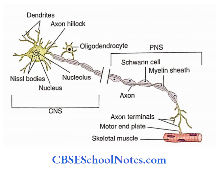
A neuron consists of a cell body or soma and neuronal processes. The neuronal processes that emerge from the cell body are called dendrites (usually multiple) and axons (single).
Nerve Cell Body Or Soma
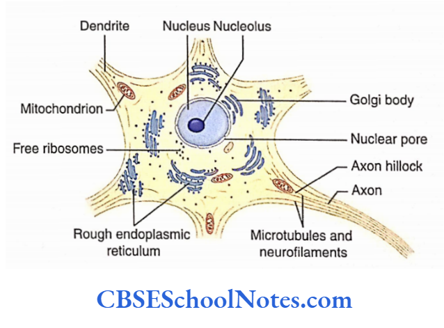
The nerve cell body has a typical pale staining euchromatic nucleus with a prominent dark nucleolus. The cytoplasm of a neuron contains prominent basophilic material called Nissl bodies.
The cell body also contains the Golgi apparatus, mitochondria, lysosomes, and smooth endoplasmic reticulum.
Normally, neurons after birth are unable to replicate their DNA and thus do not undergo cell division.
Read and Learn More Neuroanatomy
Nerve Cell Processes
The elongated cytoplasmic processes take origin from the cell body. These processes may travel long distances from the neuron. They are of two types:
Dendrites and Axon. Usually, a neuron consists of a single axon and multiple dendrites.
Dendrites
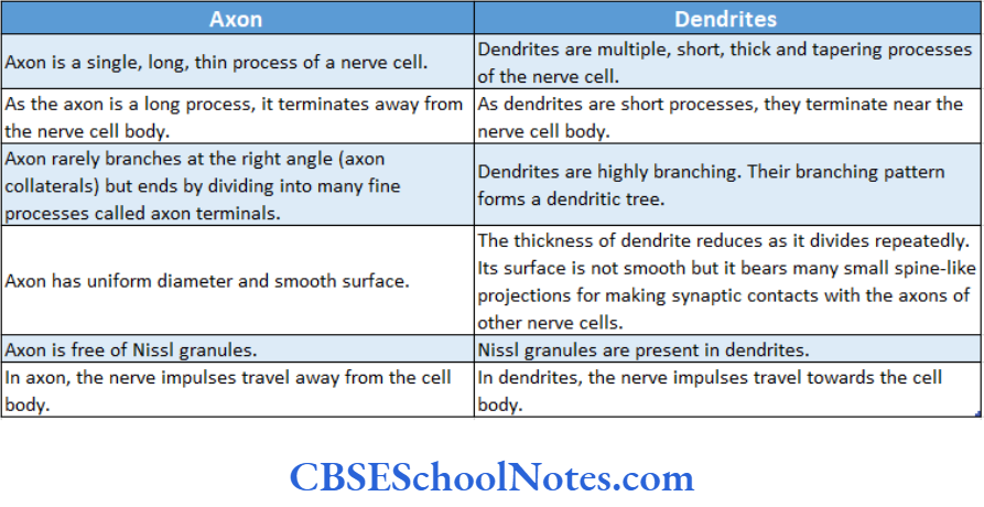
Neurons usually have short, multiple dendrites, and each of these may branch extensively to form a ‘dendritic tree’. Dendrites are involved in receiving information from other cells.
The cytoplasm of dendrites contains Nissl bodies, microtubules, microfilaments, and other organelles.
The cytoskeleton of a neuron is formed by microtubules, neurofilaments, and microfilaments.
Axons
- Axons are the nerve cell processes that send information in the form of electrical signals away from the nerve cell body to other cells.
- Usually, there is a single, extremely long axon (in some neurons, the length may reach up to 1 m) for each neuron.
- It originates at a conical region of the cell body known as the axon hillock.
3 Types Of Neurons And Functions
Classification Of Neurons
Neurons are classified based on their structure, size, and function.
Classification Based On Structure of Neurons
The structure and shape of a cell body are dependent on the number and orientation of cell processes arising from it.
Depending on the number of processes emerging from the cell body, the neurons can be classified as follows:
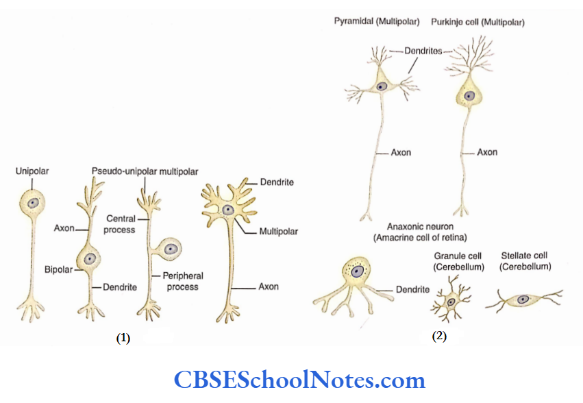
- Multipolar
- Bipolar
- Unipolar or pseudo-unipolar
Multipolar neurons: Multipolar neurons have a single axon and many dendrites extending from the cell body. Most of the neurons present in the CNS are multipolar.
Bipolar neurons: Bipolar neurons have two processes emerging from the cell body (e.g. bipolar neurons of the retina and neurons of olfactory neuroepithelium).
3 Types Of Neurons And Functions
Unipolar or pseudo-unipolar neurons: Unipolar or pseudo-unipolar neurons are found in the dorsal root ganglion of spinal nerves (sensory ganglion) and sensory root ganglia of cranial nerves.
Such a neuron has a single process that extends from the cell body; this process then bifurcates to form a T-shaped process.
According to some authors, true unipolar neurons have only a single process extending from the cell body, usually the dendrite.
These kinds of neurons are present in the mesencephalic nucleus of the trigeminal nerve.
Classification Based On Size of Neurons
The neurons can also be classified based on their size.
According to this classification, the neurons are as follows:
- Golgi type1 neurons
- Golgi type2 neurons
Golgi typeI neurons: Golgi type1 neurons have a single, long axon, sometimes more than a meter in length.
The axons of these neurons (e.g. pyramidal cells, Purkinje cells) extend long distances within the CNS (as part of the tract) or in the PNS (as part of peripheral nerves, e.g. sciatic nerve;
Golgi type 2 neurons: Golgi type 2 neurons have short axons and these terminate close to the cell body (e.g. stellate and granule cells of the cerebellar cortex).
3 Types Of Neurons And Functions
Classification Based On Function of Neurons
Functionally, neurons are classified into sensory”, motor, and interneurons.
Sensory or afferent neurons: Sensory neurons are specialized cells capable of detecting various kinds of stimuli, i.e. pain, touch, temperature, light, pressure, and chemicals.
Motor (efferent) neurons: Motor neurons carry efferent to the sensory stimuli.
Motor neurons are of two types:
Somatic and visceral. These neurons send motor impulses to the muscles, which results in their contraction.
Somatic motor neurons send impulses to the skeletal muscles while visceral motor neurons stimulate the smooth and cardiac muscles.
The visceral motor neurons also send signals (secretomotor) to exocrine glands, which results in secretion from these glands.
Interneurons: Interneurons are located in between sensory and motor neurons. These neurons interconnect sensory and motor neurons. These neurons carry out the integrative function.
3 Types Of Neurons And Functions
Neuroglia
Besides neurons, the nervous tissue also consists of supporting cells, known as neuroglia (glial cells). These cells and their processes fill up the space between neurons.
Neuroglia provide insulation to neurons and their processes by completely enveloping them.
The size of neuroglial cells is much smaller than that of neurons. However, in the CNS, their number is 5-50 times more than that of neurons.
The neuroglial cells are capable of multiplying in mature nervous tissue. Brain tumors may have their origin in glial cells and are called gliomas.
In contrast to the nerve cells, neuroglial cells are unable to enerate or transmit the impulse.
Neuroglial cells are classified as follows:
1. Neuroglial cells of the CNS
- Ependymal cells
- Astrocytes
- Oligodendrocytes
- Microglia
2. Neuroglial cells of the PNS
- Schwann cells
- Satellite cells
Neuroglial Cells Of The CNS
Ependymal Cells
The ependymal cells are arranged in a single layer. They are cuboidal or columnar. The microvilli/cilia are present on their apical surface. Ependymal cells line the ventricles of the brain and the central canal of the spinal cord.
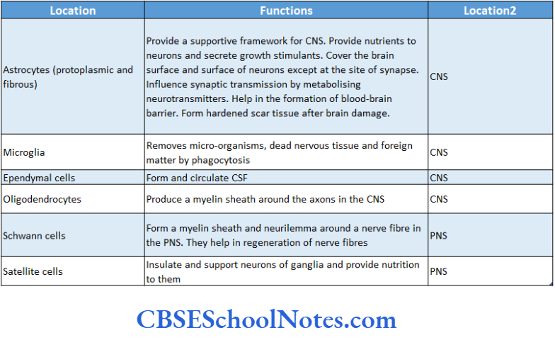
Function. Ependymal cells help in the formation and circulation of CSF.
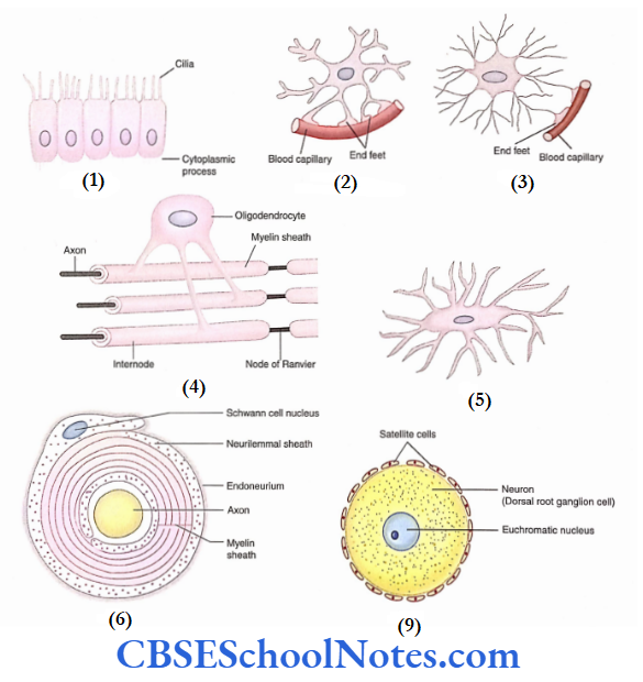
Astrocytes
Astrocytes are so-called because they are star-shaped They have many star-like radiating processes.
Astrocytes are of two types: Protoplasmic astrocytes and Fibrous astrocytes.
Protoplasmic astrocytes have thick processes with abundant granular cytoplasm.
They are mainly found in grey matter. Some of these are attached to the neighboring blood capillaries by bulbous ‘end feet’ or ‘vascular feet’.
Fibrous astrocytes have long, thin, and straight processes These cells are present only in the white matter.
Astrocytes Functions. Astrocytes completely cover the brain surface and nonsynaptic regions of the neurons. The blood-brain barrier is formed by astrocytes.
Oligodendrocytes
- Oligodendrocytes are small, round, or oval cells with only a few cytoplasmic processes.
- These cells are only present in the CNS. Embryologically, oligodendrocytes develop from the neural tube.
- Functions Oligodendrocytes produce myelin sheaths around the axons of multiple neurons in the CNS.
Microglia
Microglial cells are involved in phagocytic activities within the CNS. These cells are small with few tortuous processes.
Neuroglial Cells Of The PNS
Schwann Cells
Schwann cells are flattened cells with a flattened nucleus that is surrounded by abundant cytoplasm. These cells are present in the PNS only.
Function. Schwann cells produce a myelin sheath along the axon of a neuron, in the PNS. They also participate in the regeneration of the PNS axon.
Satellite Cells
Satellite cells surround the nerve cells of the ganglia (spinal and with prominent nuclei They are present only in the PNS.
Function. Satellite cells insulate and support neurons of the ganglia (in the PNS).
The functions of glial cells.
Nervous Tissue Summary
- The nervous tissue is composed of neurons, nerve cell processes (axons and dendrites), and neuroglia.
- Neurons are highly specialized cells and carry electrical signals from one cell to another.
- These cells have a typical pale staining achromatic nucleus, dark nucleolus, and Nissl granules in the cytoplasm.
- The elongated cytoplasmic processes take origin from the nerve cell body. They are of two different types:
- Axon and Dendrites.
- Neurons usually have short, multiple, branching dendrites. They receive information from other cells and usually have only one extremely long axon.
- Axons send information away from the nerve cell body to other cells.
- The proteins (neurotransmitters) produced in the cell are transported to the distal region of the axon and back. This is known as axonal transport.
- The classification of neurons is based on their structure (multipolar, bipolar, etc.), their size (Golgi types 1 and 2), and their functions (sensory neurons, motor neurons, and interneurons).
- Neuroglia (glial cells) are supporting cells of nervous tissue. They also provide insulation to neurons (satellite cells)
and their processes (oligodendrocytes and Schwann cells). - Ependymal cells help in the formation and circulation of the cerebrospinal fluid. The blood-brain barrier is formed by astrocytes while microglia are involved in phagocytic activities within the CNS.
Multiple Choice Questions
Question 1. Which of the following statements is false?
- The neuron is specialized to carry information in the form of electrical signals
- Dendrites are multiple, short processes while an axon is a single, long process
- The cytoplasm of neurons contains prominent eosinophilic material called Nissl bodies
- The nucleus of a neuron is euchromatic
Answer: 3. Cytoplasm of neurons contains prominent eosinophilic material called Nissl bodies
Question 2. Which of the following statements about axons is false?
- Axon begins at Axon Hillock
- The cytoplasm of the axon (axoplasm) contains microfilaments and microtubules
- Axon sends electrical signals (impulses) in one direction only, i.e. away from the nerve cell body
- Axonal cytoplasm (axoplasm) carries proteins from the cell body towards the distal end of the axon, i.e. in one direction only.
Answer: 4. Axonal cytoplasm (axoplasm) carries proteins from the cell body towards the distal end of the axon, i.e. in one direction only.
Question 3. Which ofthe following statements about Nissl substance is false?
- It has an acidic component
- Nissl bodies are aggregations of rough endoplasmic reticulum
- Nissl bodies are also the aggregation of ribosomes
- Axons are free ofNissl granules but dendrites contain Nissl granules
- None of the above
Answer: 5. None of the above
Question 4. Which of the following statements about the transport of tetanus toxin is true?
- By retrograde axonal transport
- By anterograde axonal transport
- By transport through endoneurial space
- None of the above
Answer: 3. By transport through endoneurial space
Question 5. Which of the following statements about the location of bipolar neurons is false?
- Neurons in the inferior ganglion of the vagus
- Neurons in retina
- Neurons ofvestibular and spinal ganglion
- Neurons olfactory neuroepithelium
Answer: 1. Neurons in the inferior ganglion of the vagus
Question 6. Which kind of neurons is found in the dorsal root ganglion of spinal nerves?
- Multipolar
- Bipolar
- Unipolar or pseudo-unipolar
- Anaxonic
Answer: 3. Unipolar or pseudo-unipolar
Question 7. Which of the following cells is unable to propagate nerve impulses?
- Sensory neurons
- Motor neurons
- Neuroglia
- Interneurons
- Golgi type2 neurons
Answer: 3. Neuroglia
Question 8. Which of the following neuroglial cells is not present in the CNS?
- Ependymal cell
- Fibrous astrocytes
- Oligodendrocytes
- Satellite cells
- Microglial cells
Answer: 4. Microglial cells
Question 9. Which of the following statements about astrocytes is false?
They secrete growth stimulants
- They cover the brain surface and the surface of neurons in the ganglia
- They are metabolic neurotransmitters
- They help in the formation of the blood-brain barrier
- They form scar tissue after brain damage
Answer: 2. They cover the brain surface and the surface of neurons in the ganglia
Question 10. Which of the following statements about Schwann cells are true?
- They are present in the peripheral nerve system only
- Embryologically, they are derived from neural crest cells
- They form a neurilemmal sheath around axons in the PNS
- They help in the regeneration of nerve fibers
- All of the above
Answer: 5. All of the above