
Human Osteology Introduction
The subject of “Human Anatomy” is mainly studied with the help of the dissection of cadavers (dead bodies).
The structure of various parts of the body (i.e., muscles, blood vessels, nerves, joints, and organs) and their interrelationship can be easily seen by the dissection of dead bodies.
All these soft structures are arranged around bones, i.e., muscles and ligaments are attached to bones while, nerves, vessels, and other soft structures are related (or present) close to the bones.
It should also be realized that bones form the skeletal framework of the body and, therefore, give shape to the body.
Hence, for a proper understanding of the gross anatomy of any region, it is essential to learn the structure of bones of that region first, i.e., before starting the dissection of that region.
Students should note that gross anatomy can’t be learned without a sound knowledge of osteology.
What is Osteology?
Osteology is a branch of gross anatomy, which deals with the study of bones.
Parts Of The Skeletal System
The skeletal system consists of bones and cartilage. The cartilage is present at the ends of the bone. The skeletal system consists of two main parts, i.e., axial and appendicular skeleton.
The Axial Skeleton consists of bones lying close to the central axis of the body, e.g., skull, vertebrae, sacrum, coccyx, hyoid bone, sternum, and ribs.
The Appendicular Skeleton It consists of the bones of the upper limb and lower limb including bones forming shoulder and pelvic girdles.
Read and Learn More Human Osteology Notes
How many bones are present in a human skeleton? There are approximately 206 bones present in an adult human. Out of these 80 are present in the axial skeleton and 126 in the appendicular skeleton.
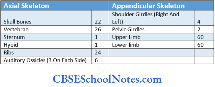
The total number of bones in the human body may exceed 206. This is because some accessory (supernumerary) bones may appear especially in the skull and lower limbs.
Bone Structure
Bone is a dynamic living tissue of the body. It is a constantly changing tissue, i.e., old bone tissues are constantly replaced by new ones.
Bone is composed of cells and intercellular matrix. Bundles of collagen fibers are embedded in the matrix. It is hard because of the deposition of calcium salts in the matrix.
Gross (Macroscopic) Structure of An Adult Living Long Bone.
A typical long bone (bone of limb) consists of a shaft or body and two ends. Both the ends of a bone are knobby (enlarged) and are covered by articular cartilage. The shaft of a long bone lies between two ends.
It is narrow in the middle and expanded towards each end. It encloses a cavity called a marrow cavity, which in an adult is filled with yellow bone marrow.
The entire bone is covered with a fibrous membrane (periosteum) except for the areas covered by articular cartilage.
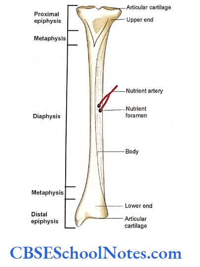
Blood vessels and nerves enter the bone through numerous foramina present near the ends and at the middle of the shaft. The prominent foramen of the shaft is called as nutrient foramen.
Further details of a living long bone may be studied by observing a longitudinal section of any long bone from the upper or lower limb.
The section reveals two different kinds 0f bones, i.e., compact and spongy.
Compact Bone
The compact bone is a dense bone in which no spaces are visible on naked eye examination. Its texture is like an ivory.
Though the compact bone covers the entire bone it is well developed in the shaft or body. The compact bone of the shaft encloses the marrow cavity filled with bone marrow.
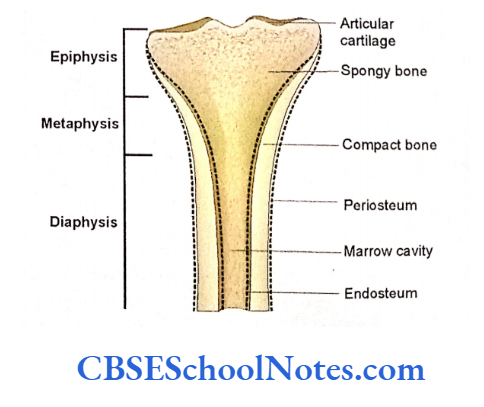
Spongy or Trabecular Bone
It is also known as cancellous bone. Spongy bone is a meshwork of bony spicules (small rods and thin curved plates), which enclose large spaces.
The spongy bone occurs at the ends of a long bone. On the outer surface, the spongy bone is always covered with a thin layer of compact bone. The spaces between spicules are filled with red bone marrow
Parts of a Living Long Bone
From the above description it becomes evident that a living long bone consists of the following parts:
- Shaft or body (Diaphysis and Metaphyses).
- Two ends (Epiphyses).
- Articular cartilage.
- Periosteum.
- Endosteum.
- Medullary cavity.
- Bone marrow.
1. Shaft or Body
The shaft of a long bone is a long, cylindrical (tubular) structure, made up of compact bone. It is also called diaphysis.
It encloses a tubular space called a marrow cavity. The compact bone of the shaft provides support and bears the weight. It is also capable of resisting the various stresses produced by movements.
The expanded distal ends of the shaft or body, where they meet with the articular ends (epiphyses), are called metaphyses.
2. Two Ends of Proximal and Distal
Two ends (proximal and distal) are expanded and made up of spongy bone covered with a thin layer of compact bone. At the ends, a long bone forms synovial joints and thus comes in contact with other bones. The ends are covered by hyaline cartilage. This particular end of a long bone is also called an epiphysis.
3. Articular Cartilage
As stated earlier the ends of a long bone are covered by a thin layer of hyaline cartilage at the site where bone comes in contact with other bone (forms a joint). The articular cartilage provides a smooth surface thus reducing friction between two bones during movements. It also helps in the absorption of shock during movements.
4. Periosteum
The entire outer surface of the bone (except where it is covered by articular cartilage) is covered with a tough sheath of dense connective tissue. This membrane is attached to the bone tissue by Sharpey’s fibers.
The periosteum consists of an outer layer of collagen fibers and fibroblasts and an inner layer of fibroblasts-like cells called osteoprogenitor cells.
The osteoprogenitor cells have the potential to divide and change to osteoblasts (bone-forming cells).
The periosteum serves the following functions:
- It forms an outer limit of bone and thus maintains its shape. It also protects the bone.
- The periosteum contains bone-forming cells, which help the bone to grow in diameter.
- It is richly supplied with blood vessels thus helping in providing nutrition to bone and assisting in fracture repair.
- It is also richly supplied with sensory nerves, making it sensitive to pain.
- Periosteum provides attachment to ligaments, tendons, muscles, intermuscular septa, and articular capsule of a joint
5. Endosteum
The endosteum is a cellular membrane that lines the medullary cavity of the shaft and the medullary spaces of spongy bone at the ends. It is composed of a single layer of flattened osteoprogenitor cells and a very small amount of connective tissue.
6. Medullary or Marrow Cavity
It is a space within the shaft or body of a long bone. The marrow cavity of the shaft is continuous with the spaces of spongy bone at the ends.
7. Bone Marrow
The marrow cavity of a newborn is filled with red bone marrow that is actively involved in the formation of blood. Red bone marrow consists of adipose and hemopoietic (blood-forming) tissues.
However, with the advancing age, the red marrow in the shaft of bone is replaced by yellow marrow (fatty marrow), which is unable to produce blood cells.
In adults, red marrow only remains at the ends of long bones, sternum ribs, skull bones, and vertebrae. At these places, red marrow is actively involved in the production of blood cells throughout life.
Some important facts about the bone
Students should note that the above description is of a living bone (bone present in a living person).
They should realize that the bones, that they handle in the classroom, are dry and I devoid of many structural components of a living bone.
For example, a dry bone is not covered by hyaline cartilage at its epiphyseal ends. Similarly, it is also devoid of periosteum, endosteum, bone marrow, blood vessels, and nerves.
Bone is not only a living tissue but it is also a dynamic tissue. It is continuously engaged in building new bone and breaking down the old bone.
Students should also realize that each living bone is not just a bone tissue but somewhat similar to an organ.
It is evident by the fact that a bone consists of not only bone tissue proper but also many other tissues like fibrous membranes (periosteum and endosteum), cartilage (articular cartilage), bone marrow (adipose and hemopoietic tissues), nerves and blood vessels.
Similar to any other organ of the body, bone is also involved in various functional activities (locomotion, support, and protection of delicate organs, formation of blood, and storage of calcium).
Features on the surface of dry bone (bone markings)
In your osteology classroom, you will handle the dead dried bones to learn their general features. The structures, which are attached to a bone or are in close contact with it, in living conditions, leave marks on the bone surface.
The following kinds of bony features (smooth areas, surface elevations, surface depressions, and foramina) can be observed on the surface of dried bones:
Smooth Areas
The smooth areas on a bone are found at places where it gives attachment to muscle fibers; where it is covered by articular cartilage; and where it lies directly beneath the skin in living conditions.
Facet
It is a flat and smooth area on the bone. In a living state, it is covered with articular cartilage, e.g., articular facets on the surface of carpal and tarsal bones.

Surface Elevations
These are of many types, i.e., crest, line, lip, and ridge are elongated surface elevations. While tubercle, tuberosity, epicondyle, and trochanter are irregular elevations.
These surface elevations are mostly due to the attachment of tendons, ligaments, or aponeurosis. Some surface elevations are sharp like the spine, cornu, or styloid process.
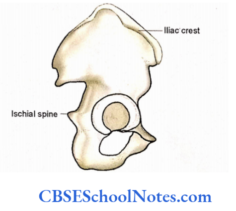
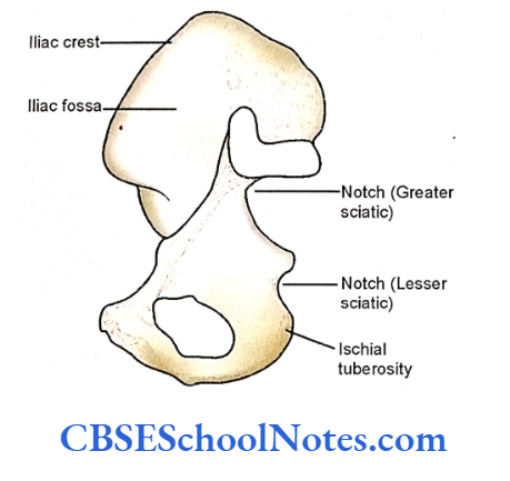
Crest: It is a bony ridge, which may be quite wide, e.g., the iliac crest of the hip bone or narrow, e.g., the external occipital crest.
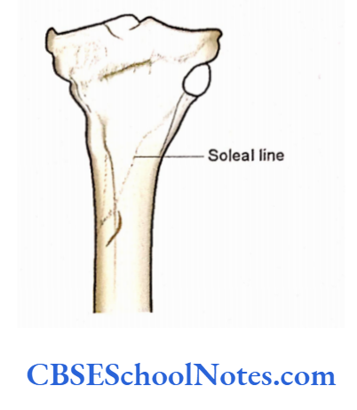
Line: An elevated line on the surface of the bone, e.g., the sole line of the tibia and superior nuchal line of occipital bone.
Condyle: A bony mass, that may have a somewhat rounded or circular articular area, e.g., condyles of the femur.
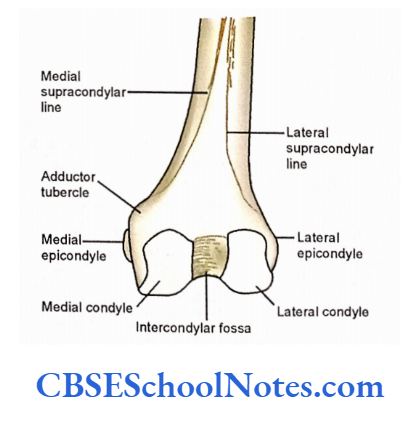
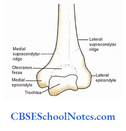
Epicondyle: Bony eminence (protuberance) situated on the surface of the condyle, e.g., lateral and medial epicondyle of the femur.
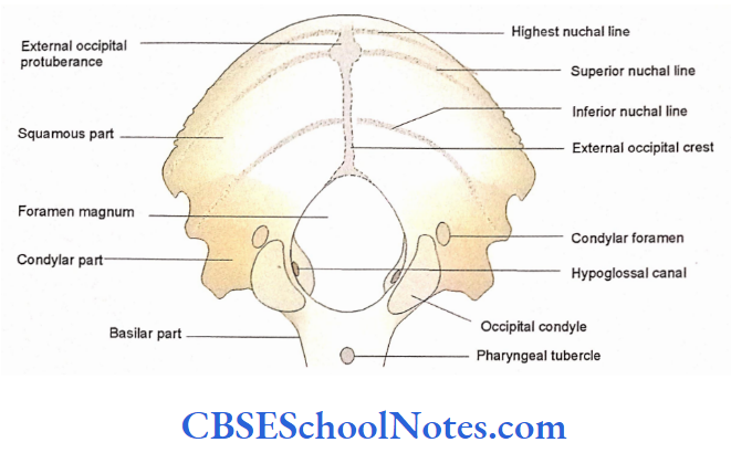
Protuberance: A projection from the surface of the bone, e.g., the external occipital protuberance.
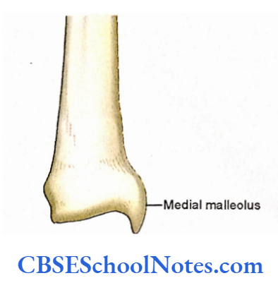
Malleolus: It is a rounded bony process, e.g., the medial malleolus of fiber
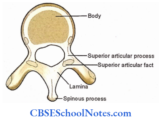
Spinous process: A spine-like projection, e.g., the spinous process of a vertebra
Trochanter: It is a large blunt elevation from the surface of the bone, e.g., greater and lesser trochanters of the femur.
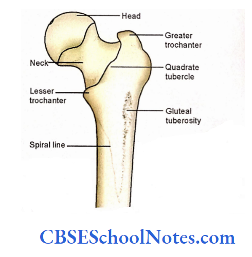
Tuberosity: A large rounded elevation, e.g., the ischial tuberosity
Tubercle: A raised elevation, but smaller than trochanter, e.g., adductor tubercle of femur
Surface Depressions and Foramina
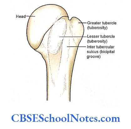
Groove or is an elongated depression on the sulcus: the surface of the bone, e.g., the bicipital groove of the humerus.
The groove or sulcus on a dry bone indicates that the bone in living condition was concerning blood vessels or tendon relation to blood Iliac crest iliac fossa vessels or tendon
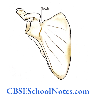
Notch: It is a deep indentation at the border of a bone, e.g., greater and lesser sciatic notches of the hip bone and suprascapular notch of the scapula.
Fossa: A depression on the surface of the bone, e.g., the iliac fossa of the hip bone and the olecranon fossa of the humerus.
Foramina: Bones show many openings on the surface, i.e., canals and foramina. A canal is a tunnel-like passage with one opening at each end. These are usually for the passage of blood vessels and nerves.
A bone may also show some other surface features, which you must learn with the help of your teachers.
How to study a bone?
Following guidelines will be of help when you are learning any particular bone for the first time.
While you are studying a bone you should have the same bone before you. There is no sense in reading about a bone, from a book, without having that bone in your hand.
The first step in learning a bone is to identify its ends (upper end, lower end, etc.), borders, and surfaces. This can be learned with the help of your teachers or with the help of diagrams from this book.
The second step is to identify some of the important projections (e.g., spine, tubercle, ridge, condyle, etc.) and depressions (e.g., fossa, foramen, notch, etc.), if present on the bone.
Side Determination of the Bone
Once you have identified the ends, borders, and surfaces and some of the bony features you will be able to determine the side of the bone.
Take the help of your teachers to know which are the bony features of that particular bone which will help to determine the side of bone.
(In the case of bones belonging to the axial skeleton there is no need to determine the side as many of them are unpaired, i.e., some bones of the skull, vertebrae, etc.). Side determination of the Lateral bone is very important.
Bone Anatomical Position
The next step is to learn to hold the bone in an anatomical position. Once you have determined the side of the bone hold it in the position it occupies in the body (as in anatomical position).
All the description of the bone in the textbooks is given considering the position of the bone in an upright posture (anatomical position).
If the bone, while studying, is not held in an anatomical position, confusion will arise regarding its borders and surfaces.
Bone General Features
Now learn the bony features (“General Features”) of bone in detail. After understanding the general feature it will become very easy to know the side of the bone and to keep it in the anatomical position.
Bone Particular Features
Once you have understood the general features of a bone it is now time to study the “particular features” (attachments of muscles and ligaments and relations of nerves and vessels).
This can be practiced by marking the area of origin of the muscle with red chalk and insertion of muscle with blue chalk on the surface of the bone. All the sites for attachments of ligaments should be marked with green chalk.
Mark the area of bone, which is with the nerve, by a solid line with yellow chalk. Similarly, the relation of artery and vein on the surface of a bone can be marked with the red and blue lines respectively.
Bone Anatomical Position Ossification
You may also study the ossification of bone if needed. (Ask your teacher whether you are supposed to know the ossification or not?).
Note the age of appearance of primary centers, secondary centers, and the time of fusion of primary with secondary centers.
Also, note which ends of a long bone is a “growing end”. Students are suggested to learn about the “ossification” and “growing end of bone” from a book of “General Anatomy.
Bone Anatomical Position Clinical Importance
Lastly, you should also learn the “applied” or “clinical importance” of the bone, if any.