
Integumentary System (Skin) Introduction
The skin is the outer covering of the body. The hair, sebaceous glands, nails and sweat glands are considered as derivatives or appendages of skin. Skin is the largest organ of the body. It consists of 16% of the body weight. The skin and its appendages constitute the integumentary system.
1. Skin Functions
- It acts as a protective shield for the body and protects us from injury, microorganisms, ultraviolet irradiations and chemical injuries.
- It provides a water barrier. Water cannot be absorbed or lost through superficial layers of epidermis, i.e., stratum corneum
- Sweat glands and unique vascular supply help in heat regulation. Sweat glands also excrete waste like urea
- Skin is an important sense organ for sensations like pain, touch, temperature and pressure
- It helps in the absorption of ultraviolet radiation from sun for the production of vitamin D
Skin Remember:
The skin along with its accessory structures is considered an important body system, which serves many important functions
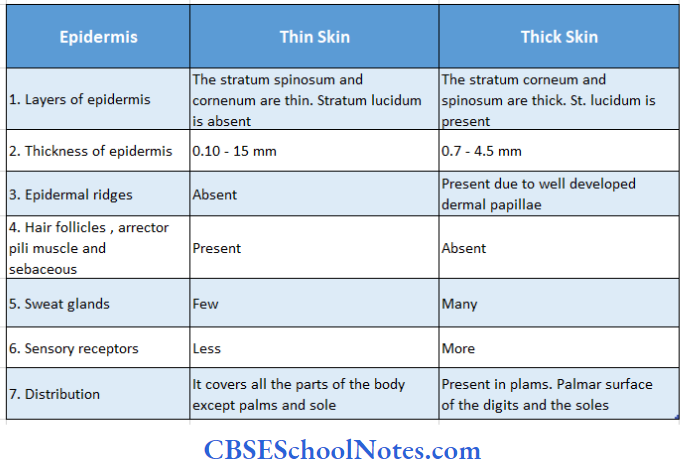
2. Thick and Thin Skin
The skin on the surface of the palm and sole is thick. Here, the epidermis is much thicker than elsewhere. Thick skin is hairless skin in other places in the body, skin is thin and hairy, for details on thick and thin skin.
Differences between thin and thick skin:
Microscopic Structure Of Skin
The skin consists of two layers
- Epidermis
- Dermis
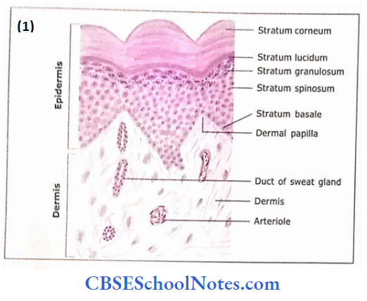

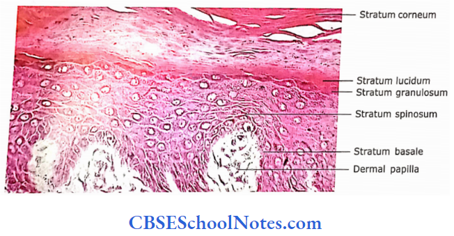
1. Epidermis:
It consists of stratified squamous (keratinized) epithelium. Following live layers can be distinct-guessed in thick skin from deep to superficial surface.
- Stratum hostile: It consists of a single layer of cuboidal cells, which are situated on the dermis. A thin basement membrane is situated between the stratum basale and der¬ mis. Basally located hemidesmosomes attach the cell to the basal lamina. Cells of this layer show high mitotic activity. The newly produced cells move towards the superficial layer
- Stratum spinas am: It consists of several layers of polygonal cells, which are held together by desmosomes. These cells contain tonofilaments and tonofibrils in their cytoplasm. The presence of tonofibrils causes the cytoplasm to become eosinophilic. The artefact of fixation causes shrinkage of the cell membrane except at the desmosomes, giving the cell a spiny appearance. Because of this reason, this layer is called stratum spinosum
- Stratumprumdusunr: This layer is made up of 3 5 layers of flattened polygonal cells. These cells are filled with keratohyalin granules
- Stratum lucidum: This layer is seen only in thick skin. Cells in this layer are flattened, translucent, eosinophilic and without any organelles including the nucleus. These cells are filled with proteins called keratin and eleidin (a product of keratohyalin)
- Stratum corneum: It is the most superficial layer of the epidermis. It is composed of structureless dehydrated dead cells. The interior of the cell is filled with keratin. The thickness of the stratum corneum is much higher in thick skin compared to thin skin. The superficial layer ot the stratum corneum is continuously sloughed oil. This process takes 20- 30 days
Stratum Remember:
The appearance of the cells of stratum spinosum is due to their shrinkage during fixation in formaldehyde during the preparation of the slide. In living conditions, cells are almost rounded and without spines. Students should also remember that layer “stratum lucidum” is only present in thick skin. They should not try to find this layer while observing a slide of thin skin
2. Dermis
The dermis is predominantly made up of collagen bundles.It also contains elastic fibres, connective tissue cells, nerves, lymphatics and blood vessels. Dermis is usually divided into two layers
- Papillary layer: It is a narrow band of loose connective tissue in contact with the basement membrane of the stratum basale. This layer shows finger-like processes projecting into the undersurface of the epidermis. These projections are called as dermal papillae and serve to interlock the dermis and epidermis. The papillae contain type 3 collagen and elastic fibres, nerves, blood vessels and various types of connective tissue cells
- Reticular layer: The reticular layer of skin is an example of dense irregular connective tissue. It contains coarse bundles of type 1 collagen, thick elastic fibres, nerves, and blood vessels, but few connective tissue cells (fibroblasts, mast cells, lymphocytes, macrophages and fat cells
Skin Pigmentation:
The colour of the Skin depends on the following factors:
- The pigment carotene of epidermal cells is responsible for the yellow colour of skin.
- Carotene is an exogenous or ange pigment taken up from food and concentrated in tissue containing fat
- The pigment melanin of epidermal cells gives a black colour to the skin
- The blood vessels of the dermis are responsible for the pink colour of the skin
The colour of the skin of an individual depends on a combination of the above factors.
Cells of Epidermis
The following types of cells are seen in the epidermis
- Keratinocytes: ‘Ninety per cent of cells in the epidermal layer arc epithelial cells, which are sometimes called keratinocytes (because of their capacity to produce protein keratin). The process of formation of keratin filaments is a continuous process, while the keratinocytes pass from stratum basale through st. spinosum and st. granulosum to the st. corneum
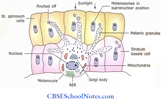
- Melanocytes: The melanocytes are rounded cells with dendrite-like branches. These cells are present in the stratum basale. Melanocytes produce melanin pigment, which is responsible for the colour of skin.
- They transfer melanin pigment to epidermal cells by “cytocrine secretion” (cell-to-cell transfer, i.e.„ it is a process in which keratinocytes phagocytosis the tip of melanocyte process) through their long dendrite-like branches.
- These cells are derived from neural crest cells.
- In white people, melanin is degraded by lysosomes, while in black people this pigment is more stable.
- Melanin saves the nuclei from the ultraviolet rays of the sun
- The melanin pigments arc present in the supranuclear region to protect the nucleus from ultraviolet rays from the sun
- Langerhans Cells: These cells are also called non-pigmented granular dendrocytes.
- These cells are present in the stratum spinosum and normally constitute 2 to 4% of the epidermal cell population (their number may reach up to 800 per mm²).
- They possess dendritic processes similar to melanocytes. Its nucleus is indented in many places and the cytoplasm contains rod-shaped granules.
- Langerhans cells are phagocytic and belong to the mononuclear phagocytic system.
- These cells protect the skin from foreign invasions, i.e., antigens, micro-organisms etc.
- Therefore Langerhans cells are also known as antigen-presenting cells
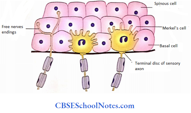
- Merkel Cells: Merkel cells are sensory cells of the epidermis.
- They are present in stratum basale and innervated by sensory nerves. Merkel cells are abundant in the fingertips, oral mucosa and hair follicles.
- It is believed that Merkel cells function as mechanoreceptors. These cells are derived from the neural crest.
Cells of the epidermis Remember:
The cells of the epidermis are also known as keratinocytes The 90% of the cells of the epidermis are keratinocytes, while the remaining are melanocytes, Langerhans and Markel cells. Melanin saves the nuclei from the ultraviolet rays of the sun. Langerhans cells are known as antigen-presenting cells, while Merkel cells function as mechanoreceptors.
Skin Clinical Applications
- Albinism: Albinism is a kind of inborn error of metabolism. Here, melanocytes are unable to synthesize melanin pigments due to the absence of the enzyme tyrosinase. Thus, skin remains unprotected from sunlight and may develop skin cancers (basal and squamous cell carcinoma).
- Vitiligo: In vitiligo, there occurs the degeneration and disappearance of already existing melanocytes. This results in patchy de-pigmentation of the skin
- Warts: Warts are small round growths from the epidermis caused due to infection of epidermal cells by papillomaviruses. The virus invades skin cells and forces them to multiply, resulting in thickened areas of skin. Warts usually occur on hands and feet and mostly are harmless. Warts may also occur on the sole
- Skin Cancer:
- Chronic exposure to excessive solar ultraviolet radiation may damage the DNA leading to basal cell carcinoma or malignant melanoma
- In adults, most of the skin tumours are derived from basal cells, squamous cells of the stratum spinosum and melanocytes. They produce basal cell carcinoma, squamous cell carcinoma and melanomas, respectively
- Malignant melanoma is an invasive tumour of melanocytes. This tumour may penetrate the basal lamina to enter the dermis. From here it may invade the blood and lymph vessels to gain wide distribution throughout the body
Question 1. Why does skin become wrinkled in old age?
Answer:
The suppleness of the skin, in young and adults, depends on the adequate presence of collagen and elastic fibre in the dermis. However, in old people there occurs the loss of these fibres due to decreased production. The overexposure to the sun also causes the degeneration of these connective tissue fibres. Both these conditions result in the wrinkling of the skin
Derivatives Of Skin
The following are the derivatives of the skin:
- Nails
- Hair
- Sebaceous glands
- Sweat glands and
- Mammary glands
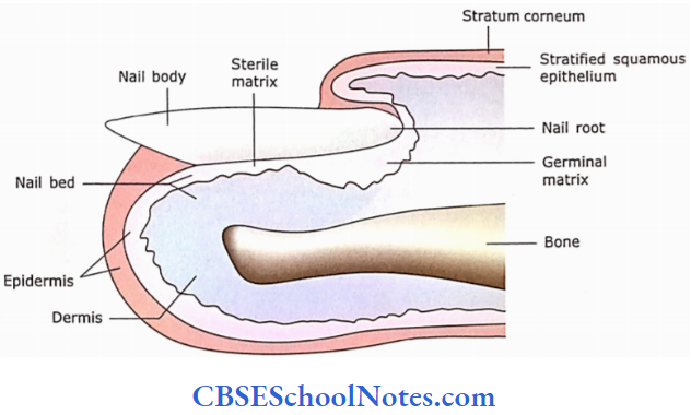
1. Nails
Various parts of nails are the inferior surface of the nail sits on a nail bed, which is made up of the stratum basale and stratum spinosum ofthe epidermis. The germinal matrix is the portion of the nail bed involved in the growth of the nail. The body and root ofthe nail are modified stratum corneum. The body of the nail corresponds to the upper cornified layer of the skin
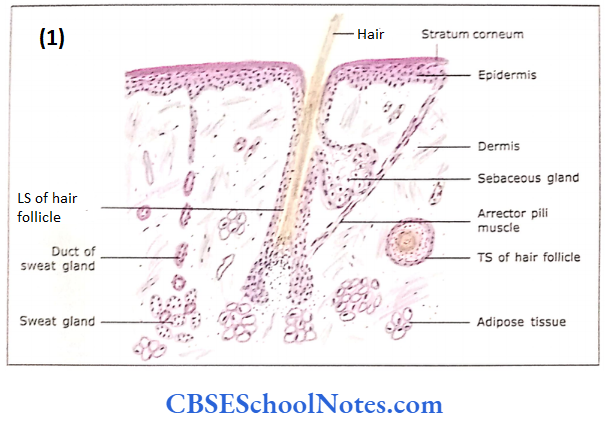
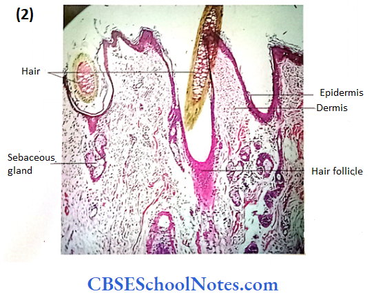
2. Hair
A hair has a shaft, which projects above the surface of the skin, and a root which is enclosed by a hair follicle. The hair follicle is a tubular invagination that is partly epidermal and partly dermal origin
- Structure of Shaft and Root of Hair: The structure of the shaft and root of hair consists of epidermal cells that are for the most part keratinized. These cells contain hard keratin and melanin granules. In a cross-section, various layers are seen such as the medulla, cortex and cuticle
- Structure of Hair Follicle: The hair follicle is a tubular invagination of the epidermis and dermis in which the hair root resides Epithelial root sheath is derived from the epidermis. It has two parts
- Outer epithelial root sheath: This layer is the continuation of the skin and corresponds to the stratum basale and stratum spinosum
- Inner epithelial root sheath: This is a keratinized sheath that arises from cells in the hair matrix
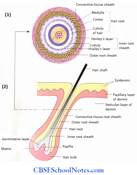
Connective tissue root sheath:
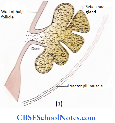
- The connective tissue root sheath is derived from the dermis. It contains nerves vessels, etc. The arrector pili muscle (smooth muscle) extends from the papillary layer of the dermis to the connective tissue sheath surrounding the hair follicle. The contraction of the muscle depresses the skin and elevates the hair shaft and skin around the hair shaft. This leads to the
- Formation of “goosebumps” on the surface of skin. The Goosebumps are seen when a person is frightened (sympathetic overactivity) or chilled.
Hair bulb:
- The lower end of the hair follicle is expanded and this expansion is called a hair bulb. The cells of the hair bulb correspond to those of the stratum spinosum and form a germinal matrix or hair matrix. These cells are concerned with the growth ofthe hair.
- The cells of the germinative layer make up a single layer that rests directly on the dermis (the hair papilla). New cells are produced here and push older cells up. Cells acquire melanin and become keratinized.
Hair papilla (dermal papilla):
- The dermal papilla fills the indentation at the base ofthe hairbulb.
- It consists of highly cellular connective tissue, which is rich in cells, capillaries, and nerves and contains melanocytes.
Hair Remember: Hairs consist of keratinized cells that develop from hair follicles
3. Sebaceous Glands
These glands are present in the dermis in association with hair follicles.
- It secretes oily substances called sebum, which consists of cellular debris and various types of lipids.
- The sebum functions as a lubricant of the skin and hair shaft (prevents their dryness).
- The sebaceous gland is a solid mass of cells (thus no lumen is present).
- The basal cells undergo mitosis and polyhedral daughter cells are pushed towards the centre ofthe glands. The centrally located cells are degenerating.
- As the cells of sebaceous glands contain lipids they are either unstained or stained lightly with eosin in
- H&E preparation. The gland has a short and wide duct, which opens into a hair follicle The mode of secre¬ tion is holocrine, i.e.,
- The entire cell is lost in the process of secretion. The contraction of arrector pilgrim helps in the exudation of secretion’
Sebaceous Remember:
The sebaceous gland secretes sebum that functions as a lubricant of skin and hair shaft (prevents their dryness)
Sebaceous Clinical Applications
- Acne Vulgaris: At puberty, under the influence of sex hormones, sebaceous glands grow In sire and increase their production of sebum.
- If the normal secretion of sebum is obstructed, then it may result in acne.
- Acne Is the inflammation of sebaceous glands due to Infection by bacteria.
- Acnes are small elevations of skin (pimples) that may contain pus and are usually confined to the face.
- This occurs predominantly in teenagers. The disease is self-limiting and dis¬ appears after some time.
4. Sweat Glands
It is a simple (unbranched) tubular gland of epidermal origin. Sweat glands extend from the surface of the epidermis to the subcutaneous layer. It has two parts, i.e., the secretory portion and the excretory duct
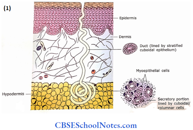
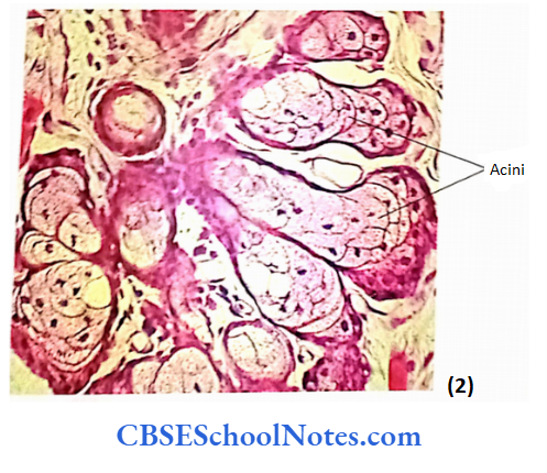
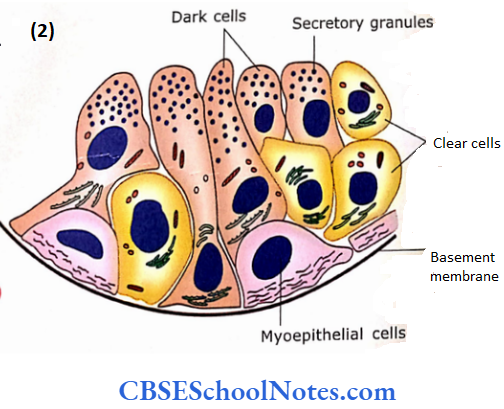
The secretory portion is present in the deep dermis or subcutaneous tissue in the form of a twisted coil. The epithelium is simply cuboidal or columnar. It has three types of cells, i.e., clear cells, dark cells and myoepithelial cells In H and E preparation, clear cells look light due to abundant glycogen in their cytoplasm and dark cells contain large amounts Of RER.
Clear cells secrete watery components and dark cells secrete glycoprotein of sweat. The contractile myoepithelial cells are located between the base of secretory cells and basal lamina. Contraction of these cells helps in the expulsion of secretory products to Duct
The excretory duct is long and extends From the secretory portion to the surface of the epidermis. The epidermal portion is spiral and has no lining of its own it is bordered by epidermal cells of epidermis. The portion of duct lying in the dermis is lined by stratified cuboidal epithelium. These cells are involved in the reabsorption of sodium from the sweat. The secretion of the sweat gland is clear and watery and may contain electrolytes, urea lactic acid and some drugs etc, the mode of secretion is merocrine
Sweat glands are of two types:
- Eccrine and
- Apocrine
Eccrine sweat glands are widely distributed throughout the skin, but are present most densely in the skin of palms and soles (up to 450 glands per square centimetre) Apocrine glands are found in the skin ofaxilla, groin, areola of the breast, labia minora and perianal region.
- The secretion of eccrine glands is watery, while apocrine glands secrete viscous secretion containing protein and lipids ceruminous glands are modified apocrine sweat glands. These glands are present in the skin ofthe external acoustic meatus (external car canal) and produce a wax secretion.
- This secretion is called cerumen, which along with the hair of the ear canal provides a sticky barrier for foreign bodies. Sometimes, cerumen may accumulate in the ear canal and prevent the sound waves from reaching the tympanic membrane.
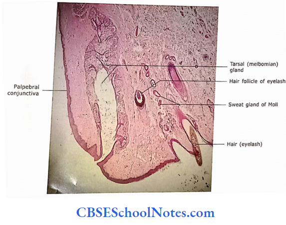
- The modified apocrine glands of eyelashes are called as glands of Moll Similarly, the modified sebaceous glands of fluid are known as Meibomian glands (tarsal glands). These glands are large and embedded in the tarsal plate
5. Mammary Glands
Mammary glands are modified (specialized) sweat glands that secrete milk in females, for histology of mammary glands.