
The Cell Structure
The Light and Electron Microscopic Structure of Cell
A cell is a basic structural unit of any living organism and is involved in vital functions to maintain its life. Though the various cells of the body differ in their structure and function, most of them have many common structural components.
They all are surrounded by plasma membranes, possess organelles, and produce macromolecules and energy. This chapter will deal with the common structure of a cell.
The Cell
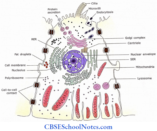
A cell is bound by the cell membrane. The cell membrane encloses the cytoplasm and nucleus. The cell varies in shape (flat, cuboidal, columnar, pyramidal, fusiform, multipolar, etc) and size (5-50 μm).
We shall study the structure of a cell under the following three headings:
- Cell membrane
- Cytoplasm
- Nucleus
The Cell Remember
A cell is a structural and functional unit of any living organ¬ ism. It is the smallest and independently living part of a living organism. It varies in shape (flat, cuboidal, columnar, pyramidal, fusiform, multipolar, etc) and size (usually 5-50 μm).
Cell Membrane
The cell membrane is also known as the plasma membrane or plasmalemma. It forms the boundary of the cell and acts as a barrier between the cytoplasm and the surrounding environment of the cell.
- The cytoplasm contains many organelles that are also made up of membranes (For example, endoplasmic reticulum, Golgi apparatus, mitochondria. etc).
- Although the cell membrane and membranes surrounding cytoplasmic organelles differ slightly in their thickness and protein contents, they all have the same basic molecular organization and are also known as unit membranes. The cell membrane is made up of lipids, proteins, and carbohydrates.
Structure Of Cell Membrane
- The total thickness of the cell membrane is just about 8-10 nm. Hence, it is not visible with a light microscope.
- When viewed under an electron microscope the plasma membrane seems to be made up of three layers i.e., trilaminar.
- The trilaminate appearance is because two dark lines are separated by a clear (unstained) intermediate zone.
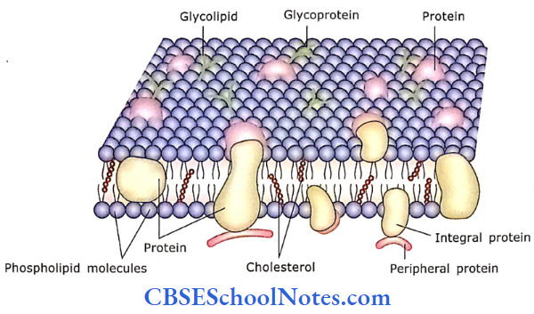
- This appearance is due to the arrangement of phospholipid molecules in two different layers.
- A phospholipid molecule has a head and a tail end. These molecules are so arranged that their head ends face the outer and inner surface of the membrane while their tail ends face each other.
Cell Membrane Remember
The cell membrane (plasma membrane) is about 8-10 μm in thickness. It is visible only under an electron microscope and seems to be made up of three layers (trilaminate).
The trilaminate appearance of the membrane is because the heads of the phospholipid molecules form the dark-staining part of the membrane while the light-staining intermediate zone is formed by the tails of the molecules.
- Besides the molecules of phospholipids, the membrane also contains several types of proteins in the form of globular masses. These proteins are present within the thickness of the cell membrane or may project through its outer or inner surfaces.
- Many integral proteins pass through the entire thickness of the membrane (transmembrane proteins).
- On the outer surface of the cell membrane, carbohydrates may attach to proteins forming glycoproteins. Although at certain places carbohydrates may also attach to lipid-forming glycolipids.
- Thus, the carbohydrates are only present on the outer surface of the plasma membrane. The coat of glycoprotein and glycolipid, on the outer surface of the plasma membrane is called a cell coat (glycocalyx).
- The glycocalyx is formed by carbohydrate chains and protects the cell from interaction with inappropriate proteins, chemicals, and physical injuries.
The cell membrane is composed of lipids as well as proteins. Protein molecules are about half of the total mass of the membrane, i.e., lipids and proteins are usually in 1:1 proportion by weight.
- Besides phospholipids and glycolipids, the plasma membrane also contains cholesterol that is present among the fatty acid tails of phospholipids.
- The lipid bilayer behaves like a fluid within which the globular proteins are free to move laterally if not attached to filaments in the underlying cytoplasm.
- Neighboring lipid molecules may exchange places about 10 million times per second and may wander completely around a cell in a few minutes. The globular proteins of the cell membrane can float like icebergs in the sea of phospholipids.
The above description of the molecular organization of cell membrane is called as “fluid mosaic model”.
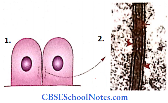
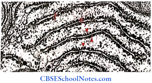
- The cell membrane of two adjacent cells
- Electron micrograph of two adjacent cell membrane
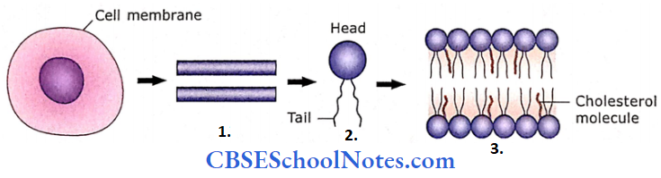
- The cell membrane when seen under an electron microscope shows the tri-laminar appearance
- Structure of a phospholipid molecule.
- Plasma membrane
There are three types of lipids in the plasma membrane i.e., phospholipids (most abundant), glycolipids, and cholesterol.
The globular proteins of the cell membrane can float like icebergs in the sea of phospholipids therefore, this model of the cell membrane is called “fluid! mosaic model.”
Cell Membrane Further Details
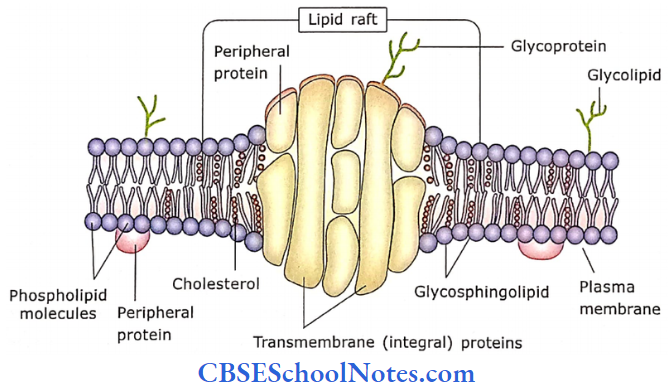
Lipid Raft
In the plasma membrane, there are regions containing high concentrations of cholesterol and glycosphingolipids. These regions are called “lipid rafts.”
- Glycosphingolipids are highly saturated fatty acid chains. These along with high concentrations of cholesterol make the lipid raft area thicker than the surrounding area.
- Because of the thickness of the raft, its fluidity is less. Raft contains a large number of integral and peripheral membrane proteins, which are involved in receiving and conveying cell-specific signals.
Proteins of the Cell Membrane and their Functions
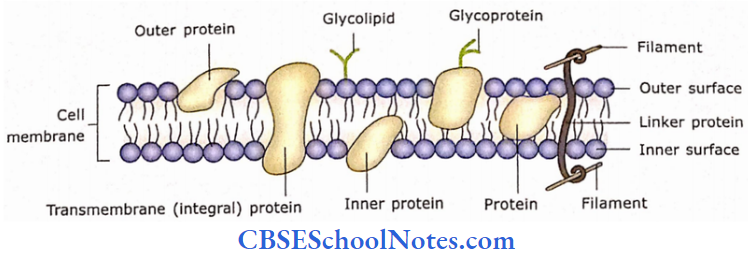
Proteins of the cell membranes are divided into two groups, that is, integral and peripheral proteins.
- The integral proteins are incorporated in the lipid bilayer, while peripheral proteins are present on the membrane surface.
- Many integral proteins pass through the entire thickness of the lipid bi-layer (Transmembrane integral proteins), while other integral proteins are embedded in the outer or inner leaflets of the liquid bilayer.
Some of these transmembrane proteins are very long and many pass through the membrane many times and are thus known as multipass proteins. The peripheral proteins are present on the membrane surface.
- Six different functional types of proteins are present in the cell membrane. These are structural, transport or carrier (pumps, channel), enzyme, receptor proteins, etc.
- Structural proteins are part of the structure of the cell membrane. These proteins are present especially where they form junctions with neighboring cells, i.e., tight junctions.
- Some proteins are? involved in the active transport of ions across the cell membrane and are called pumps. They transport the ions (Na+) and macromolecules such as amino acids and sugars from one surface of the membrane to another surface by their movement within the fluid lipid bilayer.
- Some proteins form transmembrane channels that control the entry of specific ions through the cell membrane. These channels are capable of regulating the passage of ions and molecules by closing and opening their lumen. Most channels are ion channels. There are more than 100 different types of channels.
Most common ion channels are forK+ (potassium ions) orCl (chloride ions), and fewer channels are for Na+ (sodium ions) or Ca2+ (calcium ions). Most of these channels are open all the time but the opening and closing of many more channels are guarded.
- These channels are known as “gated” channels as their opening and closing are regulated by the chemical or electrical changes occurring inside or outside the cell. When the gates are open their ions diffuse in or out of the cells.
- Proteins of cell membranes also act as receptors for specific hormones and other signaling molecules that affect the activity of cells. For example, antidiuretic hormone is the activity of cells. For example, antidiuretic hormone permeability of cell membrane. Receptor proteins are likely to be glycoproteins in nature.
- Some proteins of the membrane act as enzymes, e.g., certain ATPases are membrane-bound. The enzyme catalyzes the chemical reactions outside or inside the cell membrane.
Proteins of the Cell Membrane and their Functions Remember
Proteins of the cell membranes are divided into two I groups, that is, integral and peripheral proteins. Functionally they are divided into many types, i.e., structural, transport or carrier (pumps, channel), enzyme, and receptor proteins.
- Glycoproteins and glycolipids of cell membranes may act as cell-identity markers. With the help of this marker, a cell can recognize whether other cells are of the same kind or foreign entity, For Example., ABO blood group markers, and major histocompatibility (MHC) proteins.
- Some proteins act as linker proteins. They anchor filaments (actin) inside and (collagen) outside of the cell membrane. This helps in providing shape and stability to cells.
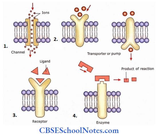
- Some proteins act as channels for the transport of ions
- Transporter proteins or pumps transport specific substances across the membrane by changing their shape
- Receptor proteins are capable of recognizing specific ligands, which in turn may alter the cell’s function
- Enzyme proteins can catalyze reactions occurring on the cell surface or within the cell (a ligand is a molecule that has a high affinity for receptors).
Lipid Raft Clinical Application
Defective Receptors
The defect in the receptors may lead to various kinds of diseases. A defective receptor becomes non-functioning and does not respond to its respective hormones and other signaling molecules.
For example, when growth hormone receptors are defective they do not respond to the growth hormone resulting in a type of dwarfism (abnormal short height).
Transport across the cell membrane by formation of vesicles: Endocytosis and Exocytosis
Cells are surrounded by extracellular fluid from which they derive their nutrition and release metabolites. The cell membrane permits diffusion and active transport of ions and gases into and out of the cell but prevents passive entry of most large molecules.
- The method by which large molecules or particulate matter (bacteria, red blood cells, and molecules of polysaccharides and proteins) can go in or out of the cell is called endocytosis and exocytosis respectively.
- The cell membrane takes an active part in the process of endocytosis and exocytosis. The process of endocytosis involves the formation of membrane-bound vesicles.
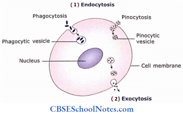
Endocytosis
Endocytosis is of three types, i.e., receptor-mediated endocytosis, phagocytosis, and pinocytosis.
- Receptor-mediated endocytosis is a highly selective type of endocytosis in which receptor protein in the plasma membrane recognizes and binds to specific ligands in the extracellular fluid. This receptor-ligand complex is pinched off and is taken in the form of a membrane-bound vesicle.
- The process of ingestion of solid particulate matter (e.g., bacteria, pigments, or other solid particles) is called phagocytosis. It is the process of eating by the cell.
- In the process of phagocytosis when particles come in contact with the outer surface of the cell membrane, the membrane throws pseudopodia to enclose the particles.
- When enveloping pseudopodia meet they fuse and the particles are drawn inside the cell. The invaginated membrane is then pinched off from the rest of the cell membrane and forms a phagocytic vacuole.
The process of ingestion of fluid or other small molecules is called pinocytosis. It is the process of drinking by the cell. In the process of pinocytosis when fluid molecules come in contact with the outer surface of the cell membrane, they become indented and form a pinocytic vesicle by pinching off from the rest of the cell membrane.

Exocytosis
In the process of exocytosis, there occurs the discharge of substance from the cell. The membrane-bound secretory granules of vesicles come in contact with the inner surface of the cell membrane and fuse with it. Then there is a rupture of the fused portion and the contents of the vesicle are released into the extracellular space.
Exocytosis Remember
Endocytosis is the process by which a cell ingests macro¬molecules, particulate matter, liquids, and other substances. During exocytosis, substances are discharged from the cell. Endocytosis is of three types, that is, receptor-mediated endocytosis, phagocytosis, and pinocytosis.
Exocytosis Clinical application
Receptor-mediated Endocytosis and HIV Infection
- Although receptor-mediated endocytosis is used by cells to import needed material from outside the cell, some viruses may enter the cell by this process of endocytosis.
- The human immunodeficiency virus (HIV) usually gets attached to the CD4 receptor on the plasma membrane of helper T cells (a kind of white blood cell) and enters the cell by receptor-mediated endocytosis.
- In this way, the cell gets infected with HIV which causes acquired immunodeficiency syndrome (AIDS).
Functions of Cell Membrane
- It maintains the shape (structural integrity) of the cell.
- Acts as an interface between the cytoplasm and the outside milieu (tissue fluid).
- It controls the movements of substances in and out of the cell (i.e., only selected substances are permitted to cross).
- It is capable of recognizing foreign bodies.
- It transmits the chemical signals across the membrane to elicit intracellular events. Cell signaling is the communication that occurs when signaling cells release signaling molecules that bind to the cell surface receptors of target cells.
- The cell membrane regulates the cell-to-cell interactions
Cytoplasm
In a cell, the cytoplasm extends between the plasma membrane and the nuclear envelope. The cytoplasm consists of a cytoplasmic matrix or ground substance that is also known as cytosol. The cytosol contains organelles, inclusion, and cytoskeleton.
Cytosol
The cytoplasm ground substance (cytosol) is made up of a fluid base containing ions (Na, K, Ca), various organic molecules (carbohydrates, lipids, proteins, and RNAs), and a three-dimensional network of the trabeculae.
The cytosol contains various structural elements like organelles, inclusions, and cytoskeleton.
- The organelles are the small organs of the cell. They have their distinctive structure and are involved in various biochemical processes necessary for the metabolism of the cell (For Example., endoplasmic reticulum, Golgi complex, mitochondria, lysosomes, ribosomes, and centrioles).
- The inclusions, on the other hand, are non-functioning elements of the cytoplasm. They are involved in the storage of nutrients such as glycogen and lipids, pigment, and secretory granules.
- The cytoplasm also contains a cytoskeleton that is made up of microtubules and microfilaments.
Cytosol Remember
The cytoplasm consists of the cytoplasmic matrix (cytosol), which itself contains various Structural elements like organelles, inclusions, and cytoskeleton (Microtubules and microfilaments).
Cytoplasmic Organelles
Most of the organelles are membrane-bound (this membrane is similar to the plasma membrane). Examples of membranous organelles are the endoplasmic reticulum, Golgi complex, mitochondria, lysosomes peroxisomes, and endosomes. Thus their contents and functions are confined within the membrane.
On the other hand, some organelles are not bounded by a membrane and thus come in direct contact with cytosol, For Example., ribosomes and centrosomes.
Cytoplasmic Organelles Remember
Most of the cytoplasmic organelles are bound by a membrane. This membrane is similar to the plasma membrane.
Endoplasmic Reticulum
Endoplasmic reticulum (ER) is the network of membranes that may be in the form of branching and anastomosing flattened tubules and vesicles. The lumen of these tubules and vesicles is known as a cistern.
The ER is found almost throughout the cytoplasm but predominantly present near the nucleus to which it is attached.
The ER is of two different types, i.e., rough endoplasmic reticulum (RER) and smooth (SER).
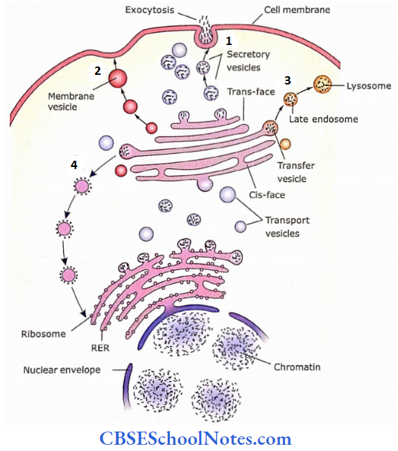
- Secretory vesicles are transported toward the plasma membrane for exocytosis
- Membrane vesicles that contain membrane protein
- Some storage vesicles contain lysosomal enzymes
- Some vesicles also originate in the Cis-face of Golgi and are retrogradely transported to RER.
Endoplasmic Reticulum RER
RER is prominent in cells that are involved in protein synthesis i.e., exocrine pancreas (secretes digestive enzymes), plasma cells (secretes antibodies), and fibroblasts which synthesize collagen.
- The RER is usually in the form of flattened sacs (cisterns) arranged one upon the other. These cisterns are connected and form a continuous system of membrane-limited cavities.
- The membrane of RER is connected with the nuclear membrane.
- On the outer surface of its membrane, numerous granules (25-30 nm in size) are attached. These granules are ribosomes that are responsible for the basophilic stain of RER.
Functions of RER
- Proteins synthesized by ribosomes enter the cavity of RER for processing and storage.
- RER is also involved in the synthesis of phospholipids.
- In the cavity of RER, the protein may combine with the carbohydrates to form glycoprotein or it may combine with phospholipids. These reactions take place under the influence of enzymes present in RER.
- Thus, RER is involved in the synthesis of secretory proteins and molecules used in the formation of plasma membranes (lipids and integral proteins).
Endoplasmic Reticulum SER
The SER also consists of short anastomosing tubules (Fig.1.9b). As the ribosomes are not attached on its surface
Functions of SER
Though SER is not involved in the synthesis of proteins, similar to RER it is also involved in the synthesis of phospholipids.
SER is involved in the synthesis of fat (cholesterol, triglycerides) and steroid hormones (estrogen, testosterone, etc.).
In the liver cells, it is involved in the detoxification of drugs and other chemicals (breakdown of alcohol and barbiturates, etc.).
SER is specialized in skeletal muscle fibers and is known as the sarcoplasmic reticulum, which helps in the control of muscle contraction.
Endoplasmic Reticulum Remember
- The endoplasmic reticulum (ER) is of two different types, i.e., rough endoplasmic reticulum (RER) and smooth endoplasmic reticulum (SER). The RER is called rough ER because of the presence of granules (ribosomes) on its surface.
- while SER is called smooth ER because ribosomes are not attached to its surface. RER is involved in the synthesis of protein, while SER in the synthesis of fat and steroid hormones.
Golgi Complex
- This cytoplasmic organelle is present in almost all cells but is well-developed in the secretory cells.
- In the glandular cells, it is present between the nucleus and the apex of the cell. It is 0.5 to 2 μm in diameter.
- It is made up of 3-20 flattened membranous sacs (cisternae) that are curved. Thus, the shape of the Golgi complex is like a shallow cup.
- The Golgi complex has a convex and a concave surface. The convex surface faces towards the RER and nucleus, while the concave surface faces towards the cell membrane.
- The convex surface is also called as forming face (cisface) and the concave surface is called the maturing face (trans-face).
- The small vesicles that bud off from the RER are transported towards the cis-face of the Golgi complex where they Rise with the outermost convex cistema of forming face.
- The secretory product then moves from the cistema of forming face to maturing face through vesicles that bud off from the periphery of one cisterna and fuse with the next.
- While the secretory products are moving from his face to fransface of Golgi, they are modified.
- At the trans-face or maturing face, the products are accumulated & concentrated in cisterna.
- Membrane-bounded secretory vesicles are formed at the trans-face that leave the Golgi and move towards the apical end of the cell for secretion.
Golgi Complex Functions
- One of the important functions of the Golgi complex is to sort proteins for their respective pathways, that is, to the plasma membrane, secretory granules, or lysosomes.
- Golgi is also involved in membrane synthesis (by forming the membrane vesicles).
- It forms secretory vesicles for exocytosis.
- It is also involved in the production of lysosomes with RER.
- Enzymes of the Golgi modify the proteins to form glycoproteins and lipoproteins. They also modify glycolipid.
![]()
Golgi Complex Remember
The Golgi apparatus is made up of a series of flattened membrane-bound cisternae. It consists of a cis-face and a trans-face. Its function is in the synthesis of carbohydrates and the modification and sorting of proteins.
Lysosomes
- Lysosomes are electron-dense, membrane-bounded bodies (vesicles), measuring 0.2-0.8 μm in diameter.
- They are formed in the Golgi complex and are called primary lysosomes. When a primary lysosome fuses with the endocytic vesicle (the contents of which are to be digested), it is called a secondary lysosome.
- Lysosomes contain 40 types of powerful hydrolytic (digestive) enzymes that are capable of breaking down various kinds of molecules.
- Lysosomal enzymes work best at acid pH. Therefore, the lysosomal matrix is 100 times more acidic than cytosol. The lysosomal membrane possesses proton pumps that actively transport H+ ions into the lysosome, maintaining its lumen at pH 5.
- Although lysosomes contain hydrolytic enzymes, their membrane is resistant to hydrolysis by their enzymes. This is because their membrane has an unusual phospholipid structure.
The membrane proteins in lysosomes are highly glycosylated. Sugar molecules cover the cytoplasmic surface proteins that protect them from digestion by lysosomal enzymes.
Lysosomes Functions
- Lysosomes are involved in the digestion of substances or particles (bacteria, etc), which are brought into the cell using endocytosis. This process is called heterophagy.
- Lysosomes are also capable of digesting the old (worn out) organelles of cytosol and returning the digested components to the cytosol. This process is called autophagy.
- In some pathological conditions, lysosomes may also destroy their cells. This process is known as autolysis.
Lysosomes Remember
Lysosomes have an acidic pH and contain 40 types of powerful hydrolytic (digestive) enzymes that are capable of breaking down various kinds of molecules. Lysosomes are involved in the digestion of substances or particles (bacteria, etc) that are brought into the cell using endocytosis.
Peroxisomes
- Peroxisomes are small (0.2-1 μm in diameter) ovoid organelles. This structure is almost similar to lysosomes. These are also membrane-bounded bodies containing more than 40 oxidative enzymes.
- Peroxisomes contain enzymes that oxidize amino acids and fatty acids as part of normal metabolism.
- The enzymes of peroxisomes also oxidize toxic substances like alcohol.
- In the process of oxidation, hydrogen peroxide (H202) is released as a by-product that is toxic to cells.
- Peroxisomes contain an enzyme called catalase that detoxifies H202 within the cell.
Peroxisomes Clinical Application
Tay-Sachs Disease
- This is a lysosomal storage disorder.
- This is an inherited (genetic) disease. It is due to the absence of a single lysosomal enzyme (B-hexosaminidase).
- In the absence of this enzyme, the glycolipid (ganglioside) cannot be broken down and thus accumulates in nerve cells.
- This leads to inefficient working of nerve cells resulting in seizures, muscle rigidity, blindness, and death before 5 years of age.
Mitochondria
- Mitochondria are called “powerhouses” of the cell because they generate ATP (a stable storage form of energy).
- These are present in all types of cells, except red blood cells.
- Their number within a cell may vary from a few to several thousand (about 2000 in each liver cell).
- A large number of mitochondria are present in highly active cells, e.g., muscles, liver, and kidney cells.
- This membrane-bounded organelle is elliptical in shape and measures about 0.5-3 μm in length.
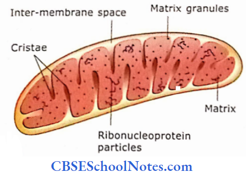
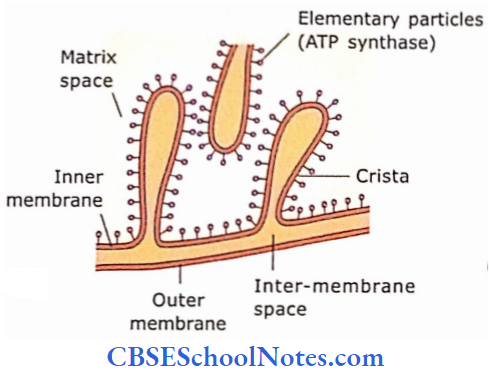
- Unlike other membrane-bound organelles, mitochondria are made up of two parallel membranes each of which is structurally similar to the plasma membrane.
- The outer membrane is smooth while the inner membrane is arranged in a series of folds called cristae.
The inner membrane is rich in enzymes (ATP synthase) that are present in spherical bodies (elementary particles) attached to its inner surface. Intramembranous space contains specific enzymes.
- One of them is cytochrome C, which is important in initiating apoptosis (programmed cell death).
- The matrix is the central fluid-filled cavity of mitochondria enclosed by the inner membrane and cristae.
- Matrix contains matrix granules, mitochondrial DNA filaments, mRNA, tRNA, and rRNA.
- Mitochondrial DNA consists of a double helix in the form of a circle that contains 37 genes.
- It is believed that mitochondrial genes are inherited only from the mother as the head of sperm lacks mitochondria.
- As per the energy requirement of the cell, mitochondria may divide to increase their number. Mitochondria are present in all cells, except those lacking nuclei, i.e., RJBCs and terminal keratinocytes.
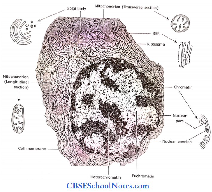
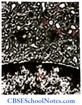
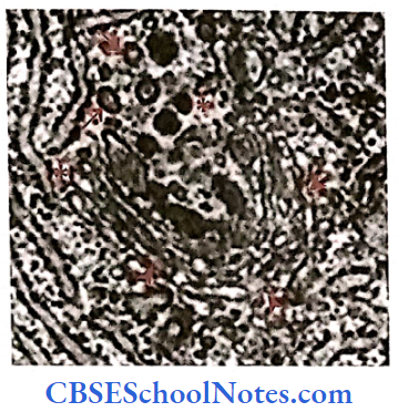
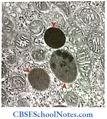
- It shows an electron micrograph of mitochondria.
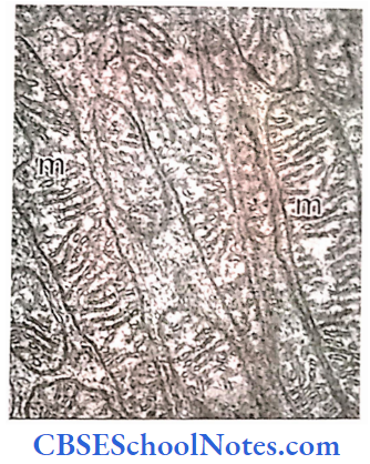
The average life span of mitochondria is about 10 days. They are self-replicating. The mitochondrion enlarges in size, replicates its DNA, and undergoes division to form two mitochondria.
Mitochondria Functions
- As the matrix of mitochondria also contains ribosomes, some protein synthesis occurs within the mitochondria, Mitochondria are capable of synthesizing their ribosomal protein.
- The remainder of the mitochondrial proteins is encoded by the nuclear DNA, which then comes from cytoplasm to mitochondria.
- Mitochondria are involved in the production of a high-energy phosphate compound, adenosine triphosphate (ATP).
- This ATP is a stable storage form of energy, which is used for many chemical reactions in the cell.
- Granules of the matrix arc cation-binding sites. Thus, the mitochondria arc is also involved in the regulation of the concentration of certain ions in the cytosol.
- Mitochondria sense cellular stress and decide whether the cell should live or die by initiating apoptosis (programmed cell death).
Mitochondria Remember
Mitochondria are present in all cells except those lacking nuclei, i.e., RBCs and terminal keratinocytes, A Large number of mitochondria is present in highly active cells, For Example., muscles, liver, and kidney cells. As per the energy requirement of the ceil, mitochondria may divide to increase their number. Mitochondria are involved in the Production of a high-energy phosphate compound, ATP.
Ribosomes
- Ribosomes are small particles about 20 to 30 nm in diameter. They contain ribonucleic acid and many types of ribosomal proteins.
- Ribosomes are made up of two subunits both of which are produced separately in nucleolus. Once produced they migrate to the cytosol and both subunits join each other.
- Ribosomes are attached in groups on the surface of RHR. This group of ribosomes is called polyribosomes. They are present in groups because they are attached to the thread of messenger RNA.
- Ribosomes are also scattered singly or in groups in cytoplasm (not attached to any organelle). These types of ribosomes are called free ribosomes or free polyribosomes, respectively.
Ribosomes Functions
- Ribosomes are the site of protein synthesis.
- Free ribosomes synthesize proteins that are used within the cell.
- Membrane-bound ribosomes are involved in the synthesis of secretory proteins. They also synthesize proteins used in the formation of new plasma membranes.
Centrosome or Microtubule Organizing Centre
- The centrosome is a small spherical area of cytoplasm situated near the nucleus.
- It consists of two parts: the pericentriolar area and the centriole.
- The pericentriolar area is made up of a dense network of smaller granular protein material.
- In the center of the pericentriolar area, two rod-shaped structures are called centrioles. The long axis of one centriole is at a right angle to the long axis of the other.
- Centrioles are hollow cylindrical structures each of which is made up of nine groups of three microtubules (triplets) arranged in a circular pattern.
- Centrioles are self-replicating organelles. Just before cell division, a new centriole is synthesized near the old one.
Centrosome or Microtubule Organizing Centre Functions
- The pericentriolar area plays an important role in the formation of mitotic spindles during cell division.
- In a non-dividing cell, this is also involved in the synthesis of microtubules.
- Centrioles are involved in the formation of cilia and flagella.
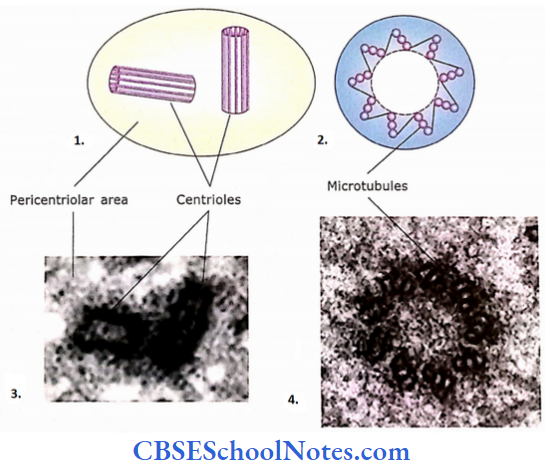
- The pericentriolar area of a centrosome surrounds two centrioles. The long axes of two centrioles are perpendicular to one another.
- Transverse section across a centriole. Each centriole is made up of 9 bundles of microtubules, with 3 microtubules per bundle.
- Electron microscopic image of the centrosome, in which two centrioles (in longitudinal section) are seen arranged at a right angle to each other.
- Electron micrograph of a centriole as seen in the transverse section. Three microtubules are visible in each 9 bundles.
Endosomes
- Endosomes are membrane-bounded compartments (between 100 to 500 nm in size) associated with endocytotic pathways. There are two types of endosomes, i.e., early and late endosomes.
- Early endosomes are located near the cell membrane. The endocytotic vesicles, which originate from the cell membrane, fuse with the early endosomes.
- The function of early endosomes is to sort the proteins received through endocytotic vesicles. Receptor proteins will go back to the plasma membrane.
- While the remaining proteins will be transported to the late endosomes through multivesicular bodies. The multivesicular bodies are structures that transport proteins between early and late endosomes.
- Late endosomes are located near the Golgi apparatus. The substances transported to late endosomes are degraded in the lysosomes.
Cytoplasmic Inclusions
Inclusions are non-living and non-functional components of a cell. They are simply the store of inert by-products of metabolism like lipids and glycogen. These are not membrane-bound.
- Glycogen: Glycogen is present in the cytoplasm in the form of dense granules that are about 25-30 nm in diameter.
- Lipid: Lipid is stored in the cytoplasm in the form of rounded droplets.
- Pigments: Some cells may show the presence of a yellowish-brown pigment called lipofuscin. These inclusions may be membrane-bound. The lipofuscin pigments are waste products of the cell, which cannot be digested completely by the lysosomal activity.
- Secretory granules: Sometimes, the membrane-bounded secretory vesicles are also classified as cytoplasmic inclusions.
Cytoskeleton
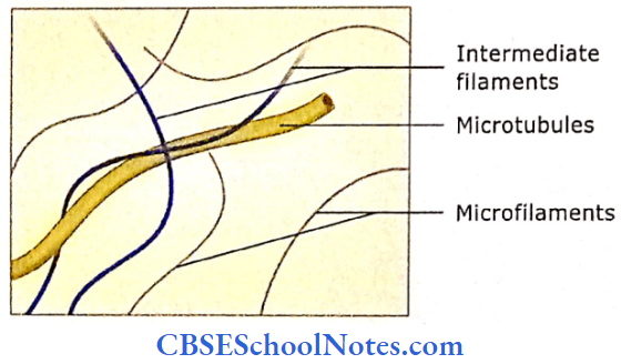
Different kinds of protein filaments and tubules form a network throughout the cytoplasm. This network is called a cytoskeleton. The cytoskeleton provides shape to the cell and organizes the cellular contents.
Cytoskeleton is also involved in the mobility of some cells, For Example., phagocytes. The cytoskeleton consists of the following three types of protein filaments:
- Microfilaments
- Intermediate filaments
- Microtubules
1. Microfilament
- Microfilaments are the thinnest (5 nm in diameter) filaments of the cytoskeleton. These filaments are mainly present near the peripheral part of the cell and are made up of proteins called actin.
- Microfilaments provide shape to the cell. They also provide the skeleton of microvilli, which are finger-like projections from the cell surface.
- Microfilaments are also involved in muscle contraction, cell division, and movement of phagocytes and other cells.
2. Intermediate Filaments
- The intermediate filaments are thicker (10 nm in diameter) than microfilaments. These filaments are found in parts of cells subjected to mechanical stress.
- The intermediate filaments of nerve cells are called neurofilaments. The tonofibrils are filaments of epidermal cells that are composed of a protein called keratin.
Cytoskeleton Clinical Application
Alzheimer’s Disease
- Failure to assemble the intermediate filaments leads to various diseases. The changes in neurofilaments lead to Alzheimer’s disease.
- In this disease, there is an accumulation of tangled masses of filaments in the cytoplasm leading to the degeneration of neurons. Patients with this disease suffer from loss of memory.
Microtubules
Microtubules are hollow cylinders about 25 nm in diameter and several microns in length. They are made up of the protein tubulin. Microtubules are assembled at the centrosome, which consists of a microtubule-organizing center.
- Microtubules are present throughout the cytoplasm where they are involved in giving shape to cells, in the intracellular transport of secretory granules, and movements of chromosomes during cell division.
- These tubules are also present in cilia and flagella and are responsible for their movements.
Nucleus
The nucleus is a membrane-bound structure. It is either a spherical or oval-shaped structure, present usually in the center of the cell.
It contains genes, which control cellular structure and the various activities of the cell. The nucleus consists of an envelope (nuclear envelope), nuclear lamina, chromatin material, nucleolus, and nuclear matrix.
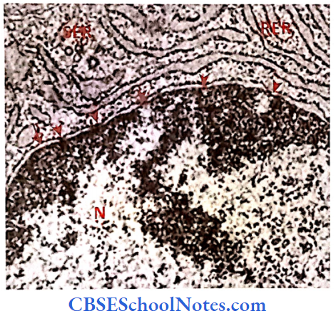
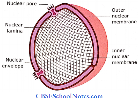
Nuclear Envelope
The nuclear envelope is made up of two parallel membranes that separate the nucleus from the cytoplasm. These membranes are similar to plasma membranes.
- The outer nuclear membrane is continuous with rough endoplasmic reticulum.
- In the nuclear membrane, there are several channels, which are called nuclear pores. Small molecules and ions diffuse passively through these pores.
- However, the transport of large protein molecules from the cytosol to the nucleus and that of RNAs from the nucleus to the cytosol is regulated by nuclear pores.
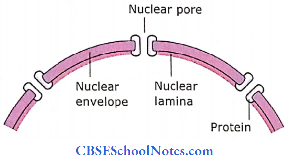
Nuclear Lamina
The nuclearlamina is a thin layer adjacent to the inner nuclear membrane. It is formed by intermediate filaments, which are arranged in a square lattice.
- The function of nuclear lamina is to provide structural stability to the interphase nucleus (nuclear envelope, nuclear pores, and chromatin).
- The nuclear lamina disintegrates during cell division but reassembles in the daughter cells.
Chromatin
The nucleus plays an important role in heredity. It contains heredity units called genes. Genes are present on chromosomes, which itself is a long molecule of DNA coiled together with several proteins.
- Innon-dividing cell (interphase cell), chromosomal material is less tightly coiled and appears as a diffuse granular mass, which is called chromatin.
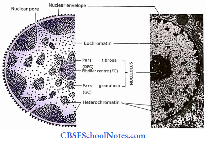
- The chromatin is stained basophilic (with hematoxylin) due to the presence of a phosphate group in DNA. In the nucleus of an interphase cell, chromatin occurs in two different arrangements, i.e., heterochromatin (condensed chromatin) and euchromatin (extended chromatin).
- Heterochromatin stains with basic dyes and hematoxylin and thus appears as irregular dark masses. Euchromatin stains lightly with basic dyes and is seen as clear areas between heterochromatin.
- Nuclei that are predominantly made up of euchromatin are called open-face nuclei, while nuclei that are made up mainly of heterochromatin are called closed-face nuclei.
Heterochromatin is regarded as metabolically inactive chromatin (For Example, chromatin in the head of sperm), while euchromatin is active chromatin (chromatin of neurons and liver cells).
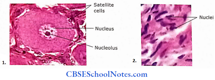
Nucleolus
The nucleolus is the spherical body within the nucleus that is stained dark with basic dyes. Some cells contain more than one nucleolus.
- It contains a protein called nucleostemin. It is a P53 binding protein that regulates the cell cycle and influences cell differentiation.
- The main role of the nucleolus is to synthesize rRNA and assemble ribosomes.
- When observed under an electron microscope, it consists of three regions.
- The innermost pale staining fibrillar center (FC) is surrounded by a dense fibrillar component (DFC) or pars fibrosis, which in turn is bounded by a granular component (GC) or pars granulosa. Pars granulosa contains maturing ribosomes.
- Transcription of DNA occurs in FC or at the FC-DFC junction. This region contains tips of chromosomes 13, 14, 15, 21, and 22 (the nucleolar organizing regions), where gene loci that encode rRNA are located.
- Most of the cleavage and modification of RNA occurs in the DFC. While steps involving protein assembly to form preribosomal particles occur in GC.
Here, small and large ribosomal subunits are organized, which migrate to the cytoplasm through nuclear pores.
Nuclear Matrix
All the material enclosed by the nuclear membrane, excluding chromatin and nucleolus, is called nucleoplasm. At present, nothing much is known about its composition but it seems that it must contain a network of fibrils (karyoskeleton), proteins, and metabolites.