
Reticular Formation
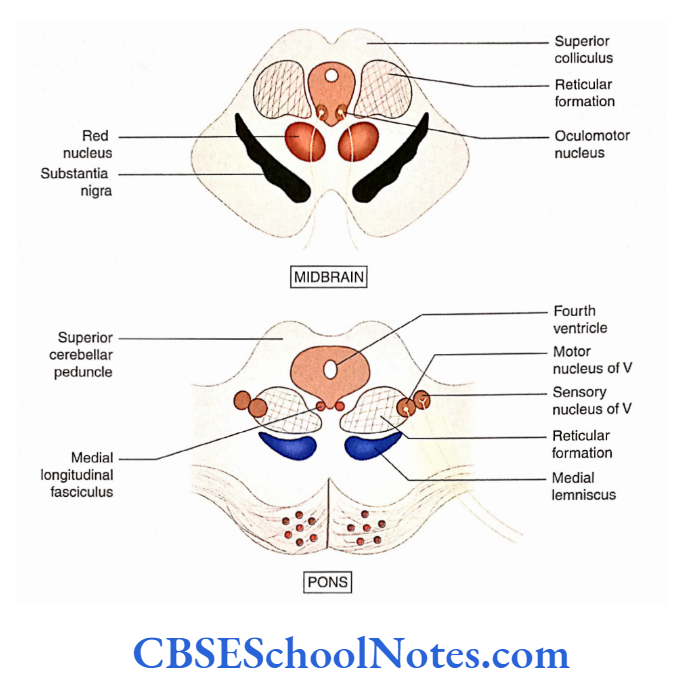
‘Reticular formation is the collection of a group of neurons and intersecting bundles of fibers present inside the brainstem.
This particular arrangement gives rise to a reticular or net-like structure that is seen in the transverse section of the brainstem.
Extent And Location Of Reticular Formation
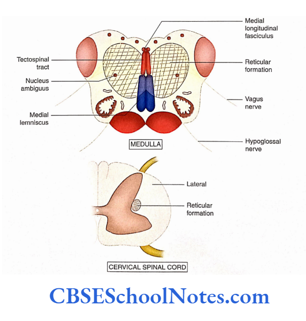
The reticular formation extends throughout the brainstem and is continuous above the subthalamus and reticular nuclei of the thalamus.
Inferiorly, it is continuous with the cervical part of the spinal cord. Students should note that the reticular nucleus of the thalamus is not a part of the reticular formation.
Read and Learn More Neuroanatomy
Composition Of Reticular Formation
The reticular formation is an ill-defined collection of neurons and fibers with multiple connections.
It also has neurons with multiple synaptic contacts with various ascending and descending tracts passing through the brainstem.
Nuclei Of Reticular Formation
Many ill-defined collections of neurons are recognized in the reticular formation and these collections are called nuclei.
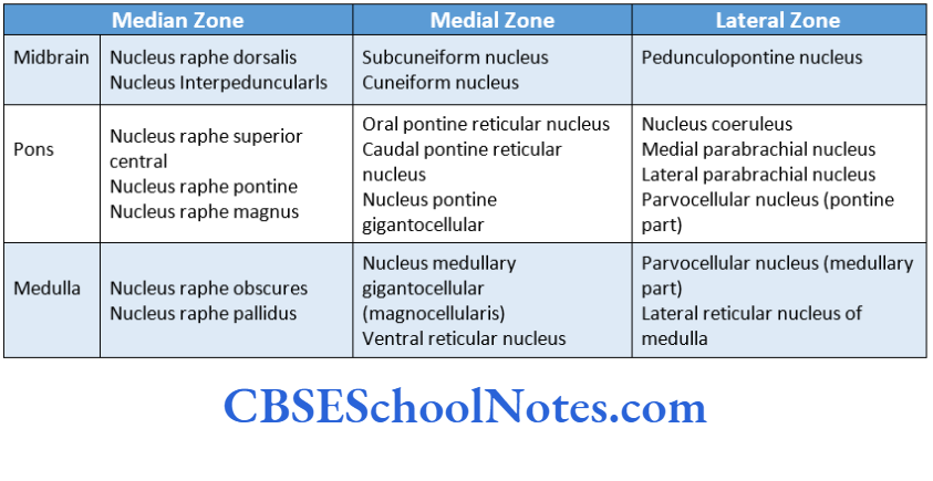
These nuclei are arranged in vertical columns and are briefly classified into the following three zones (columns):
- Median zone
- Medical Zone
- Lateral zone
Some important nuclei of all three zones are given in
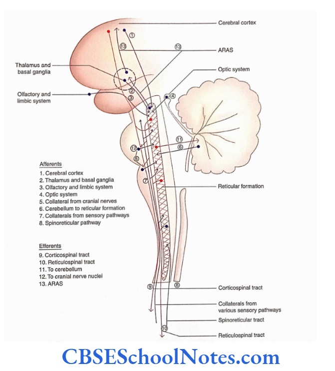
Connections Of Reticular Formation
- Reticular formation has extensive afferent and efferent connections with almost all parts of the CNS.
- The pathways originating or terminating in reticular formation are both ascending and descending. Some of these pathways are crossed while others are uncrossed.
- The nuclei in the lateral parts of reticular formation are mainly sensory while those in the medial part are mainly motor.
- Most of the connections of reticular formation, as presented here, are based on the group of nuclei secreting a specific neurotransmitter.
The connections of serotonergic raphe nuclei, cholinergic nuclei, and noradrenergic nuclei are presented separately (see in the following text).
Chemoarchitectonic Organisation Of Reticular Formation
- Although the preceding description of reticular formation has indicated collections of many nuclei, these nuclei are ill-defined.
- Modern techniques (immunocytochemistry) have defined the reticular formation as consisting of a well-defined group of nerve cells (nuclei) with specific Neurotransmitters.
Serotonergic nuclei: Serotonergic nuclei synthesize serotonin (5-hydroxytryptamine). The ‘raphe’ nuclei synthesize serotonin and use it as a synaptic neurotransmitter.
The raphe nuclei of the midbrain and upper pons project into the forebrain and help in the regulation of the wake-sleep cycle, food intake, thermoregulation, and sexual behavior.


Cholinergic nuclei: Cholinergic nuclei are involved in the synthesis of acetylcholine. Acetylcholine is used as a neurotransmitter by two nuclei.
Pedunculopontine nucleus and Lateral dorsal tegmental nucleus. These nuclei are also involved in consciousness and arousal.
Catecholamine nuclei: Chemoarchitectonically, catecholamine nuclei are of three distinct types: Noradrenergic, Adrenergic, and Dopaminergic. They synthesize noradrenaline, adrenaline, and dopamine, respectively.
Noradrenergic nucleus: The noradrenergic nucleus coeruleus’ is the largest group of noradrenergic neurons. ‘Coeruleus’ influences arousal.
Adrenergic neurons: The best-known adrenergic neurons in the medulla are within the nucleus ambiguus, the nucleus of the tractus solitarius, and the dorsal motor nucleus of the vagus.
Dopaminergic neurons: The dopaminergic neurons in the brainstem are found to be present in the midbrain (in the substantia nigra and the tegmentum of the midbrain). These neurons are involved in behavioral response.
Functions Of Reticular Formation
The reticular formation can be divided functionally into medial and lateral regions.
The medial reticular region is regarded as an ‘effector’ (motor) area while the lateral zone is referred to as the ‘sensory’ part.
Specifically, the functions of reticular formation are as follows:
1. Somatic motor function: The facilitatory and inhibitory activities of lateral and medial reticulospinal tracts (RSTs) act by influencing muscle tone to ensure smooth body movements.
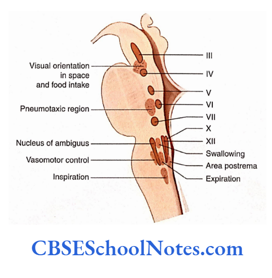
2. Motor functions of cranial nerves: The lateral zone of reticular formation has many interneurons. These interneurons form a local reflex circuit with the motor nuclei, of various cranial nerves, thereby influencing the motor activity of the cranial nerves.
Effect on the functions of the X nerve: The visceral motor functions of vagus nerves are coordinated by the ventrolateral medullary reticular formation by the reflex mechanism.
The cardiovascular responses, respiratory responses, and gastrointestinal responses of reticular formation are mediated through the vagus nerve.
Effect on the functions of 5, 7, and 12 nerves:
The neurons of the reticular formation adjacent to trigeminal, facial, and hypoglossal motor nuclei form reflex circuits with the motor nuclei of these cranial nerves.
These neurons of the reticular formation of eating involve lip movements (facial nucleus), chewing (trigeminal nucleus), and movements of the tongue (hypoglossal nucleus).
Effect on the functions of the 3 nerve: The nuclei of the reticular formation that are located close to the nuclei of the III nerve coordinate various complicated eye movements.
3. Visceral motor functions: The reticular formation of the brainstem has areas (centers) that regulate the motor functions of various systems (visceral motor functions). These systems include cardiovascular, respiratory, and gastrointestinal.
Respiratory centers: The dorsal medullary area is the inspiratory center and consists of gigantocellular reticular cells.
The expiratory center is situated medially in the parvocellular region of the medulla and includes the nucleus ambiguus. The process of inspiration and expiration is controlled by pneumatic and apneustic centers.
Cardiac and vasomotor areas: Stimulation of this gigantocellular nucleus causes slowing of heart rate and lowering of blood pressure while stimulation of the nucleus in the lateral zone (parvocellular) leads to an increase in heart rate and blood pressure.
Transmission of pain: Pain transmission through the spinoreticular tract follows the spine reticulothalamo cortical pathway.
Regulation of perception of pain: The descending pathways (reticulospinal) from the nucleus raphe magnus of reticular formation modify the perception of pain. The reticulospinal tract mostly terminates in substantia gelatinosa at all levels of the spinal cord.
Sleep and wakefulness
- Sleep is a state of temporary loss of consciousness or altered consciousness from which a person can be aroused. During sleep, the cerebral cortex is the least active.
- Sleep occurs due to diminished activity of ARAS (vide infra). Raphe nuclei contain serotonergic neurons which have an inhibitory effect on the cerebral cortex. This leads to the promotion of sleep.
Wakefulness:
- The reticular formation plays an important role in waking up from sleep and also in maintaining the state of consciousness (wakefulness).
- Stimulation of some part of the reticular formation (locus coeruleus, pedunculopontine, and oral pontine nucleus) increases the activity of the cerebral cortex which helps in the maintenance of consciousness (wakefulness and alertness).
- This part of the reticular formation is known as the reticular activating system (RAS).
ARAS:
- Many reticular formation nuclei receive the sensation from various somatic sensory ascending
- The efferents from these nuclei project to the widespread areas of the cerebral cortex. This leads to a generalized increase in cortical activity. This system of reticular formation is called ascending RAS (ARAS).
- A sleeping person can be aroused from sleep by increased activity of ARAS. ARAS can be stimulated by various somatic sensory stimuli (except a sense of smell) such as touch or pressure on the skin, painful stimuli, bright light, loud noise, or the sound of the alarm clock.
Coma
- Coma is a state of unconsciousness with little or no response to stimuli. It may be caused by extensive damage to the cerebral cortex or by lesions of the brainstem (bilateral damage to ARAS at rostral pons and midbrain).
- In the case of the lesion of the lower brainstem, unconsciousness is accompanied by respiratory and cardiovascular disturbances as these centers are situated in the medulla.
- In the case of patients with light coma, brainstem and spinal reflexes persist but in cases of deep coma, all reflexes are lost. Deep coma is usually fatal as respiratory and cardiovascular failure leads to death.
Summary
- Reticular formation of the brainstem is the collection of a group of neurons and intersecting bundles of fibers. It occupies the dorsal part of the brainstem.
- The reticular formation extends throughout the brainstem, from the thalamus to the spinal cord.
In the reticular formation of the brainstem, many ill-defined nuclei are observed. These nuclei are present in three zones (columns):
- Median,
- Medial and Lateral.
- The median zone nuclei are located in the midline, which synthesize and secrete serotonin (serotonergic). These nuclei are concerned with sleep and suppression of pain.
- The medial zone nuclei are situated on either side of the median zone. They give origin to the reticulospinal tract and central tegmental tract.
- The lateral zone nuclei are situated lateral to the medial zone. They are pedunculopontine, nucleus coeruleus, parabrachial, and parvocellular nuclei.
- The major afferent and efferent connections of reticular formation are presented.
- The medial region of reticular formation is regarded as a motor area while the lateral zone is sensory in function.
- The medial zone has long ascending and descending tracts. These tracts modulate the action of neurons involved in movements and posture, autonomic function, pain, and arousal.
- The lateral zone of reticular formation contains small interneurons that form local reflex circuits with the motor nuclei of various cranial nerves.
- Vascular, cardiac, respiratory, and gastrointestinal responses of reticular formation are mediated through the vagus nerve.
- Reticular formation plays an important role in awakening from sleep and then in maintaining the state ofconsciousness (wakefulness).
Reticular Formation Multiple Choice Questions
Question 1. Which of the following facts about reticular formation is false?
- It appears net-like in the transverse section of
- It appears net-like in the transverse section of the brainstem.
- It is a collection of groups of neurons and intersecting bundles of fibers
- It extends throughout the brainstem
- The nucleus ambiguus is located within the territory of the reticular formation
- None of the above
Answer: 5. None of the above
Question 2. Which of the following statements about the extent of reticular formation is false?
- It extends throughout the brainstem
- It is continuous above with the subthalamus and reticular nuclei of the thalamus
- Below, it is continuous in the cervical part of the spinal cord
- In the cervical part of the spinal cord, it is situated in the lateral funiculus at the junction of grey and white matter
- None of the above
Answer: 5. None of the above
Question 3. Which of the following statements about reticular formation is false?
- It is an ill-defined collection of neurons and fibers with multiple connections
- Most of these neurons are Golgi type 2 neurons
- These neurons have long dendrites and short axons cL They have multiple synaptic contacts with various ascending and descending tracts
- None of the above
Answer: 3. These neurons have long dendrites and short axons cL They have multiple synaptic contact with various ascending and descending tracts
Question 4. Following are the columns (zones) of nuclei of reticular formation except
- Median zone
- Medial zone
- Lateral zone
- Posterolateral zone
Answer: 4. Posterolateral zone
Question 5. Following are the nuclei of the median zone except
- Nucleus raphe dorsalis
- Nucleus interpeduncular
- Nucleus raphe magnus
- Nucleus raphe pontine
Answer: 3. Nucleus raphe magnus
Question 6. Chemoarchitectonically, which of the following nuclear groups exist?
- Serotonergic nuclei
- Cholinergic nuclei
- Catecholamine nuclei
- All of the above
Answer: 4. All of the above
Question 7. Which of the following statements about the reticular formation is true?
- Nuclei of the medial part of reticular formation are mainly motor
- The nuclei of the lateral part are mainly sensory
- The lateral part receives sensory information from collaterals of the spinothalamic tract, trigeminothalamic tract, and lateral lemniscus
- Giant cell area of reticular formation is the main motor area of reticular formation
- All of the above
Answer: 5. All of the above
Question 8. Following are the descending (efferent) tracts of the reticular formation except
- Lateral reticulospinal tract
- Medial reticulospinal tract
- Dorsomedial reticulospinal tract
- Raphespinal tract
Answer: 3. Dorsomedial reticulospinal tract
Question 9. Which of the following statements about reticular formation is false?
- It consists of a multisynaptic pathway
- It is confined to the midbrain and pons
- It influences the level of consciousness and alertness
- It consists of a diffuse network of fibers and matter (nuclei)
Answer: 2. It is confined to the midbrain and pons