
Functional Cerebral Areas Of Cerebral Cortex
The cerebral cortex has many areas that have been assigned to perform specific functions. These areas are usually described as sulci and gyri.
Brodmann’s Mapping
The best-known scheme to map out the functional area of the cerebral cortex is that of Brodmann. He mapped the ” cerebral cortex in 47 such areas and indicated each area by a number.
Functional Areas Of Cerebral Cortex
Animal experiments and clinical pathological studies have indicated that the human cerebral cortex possesses three different types of functional areas
- Sensory,
- Motor and
- Association areas.
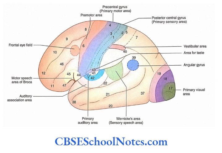
Sensory areas: These areas are concerned with the perception of general somatic sensations (pain, touch and temperature), special sensations (hearing, vision, taste and smell) and equilibrium. The sensory areas of the cerebral cortex receive fibres from the thalamus.
The primary somatic sensory area, which receives pain, touch and temperature, is located in the postcentral gyrus (areas 3, 2 and 1).
Read and Learn More Neuroanatomy
Similarly, the sensory area for vision is located in the occipital lobe (area 17) while the area for hearing is located in the temporal lobe (areas 41 and 42).
Motor areas: These areas are responsible for the contraction of skeletal muscles during electrical stimulation. The precentral gyrus (area 4) is an example of a primary motor area which sends projection fibres to the brainstem and spinal cord.
Association areas: Besides the motor and sensory areas, a large part of the cerebral cortex (up to 70% of the neocortex) is referred to as the association cortex.
Association areas are usually present adjacent to the primary sensory areas. The function of the association cortex is to integrate various types of sensory information and to direct behaviour, communication and intellect.
Primary Sensory And Association Areas Of the Cerebral Cortex
Primary Sensory And Association Areas Of the Cerebral Cortex are as follows:
- Sensory and association areas of the parietal lobe
- Sensory and association areas of the occipital lobe
- Olfactory areas
Sensory and Association Areas of the Parietal Lobe
The parietal lobe has the following sensory and association areas:
- Area for general somatic sensation
- Area for a taste sensation
- Vestibular area
- Sensory speech area
Area for General Somatic Sensation
To perceive general somatic sensations, there is a primary area and an association area defined on the cerebral cortex.
Somatic Sensory Area
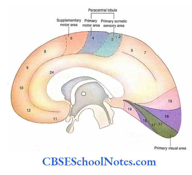
- This area is located in the postcentral gyrus on the superolateral surface and in the posterior part of the paracentral lobule on the medial surface of the cerebral hemisphere.
- This area is defined as areas 3, 1 and 2 on Brodmann’s cytoarchitecture map. The conscious perception of pain, touch, temperature and proprioception takes place here.
- The opposite half of the body is represented upside down on the primary somatic area.
- The primary sensory area receives its afferents from the ventral posterior nucleus of the thalamus through the internal capsule (as the thalamocortical tract).
Somatic Sensory Association Area
- This area of cerebral cortex corresponds to Brodmann’s areas 5 and 7 and is located in the ‘superior parietal lobule’, on the lateral surface, and in the ‘precuneus’, on the medial surface.
- This association area is concerned with the analysis and integration of general sensation based on experience.
Area for Taste Sensation
The area for taste sensation (area 43) is located at the inferior end of the postcentral gyrus, in the superior wall of the posterior ramus of the lateral sulcus. This area is located close to the general sensory area for the tongue.
Vestibular Area
The area concerned with the vestibular function, i.e. vestibular area, is located at the anterior end of the intraparietal sulcus and in the postcentral gyrus.
Functionally, the vestibular area is concerned with the motor regulation needed for the maintenance of equilibrium.
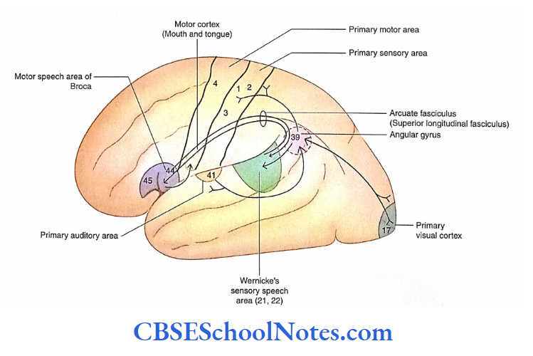
Sensory Speech Area of Wernicke
Wernicke’s sensory speech area is limited only to the posterior part of area 22 (superior temporal gyrus; The sensory speech area or Wernicke’s area is located in the dominant cerebral hemisphere.
Language functions: Wernicke’s area plays an important role in language functions.
The sensory speech centre of Wernicke’s is concerned with the interpretation of spoken and written words including signals and symbols.
Wernicke’s area (area 22) with the help of areas 39 and 40 by recognising spoken words and the meaning of writing by seeing written words interprets the meaning of speech
Language areas are located in the normally dominant left cerebral hemisphere which also contains Broca’s speech area (motor speech area).
Wernicke’s area is necessary for language comprehension while Broca’s (area 44) is concerned with language production Broca’s area activates the mouth, tongue palate and vocal cord regions of the motor cortex for articulation of speech.
Aphasia
Aphasia is a disorder affecting the ability to speak or read. A lesion of the sensory speech area of Wernicke’s leads to a person’s inability to understand both written and spoken words even though his vision and hearing are normal. This condition is called sensory aphasia.
Dyslexia
Dyslexia is a disorder of adults affecting the comprehension and use of words.
Developmental Dyslexia
- Developmental dyslexia is seen in children. These children have difficulty in reading and writing. Their writing looks uneven and disorganised.
- Sometimes, letters are written as reversed. This is due to a problem in processing visual information. These children possess normal or above-normal intelligence.
Sensory and Association Areas of the Occipital Lobe
- The occipital lobe of the cerebral hemisphere has a primary visual area (area 17) which is concerned with the ability to perceive visual impulses.
- Surrounding the visual area (areas 18 and 19) is an association area called the visual association area or visuopsychic area.
- The lesion of the visual association area leads to a condition called visual agnosia in which a person is unable to identify an object or a person seen in the past.
Sensory and Association Areas of the Temporal Lobe
- The temporal lobe of the cerebral hemisphere has an area that is concerned with hearing. This area is referred to as the primary auditory area.
- It is located on the superior surface of the superior temporal gyrus Surrounding the primary auditory area is an association area (area 22) which is concerned with the interpretation of sounds based on past experiences.
Olfactory Areas
The areas of the brain which are concerned with the function of olfaction are limen insulae, uncus, amygdaloid body and entorhinal cortex. All these structures are a part of the limbic system.
Functional Areas Of Frontal Cortex
The frontal cortex is not only concerned with motor activities but also plays an important role in judgement and foresight. This lobe also plays an important role in the development of the personality of an individual.
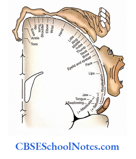
1. Primary motor area: The primary motor area is located in the precentral gyrus, in the anterior wall of the central sulcus and the anterior part of the paracentral lobule on the medial surface of the hemisphere.
This area is designated as Area 4 of Broca. The primary motor area initiates and controls the contraction of skeletal muscles that are mainly located on the opposite side of the body. The fibres arising from the primary motor area run in corticospinal, corticonuclear and corticoreticular tracts.
In the primary motor area, the muscles of various parts of the body are represented upside down. The sequence of representation in the precentral gyrus.
In addition to area 4, the cerebral cortex also has a supplementary motor area (area 6) and a cingulate motor area located on the medial surface of the cerebral hemisphere.
2. Premotor area: The premotor area corresponds to area 6 of Brodmann. It is situated in front of the primary motor area occupying the posterior-most part of the superior, middle and inferior frontal gyri.
Based on experience, the premotor and supplementary motor areas can programme skilled motor activity. The premotor area directs the primary motor area to execute the skilled movements.
3. Frontal eye field: This area is present in the posterior part of the middle frontal gyrus which corresponds to the lower part of Bradman’s area 8. It receives afferents from the visual association areas and sends efferents to the contralateral nuclei of the extraocular muscles. The frontal eye field controls the voluntary conjugate scanning movement of the eyes.
4. Prefrontal cortex: The part of the frontal lobe located anterior to the motor and premotor areas is included in the prefrontal cortex.
This roughly corresponds to Brodmann areas 9, 10, 11 and 12. The electrical stimulation of the prefrontal cortex does not elicit motor response.
Functionally, it acts as an association cortex. Its functions are as follows:
- It is the storehouse of all the past experiences.
- It controls the behaviour based on judgement and foresight.
- Through its connections with the limbic system, the area helps to develop the personality of a person.
5. Motor speech area of Broca: The motor speech area is located in the inferior frontal gyrus; this area corresponds with the pars triangularis (area 45) and pars opercularis (area 44) of the dominant cerebral hemisphere.
In right-handed individuals, the motor speech area is located in the left cerebral hemisphere, while in the case of left-handed persons this area is present in the right cerebral hemisphere.
Broca’s area, through its connections with the primary motor area (area 4), is responsible for the production of speech. A lesion of the motor speech area leads to an inability to speak (motor aphasia).
Lesion of the Prefrontal Cortex
- A bilateral lesion of the prefrontal cortex leads to a change in personality.
- In a normal subject, frustration, tension and anxiety are generated in the prefrontal cortex.
- Therefore, after a lesion of the prefrontal cortex, the patient no longer suffers from depression, anxiety and pain.
- The patient does not care for the norms of social life and becomes rude and inconsiderate to others. He becomes careless, lazy and incapable of judging the consequences of reckless actions.
- It should be noted that despite profound personality changes, the memory and intelligence of a person are not affected.
Functional Cerebral Areas Of Cerebral Cortex Summary
- The cerebral cortex consists of three different types of functional areas:
- Sensory, Motor and Association.
- Brodmann mapped the cerebral cortex in 47 areas based on its differing histological structure and function.
- The sensory areas of the cerebral cortex receive and interpret sensory impulses, the motor areas initiate movement and the association areas deal with functions such as intelligence, memory emotion, reasoning, judgment and personality.
- The primary somatic sensory area (areas 3, 1, 2) is located in the postcentral gyrus.
- The sensory speech area consists of area 22. This area is also known as the sensory speech area of Wernicke. Other areas which help in sensory speech are areas 39 and 40.
- The primary visual area is located in area 1 7 while areas 1 8 and 1 9 are known as visual association areas.
- The primary auditory area is located in areas 41 and 42 on the superior surface of the superior temporal gyrus.
- The primary motor area is located in the precentral gyrus (area 4). This area initiates and controls the contraction of skeletal muscles on the opposite side of the body.
- The frontal eye field is located in the posterior part of the middle frontal gyrus (area 8). This area controls conjugate movements of the eyes.
Functional Cerebral Areas Of Cerebral Cortex Multiple Choice Questions
Question 1. The following different types of functional areas are located in the cerebral cortex except
- Sensory areas
- Motor areas
- Association areas
- Interpretation area
Answer: 4. Interpretation area
Question 2. The following sensory areas are present in the parietal lobe except
- General somatic sensory area
- Vestibular area
- Broca’s motor area of speech
- Area of taste sensation
Answer: 3. Brocas motor area of speech
Question 3. The following functional areas are present in the frontal lobe except
- Frontal eye field
- Premotor area
- Primary motor area
- Broca’s motor area of speech
- Sensory area of speech
Answer: 5. Sensory area of speech
Question 4. Which of the following area(s) of the cerebral cortex are concerned with the recognition of painful stimuli from teeth?
- Precentral gyrus
- Postcentral gyrus
- Superior temporal gyrus
- Cingulate gyrus
Answer: 2. Postcentral gyrus
Question 5. The primary auditory cortex is located in which lobe of the cerebrum?
- Frontal
- Parietal
- Occipital
- Temporal
- Insular
Answer: 4. Insular
Question 6. Damage to the frontal lobe, just anterior to the central sulcus, would affect which of the following functions?
- Somatic motor function
- Somatic sensory function
- Vision
- Hearing
- Taste sensation
Answer: 1. Somatic motor function
Question 7. The motor speech area is located in which of the following gyrus?
- Superior frontal gyrus
- Middle frontal gyrus
- Inferior frontal gyrus
- None of the above
Answer: 3. Inferior frontal gyrus
Question 8. Lesions of uncus are associated with
- Visual hallucination
- Auditory hallucination
- Olfactory hallucination
- None of the above
Answer: 3. Olfactory hallucination
Question 9. All the following statements about the Brocas area are true except
- Lesion of Broca’s area in right-handed persons will not show a significant deficit
- It is concerned with the production of speech
- The area is located between the horizontal and ascending rami of the lateral sulcus
- Lesion results in paralysis of muscles involved in the production of speech
Answer: 4. Lesion results in paralysis of muscles involved in the production of speech
Question 10. Which of the following statement(s) about the motor area of the cerebral cortex is/are false?
- It lies in the precentral gyrus and paracentral lobule
- The motor area is supplied by the middle cerebral artery
- In this area, the body is represented upside down
- The part of the body is represented by an area proportional to the size of the part
Answer: 4. The part of the body is represented by an area proportional to the size of the part