The Digestive System 1 Oral Cavity
The digestive system consists of two groups of organs
- The gastrointestinal tract or alimentary canal: This is in the form of a tube that extends from the oral cavity to the anus. The parts of the gastrointestinal tract are: oral cavity, pharynx, oesophagus, stomach, small intestine, co¬ lon, rectum and anal canal.
- The accessory digestive organs: These are teeth, tongue, salivary glands, liver, gall bladder and pancreas. In this chapter, we shall learn about the organs of the oral cavity and salivary glands pouring their secretion in the oral cavity.
Oral Cavity
In the oral cavity, food is broken into small pieces by the teeth, moistened and mixed with the secretions (which also contain hydrolytic enzymes) of salivary glands, for its passage through the oesophagus to the stomach
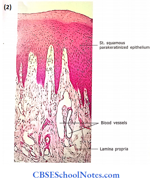
The oral cavity is lined by mucosa composed of stratified squamous epithelium, and a lamina propria of loose areolar connective tissue containing many mucus and serous secreting small glands. As this mucosa is subjected to friction of food, the epithelium at certain places in the oral cavity may show some degree of keratinization or it is para keratinized.
The para-keratinized epithelium is similar to the keratinized epithelium. Here, superficial cells do not lose their nuclei. The nuclei remain in these cells until cells are exfoliated
The lymphatic tissues (diffuse lymphatic tissue, nodules and tonsils) are present beneath the oral epithelium and provide a defence mechanism against infection as the oral cavity is the site of entry of foreign material.
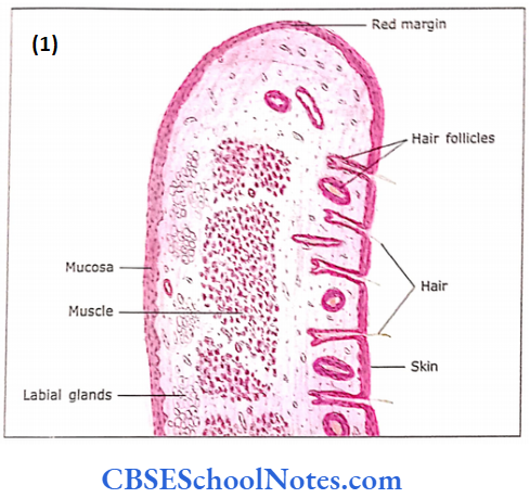
The Lip
The lip consists of an external surface lined by skin, an internal surface lined by mucous membrane and the centrally placed skeletal muscle called orbicularis oris. The external and internal surfaces meet at the free border of the lip, which is also called as “red margin” of the lip.
- The skin ofthe lip is lined by the epidermis, which is keratinized stratified squamous epithelium. In the dermis, there is the presence of hair follicles, sweat glands and sebaceous glands.
- The inner surface flip is lined by non-keratinized stratified squamous epithelium. The lamina propria is present beneath the epithelium and contains labial mucous glands and blood vessels. The secretion of these glands keeps the oral mucosa moist.
- The free border (red margin) ofthe lip is the site of the mucocutaneous junction. It shows the transition ofthe epi¬ dermis ofthe skin to the epithelium of mucosa.
- Beneath the epithelium large number of blood vessels are present. which are responsible for the pink colour ofthe red margin of the lip
The lip Remember:
The external and internal, surfaces of the are lined by the skin and mucous membranes) respectively. These surfaces meet at the free border rof the margin or vermilion zone of the lip. The central core of the lip is formed.
Teeth
The teeth are accessory digestive organs located in the sockets of alveolar processes of the mandible and maxilla. The alveolar processes are covered by the gum. The root of the tooth is attached to the alveolar socket by periodontal ligaments, which consist of dense fibrous connective tissue
General Structure of a Tooth:
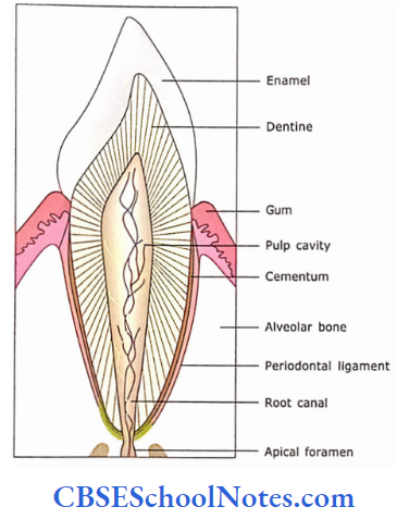
The parts of a typical tooth are. The crown is the part of the tooth above the level of the gum. The part of the tooth that lies embedded in the socket is called as root. The neck is the junction of the crown and root near the gum line
The central portion of a tooth is called as dentine. It is a calcified connective tissue that is harder than bone Dentine encloses a cavity called as pulp cavity. This cavity is filled with pulp, which consists of connective tissue containing blood vessels, nerves and lymphatic vessels. The pulp cavity continues down into the root as the root canal.
Each root canal has an opening at its base, the apical foramen, through which blood vessels, lymphatic and nerve enter. The dentine of the crown is covered by enamel. Enamel is the hardest substance in the body and consists of calcium salts. The dentine ofthe root is covered by cementum, which is a bone-like tissue. The cementum attaches the root to the periodontal ligament
Detailed Structure of Tooth:
1. Dentine:
Dentine forms the wall of the root and roof of the pulp cavity. It is composed of 80% inorganic salts (crystals of hydroxyapatite) and 20% inorganic matter (type 1 collagen and other proteins). It is a hard material similar to bone.
- Dentine is characterized by dentinal tubules radiating outward from the pulp cavity to the outer wall of the tooth.
- These tubules, in a living state, are occupied by the processes of odontoblasts. These cells line the pulp cavity and are tall columnar.
- The apical portion of the cell gives rise to a slender odontoblast process. The dentinal tubules, containing processes of odontoblasts, are parallel to each other.
- The dentine is laid down in layers that lie parallel to the pulp cavity. The layer of newly formed dentine near the apical end of odontoblasts, which is yet to be mineralized, is called dentine.
- Formation of dentine is a continuous slow process, which occurs throughout life and the pulp cavity is gradually narrowed with advancing age.
- The layers of dentine may be separated by less mineralized tissue that forms the incremental lines of Von Ebner. These lines reflect the cyclic pattern of cellular activity during the formation of dentine.
Dentine Remember:
Dentine consists of alcified tubules which are occupied by the processes of odontoblasts. the dentine of the root is covered by cementum, which is a bone-like tissue.
2. Enamel:
Enamel is made up of 99% of inorganic salts (hydroxyapatite crystals). Hence, it is considered to be the hardest substance in the body.
- Histologically, the enamel consists of enamel rods or prisms that radiate from the dentin enamel junction towards the enamel surface.
- The enamel rods are bound together by a thin layer of organic matrix called the prismatic rod sheath
- Enamel is an extracellular product of enamel organ cells. It is produced by a layer of columnar cells called ameloblasts, which covers the crown as an epithelial membrane.
- Following completion of enamel deposition (before the eruption) the ameloblasts wear off and never return.
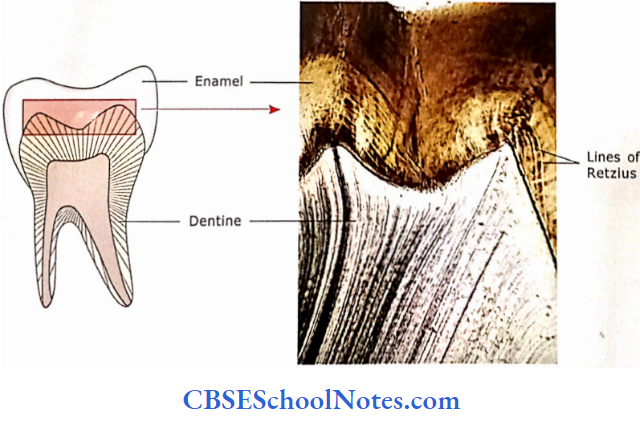
- Thus no further deposition of enamel is possible. Therefore, this layer of ameloblasts is not present in mature teeth. Enamel is produced in waves, producing growth lines.
- These lines are at a right angle to the direction ofthe enamel rods.
- These lines are called contour Lines of Retzius and reflect the wave of enamel formation. Any erosion in enamel (caries) must be repaired through filling.
Enamel Remember:
Enamel is composed of enamel rods, which are made up of calcium hydroxyapatite crystals. Enamel is the hardest substance in the body.
3. Cementum
The enamel at the neck (from the cementoenamel junction) to the apical pore
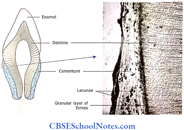
- The cementum is bone-like tissue. It is mineralized. The main organic component is collagen and proteoglycan.
- The cementum contains cementocytes in lacunae.
- Similar to an osteocyte, a cementocyte also contains many processes within canaliculi, which radiate from lacunae.
- Cementum is an avascular tissue in contrast to the bone which is vascular.
- The collagen bundles of the periodontal membrane are anchored to the cementum.
4. Periodontal Ligament:
The periodontal ligament connects the cementum to the bone of the alveolar socket.
- It is made up of both loose and dense connective tissue.
- The dense connective tissue is found in the bulk of the periodontal ligament.
- The loose connective tissue provides passage to the blood vessels and nerves.
- Besides providing attachment and support to the tooth the periodontal ligament also helps in bone remodelling during the eruption of teeth
5. Dental Pulp:
The dental pulp is present in the pulp cavity, which is bounded by mineralized dentine. The pulp on its periphery has a layer of odontoblasts.
- The central portion of the pulp consists of fibroblasts, macrophages, mast cells, collagen fibrils and a large amount of ground substance.
- The neurovascular bundles form a network in the crown portion of the pulp, just beneath the layer of odontoblasts.
- The nerve fibres forming this network contact the cell body of odontoblasts and may also travel in dentinal tubules.
- Because of this reason, dentine is highly sensitive to pain.
- The capillary loops that form the network also enter the layer of odontoblasts.
Tooth Clinical Application
- Dental Caries: If the hygiene of the oral cavity is not maintained properly, the bacteria present in the oral cavity may destroy the enamel and dentine.
- Bacteria when act on sugar present In the oral cavity produce acid. This acid gradually de-mineralizes the enamel and dentine leading to their destruction.
- The resulting condition is called dental caries. If infection is not controlled, it may reach up to the pulp cavity.
- The infection of the pulp is painful as it has sensory nerve endings
- Orthodontic Treatment: With the help of orthodontic treatment, the disposition of teeth can be gradually changed.
- As the periodontal ligament is plastic, it allows the change in the disposition of teeth in the mouth.
- Similarly, cementum has lower metabolic activity because it is not supplied by blood vessels.
- Both the above facts help in the movement of teeth by orthodontic appliances.
Tongue
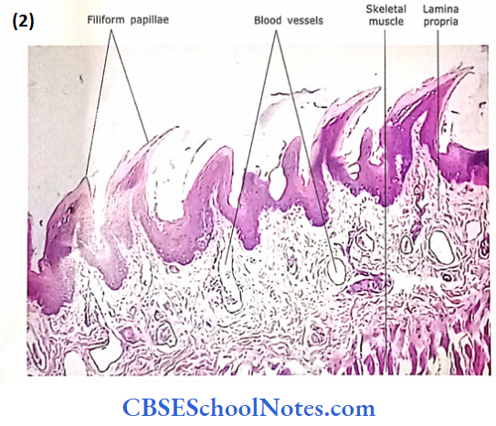
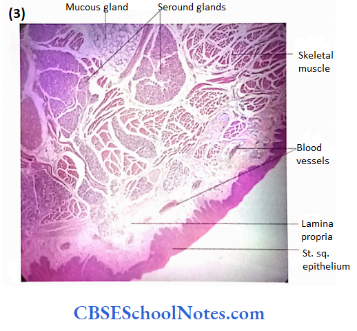
The tongue is an accessory digestive organ composed of skeletal muscle covered with mucous membranes. The lining mucosa consists of stratified squamous epithelium and a lamina propria.
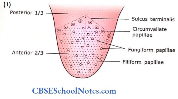
An inverted V-shaped groove, the sulcus terminalis, divides the dorsal aspect of the tongue into anterior 2/3 (body of the tongue) and posterior 1/3 (the root of the tongue) The dorsal aspect of the root of the tongue (posterior 1/3) contains many oval or rounded elevations, which are due to the lingual tonsils.
These elevations may contain lymph nodules and may show the presence of germinal centres in. Elsewhere, where lymph nodules are not present, the mucosa shows the general properties of lining mucosa. The lingual salivary glands of the posterior 1/3 of the tongue are mostly mucous and are located in the muscular layer. These glands open into the recesses of the mucosa.
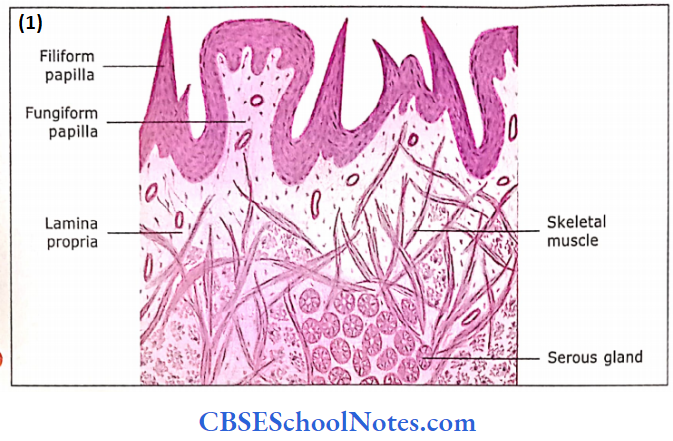
1. Lingual Papillae
The body of the tongue (anterior 2/3), on its dorsal surface, is covered with the specialization of epithelium called the lingual papillae. The lingual papillae are projections of lamina propria covered with stratified squamous epithelium, which may be keratinized. Many papillae contain taste buds.
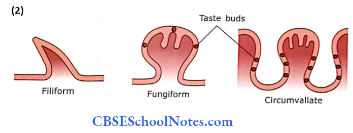
The papillae are of four types:
- Filiform
- Fungiform
- Circumvallate and
- Foliate.
The foliate papillae are not well-developed in humans
1. Filiform Papillae:
These are most numerous ofthe lingual papillae, covering most of the anterior 2/3 of the tongue. These are conical projections about 2-3 mm in length. Their tips are keratinized. These papillae are distributed in parallel rows. Filiform papillae contain no taste buds: the increase the friction between the tongue and food
2. Fungiform Papillae:
They are found distributed among the filiform papillae. Their shape is like a mushroom (narrow base with globular upper end) and they project above the filiform papillae. They have highly vascularized connective tissue cores: hence, visible on as red dots. The taste buds are present in the epithelium on its dorsal surface
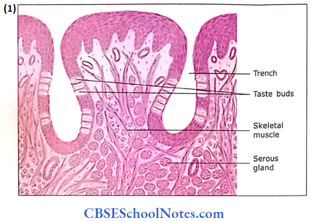
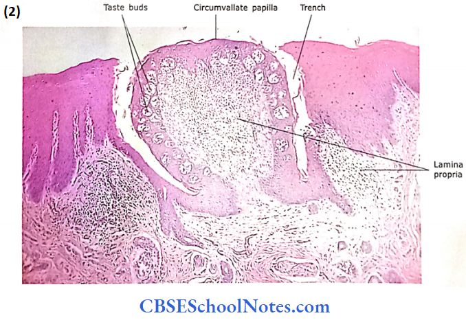
3. Circumvallate Papillae:
- Circumvallate papillae are situated just in front of the sulcus terminalis.
- They are about 8- 16 and each one measures about 1- 2 mm in diameter.
- Each papilla is surrounded by a circular sulcus (trench). The stratified squamous epithelium, covering the free surface, is smooth while the epithelium covering the walls of the sulcus (trench) contains many taste buds.
- At the bottom of the trench, there are openings in the ducts of serous glands on Ebner, which are situated in the submucosa. The secretion of these serous glands keeps the sulcus rinsed.
- A core of connective tissue, supporting vessels, lymphatics and nerves fills the papillae
Papillae Remember:
Three common papillae found in humans are filiform fungiform, and circumvallate. Taste buds are present in fungi form and circumvallate papillae. The foliate papillae are not well-developed in humans
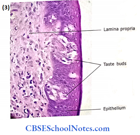
2. Taste Buds
Taste buds are neurosensory epithelial structures embedded in the surface epithelium of the tongue.
- The taste buds are also present in the epiglottis, soft palate and oropharynx. Tongue, they are present in the epithelium of fungiform and circumvallate papillae
- Taste buds appear as onion-like, oval, pale staining epithelial structures.
- 50-80 pm in length and 30-50 pm in width. The length of the taste bud extends through the full thickness of the epithelium vertically.
- The cells of taste buds are long and vertically oriented. Their tapering apical ends converge on a small depression (opening) on the surface of the epithelium. This depression (operating) js ca|iecj as taste pore
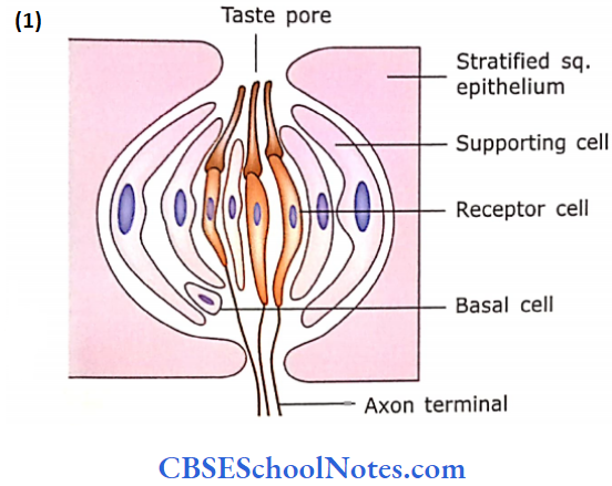
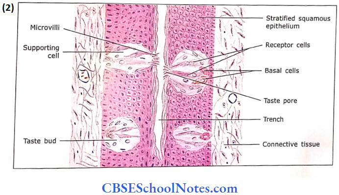
Three different types of cells are present in the taste blends:
- Neuroepithelial cells or Receptor cells
- Supporting cells and
- Basal (stem) cells
These are
- The neuroepithelial (receptor) cells:
- The neuroepithelial and supporting cells are elongated and extend from the basal lamina to the taste pore.
- Both kinds of cells send their microvilli in the taste pore. Nerve axons form a plexus around each taste bud.
- Their branches penetrate the basal lamina of the taste bud and make contact with the receptor cells. It is believed that this cell type is the principal taste receptor
- Supporting cells: The supporting cells besides providing support to receptor cells, also secrete an amorphous dense material at the taste pore.
- Near the basal lamina: Near the basal lamina there are small dark cells called basal or stem cells. The function of basal cells is to provide new receptors and supporting cells by their mitotic activity.
Taste is a chemical sensation. Various types of chemicals (tastants) stimulate neuroepithelial (receptor) cells of taste buds. We can feel sweet, salty, bitter, sour and delicious tastes through our receptor cells.
The tastants, which are with salt and sour taste act by opening and passing through ion channels of neuroepithelial cells. The sour tastants act by closing ion channels while bitter, sweet and delicious
tastes act by acting on specific taste receptors present in neuroepithelial cells.
Taste buds Remember:
Taste buds appear as onion-like, oval, pale staining epithelial structures. Three different types of cells are present in the taste buds: receptor cells, supporting cells and basal (stem) cells. We can feel sweet, salty, bitter, sour and delicious tastes through our receptor cells
3. Muscles of the Tongue
The core of the tongue (beneath the mucous membrane) contains striated muscle. The muscle fibres are arranged in transverse, longitudinal and vertical bundles. In between the muscle bundles, variable amounts of loose connective tissue, adipose tissue and lingual glands are found.
Salivary Glands
The salivary glands are accessory glands of digestion and pour their secretion (saliva) into the oral cavity.
There are two kinds of salivary glands:
- Minor salivary glands: These are small salivary glands situated in the mucous membrane ofthe lip (labial), cheeks (buccal), soft palate (palatine) and tongue (lingual).
- Major salivary glands: These are parotid, submandibular and sublingual glands. Each of these glands lies completely outside the alimentary tract and is connected to it by an excretory duct. These glands are classified as compound alveolar or tubuloalveolar.
Salivary Glands Functions
Salivary glands secrete saliva, which is composed of water, mucus, proteins, salts, salivary amylase (ptyalin immunoglobulins (particularly IgA), and lactoperoxidase
- It serves to moisten food
- Lubricates and moistens oral mucosa and lip.
- It initiates the digestion of carbohydrates.
Basic Organization of Salivary Glands
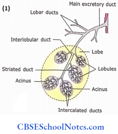
As stated earlier, the major salivary glands are compound glands that consist of multiple acini at the end of the highly branched duct system.
- An acinus is defined as a group of secretory cells that are arranged around a narrow lumen.
- The duct and acini of the salivary gland resemble a bunch of grapes in which ducts represent the branching stem and acini represent the grapes.
- Acini are made up of either serous or mucous cells.
- There may be a mix of both serous and mucous acini these are called mixed salivary glands.
- In addition, some of the mucous acini may have a cap of serous cells called the parotid gland is chiefly a serous, while submandibular and sublingual are mixed glands.
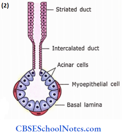
- A salivary gland contains branching ducts, which are classified as in intralobular (intercalated and striated), interlobular, lobar and main excretory ducts.
- These ducts are lined by cuboidal (intercalated), columnar (striated), pseudostratified columnar (interlobular), stratified cuboidal (lobar) and major excretory ducts are lined by stratified columnar epithelium.
Salivary glands Remember:
Major salivary glands consist of multiple acini at the end of the highly branched duct system. Acini are of three types: serous, mucous or mixed. Some of the mucous acini may have a cap of serous cells called serous demilune. Ducts are classified as intralobular (intercalated and striated), interlobular, lobar and main excretory ducts
Salivary glands other Details Of Basic Organization:
A salivary gland consists of two components
- Connective tissue component
- Parenchymal components (ducts and acini)
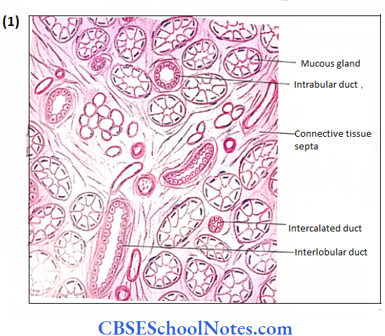
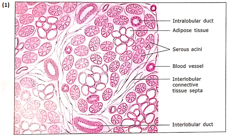
The parotid and submandibular glands are surrounded by a well-developed connective tissue capsule. Septa arising from the capsule enter the parenchyma of the gland and divide the gland into lobes and lobules.
The acini and intralobular ducts are enveloped by delicate connective tissues. The connective tissue septa form a medium through the parenchymal component of the salivary gland consisting of acini and ducts.
The acini are composed of pyramidal secretory cells, which are arranged around a narrow lumen and surrounded by myoepithelial cells. The myoepithelial cells are present between secretory cells and basal lamina The lumen of acini is continuous with the duct system
Duct System
The main excretory duct of a salivary gland divides to give lobar and interlobular ducts. The interlobular ducts further divide to give intralobular ducts. The intralobular ducts are of two types, i.e., intercalated and striated. The lumen of the intercalated duct opens directly into the acinus.
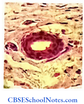
1. Intercalated Ducts:
The intercalated ducts arc of short length and serve to connect acini with the striated ducts.
- These ducts are lined by short cuboidal ceils or sometimes by simple squamous epithelium.
- The cells of intercalated ducts, similar to acini may also be surrounded by myoepithelial cells.
- Intercalated ducts are prominent in the serous glands. They secrete HCO3– into the acinar product and absorb Cl– from the product.
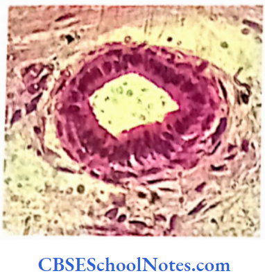
2. Striated Ducts:
The striated ducts connect interlobular ducts to intercalated ducts. These ducts are lined by simple columnar epithelium.
- These cells are stained bright pink in H&E preparation and have nuclei located near the lumen.
- These cells also show basal striations, which are due to the vertical orientation of mitochondria lodged between basal infoldings of the plasma membrane.
- This characteristic is typical of cells engaged in salt and water transport.
The intercalated and striated ducts are predominantly present in serous glands. These ducts are involved in the modification of the serous secretion (by absorbing certain constituents and secreting others, i.e., secretion of K+ and HCO3– and absorption of Na+).
As the mucus secretion is not modified while passing through the duct, the intercalated ducts are poorly developed and striated ducts are absent in mucous glands.
Striated duct Remember:
The basal plasma membrane of striated duct cells is highly folded. The longitudinally oriented, elongated mitochondria are enclosed in these foldings. Striated ducts are the sites of reabsorption of sodium and secretion of potassium
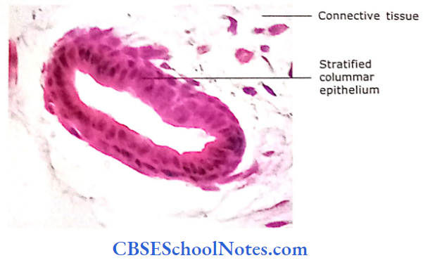
3. Interlobular, Lobar and Main Excretory Ducts:
These larger ducts are lined by epithelium that ranges from simple columnar to pseudostratified columnar (interlobular ducts) to stratified columnar or stratified cuboidal (lobar and main excretory ducts) At its opening in the oral cavity, it may be lined by stratified squamous epithelium. These ducts are surrounded by a large amount of connective tissue.
The Secretory Acinar System
The acini are described as
- Serous
- Mucous and
- Mixed

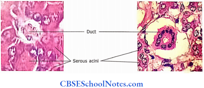
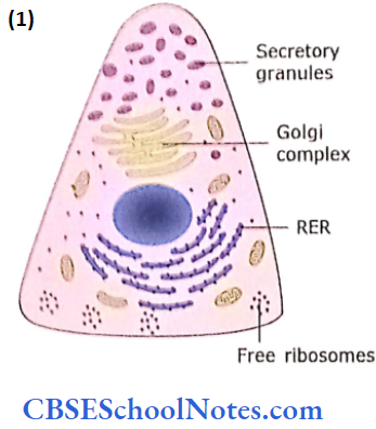
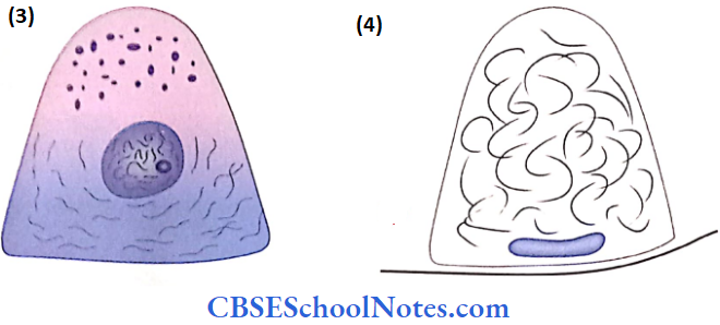
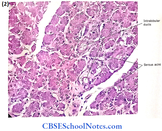
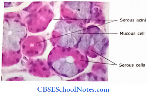
1. Serous Acini:
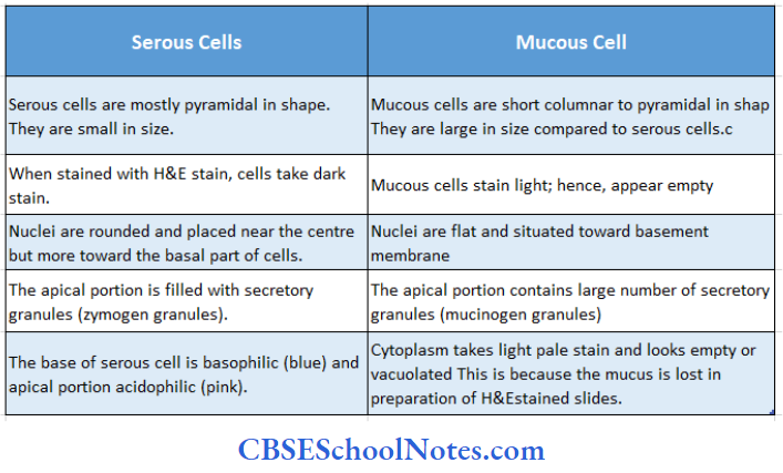
Serous acini have a small lumen and produce a clear watery secretion. Serous cells are usually pyramidal and have rounded nuclei located near the base.
- A large amount of rough endoplasmic reticulum and free ribosomes are present near the base.
- Because of this acini are stained with haematoxylin (basophilic) near the base.
- Above the nucleus and Golgi complex, apical secretory granules (zymogen granules) are present.
- The presence of secretory granules is responsible for the staining of the apical portion by eosin.
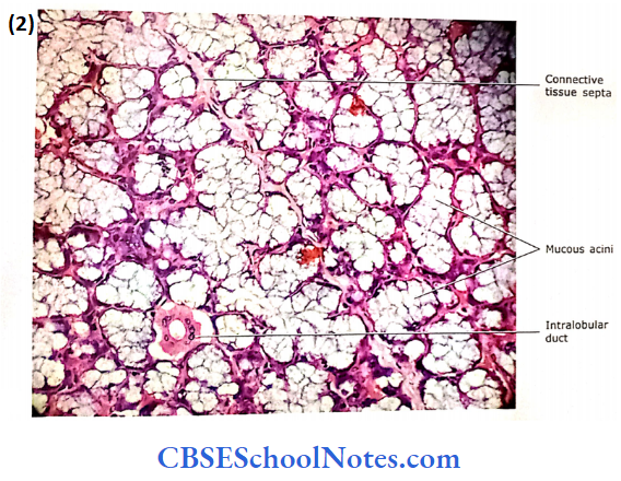
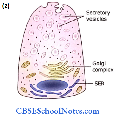
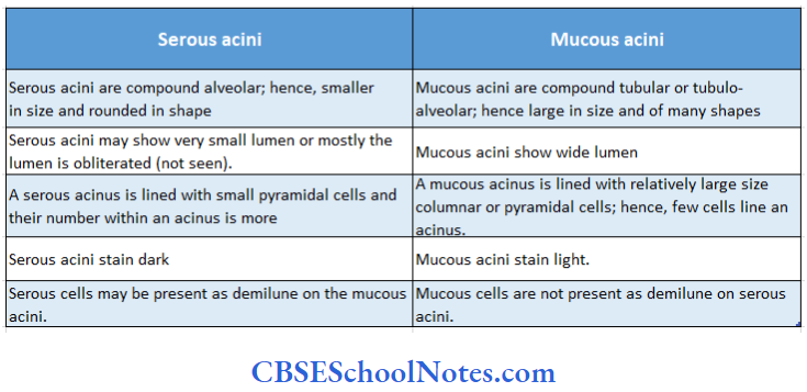
2. Mucous Acini:
The mucous acini arc is usually tubuloalveolar and has relatively wide lumens.
- Mucous cells arc roughly pyramidal or columnar and have flattened nuclei situated near their base.
- The apical portion of the cell contains mucus, which is stored in the form of mucinogen granules.
- Because the mucus is lost in the preparation of H&E-stained paraffin sections,
- The apical portion ofthe cell is not stained and shows an empty appearance
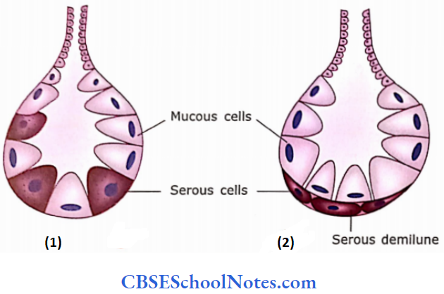
3. Mixed Acini:
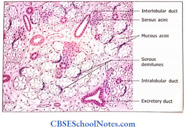
Submandibular and sublingual salivary glands contain both (serous and mucous) types of acini. In addition, some ofthe mucous acini may have the cap of serous cells called serous demilune.
Differences between serous and mucous acini:
Myoepithelial Cells (Basket Cells):
These star-shaped (branched) cells are present between the epithelium and basal lamina of the acinus and the proximal part of the duct. They contain actin filaments and hence are contractile. Their contraction is thought to help in expelling the secretion from the lumen facing.
Parotid Gland
The parotid gland is the largest salivary gland. It has a well-defined capsule. Septa divide the gland into lobes and lobules.
- Secretory acini: The acini are purely serous The gland has abundant myoepithelial (basket) cells that help to expel the secretory product from the lumen of the acinus
- Ducts: The interlobular ducts are present in the connective tissue septa. These ducts may be lined by simple columnar or pseudostratified columnar epithelium. The intralobular ducts are seen between acini. The intercalated ducts are lined by simple squamous to low cuboidal epithelium.
They are long and branching in the parotid gland. The striated ducts are lined by simple low columnar epithelium and show basal striations. The striations are responsible for the bright eosinophilic (acidophilic) staining reaction of these ducts The adipose tissue may be seen among acini and smaller ducts.
Parotid gland Clinical Applications
Mumps:
The infection of the parotid gland by mumps virus (Myxovirus) is called mumps. In this disease, there is swelling and enlargement of the parotid gland and severe pain in the throat. The infection may also spread to other salivary glands. The disease is self-limiting
Submandibular Gland
The submandibular gland is a mixed gland. It has a well-defined capsule and septa, which divide the gland into lobules.
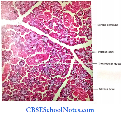
- Secretory acini: It consists of both serous and mucous acini However, sometimes the serous acini are more than mucous. At certain places, mucous acini are capped by a layer of serous cells, which may present a half-moon appearance in some sections; hence, they are called serous demilunes.
- Ducts: The epithelial lining of the main, interlobular and intralobular ducts are the same as in the parotid gland. However, the striated ducts of the submandibular gland are large and branching; hence, many are seen between acini.
Serous demilunes are, not the true histological structures:
The serous demilunes are observed in the mixed salivary gland (both under light and electron microscopes) after the tissue is fixed by the routine fixation method. However, when
- The tissue is preserved by the rapid freezing method and observed under the transmission electron microscope, a different kind of histological structure of the gland is observed.
- Here, the acini of mixed glands consists of both serous and mucus-secreting cells.
- One end of these cells rests on the basal lamina and the other end projects into the lumen of acini. Both the serous and mucous cells arc almost of the same size.
- The serous cells arc pyramidal in shape and the shape of mucous varies from short columnar to pyramidal.
- The mucous cells contain centrally located rounded nuclei and many mucinogen granules in the apical region.
- Both types of cells pour their secretion into the lumen of acinus. This appearance ofthe mucous cells seems to be the real one as the rapid freezing method preserves the tissue with minimal alteration in its morphological structure and chemical composition.
The acini, as seen in resembles the above description of tissue preserved with the rapid freezing method.
- However, when the tissue slides are prepared by using routine chemical fixative (usually formaldehyde), then the fixative causes the swelling of mucinogen granules in the mucous-secreting cells.
- This pushes the nucleus ofthe cell towards the basal lamina, which under the pressure of mucinogen granules becomes flattened. These swollen mucous cells also push the pyramidal-shaped serous cells towards the periphery of the acinus.
- It appears as if mucous acinus has having crescentic-shaped cap of serous cells, which is traditionally described as serous demilune
The tissue was obtained from an adult goat and preserved in formaldehyde.
Even though this tissue was not preserved by the rapid freezing method, its ducts showed similar features as described for rapid freezing fixed tissue.
Question 1. Why we must have found these kinds of mixed acini after routine fixation?
Answer:
The possible explanation for this may be that the goat is a ruminating animal as partially digested cud comes back in its mouth at intervals. As these animals constantly chew, the saliva is also produced constantly in large quantities.
This may be the reason for the nonaccumulation of a large quantity of mitogen granules in the mucous-secreting cells in a goat. During chemical recreation, though these granules swell they may not exert much pressure on the neighbouring serous cells. Thus, in most ofthe mixed acini serious demilunes fail to form
The above description indicates that not only the serous demi lunes but also the large size of mucous cells, the basal position and the flattered shape of its nuclei are also the artefacts of chemical fixation.
Chemical fixation Remember:
Serous demilunes and mucous cells as observed in histological slides after chemical fixation, are not the true histological structure but are artefacts of chemical fixation.
Differences between serous and mucous cells
Sublingual Gland
It has no definite capsule. The septa divide the gland into lobules
- Secretory acini: The sublingual gland is made up predominantly of mucous-secreting acini Some mixed acini with serous demilunes are also io seen. The pure serous acini are rarely seen. Thus, it is classified as a mixed salivary gland.
- Ducts: The main and interlobular ducts are present. Very’ few striated ducts are seen in this section. Intercalated ducts are difficult to find (rarely seen).