
Introduction to Connective Tissue Components
What is Connective Tissue?
Connective Tissue Definition:
Connective tissue is a term that is used for a widely dispersed group of tissues performing a variety of functions. As the name suggests, connective tissues serve as a connecting system binding, supporting, and strengthening all other tissues together.
- It protects and insulates internal organs by forming capsules, sheaths, or septas. Blood, a fluid connective tissue, acts as a major transport system within the body.
- Fat is stored in the superficial fascia, which is a major site for the storage of energy reserves and provides insulation against heat loss from the skin. Blood vessels and nerves enter or leave an organ by passing through the connective tissue only.
General Features Of Connective Tissue
Connective tissue consists of two basic components: cells and matrix. The extracellular matrix itself is made up of two components, i.e., fibers and ground substance.
- Cells in the connective tissue are widely separated from each other because of the presence of an abundant extracellular matrix. Usually, the matrix consists of a gel-like ground substance in which fibers are embedded.
- In some, connective tissue matrices may be fluid, semi-fluid, gelatinous, fibrous, or calcified. The fibers and ground substance (matrix) are synthesized by the cells of connective tissue.
- A tissue can be designated as connective tissue only when it has all three components, i.e., ground substance, fibers, and cells.
1. Ground Substance
The ground substance is colorless, sol to gel in consistency, It is highly hydrated. Fibers are embedded in it. The cells of the connective tissues are surrounded by it.
- Ground substance supports cells and provides a medium through which substances are exchanged between blood and cells. It is made up of complex molecules of polysaccharides and proteins.
- It contains many types of proteoglycans, multi-adhesive glycoproteins (laminin and fibronectin), and glycosaminoglycans (dermatan sulfate, keratan sulfate, hyaluronic acid, etc).
Ground Substance Remember
- A tissue can be designated as connective tissue only when it has all three components, i.e., ground substance, fibers, and cells.
- The extracellular matrix provides mechanical and structural support for tissue and helps in extracellular communication.
2. Fibers
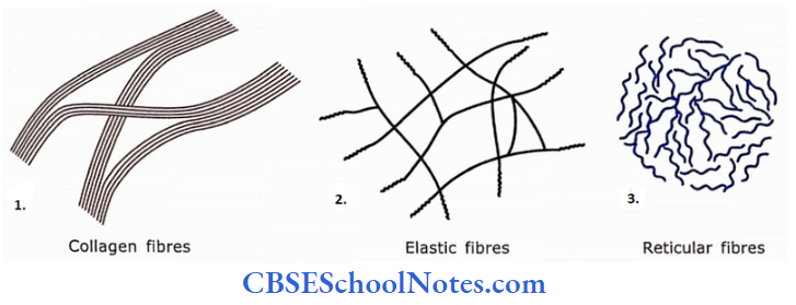
Fibers in the matrix provide strength and support to the connective tissue. Three different types of fibers may be found in the matrix: collagen, elastic, and reticular.
Collagen Fibres
Collagen fibers are the main fibers of the connective tissue. They are found in abundance in bone, cartilage, tendon, and ligament. These fibers are strong, and inelastic but flexible (20 to 300 nm in diameter).
- Collagen fibers mostly occur in bundles which may branch and anastomose with neighbouring bundles. However, an individual fiber does not branch.
- A bundle measures about 0.5 to 1 0 pm in diameter and is of indefinite length. A collagen flber shows faint transverse striations indicating that it is made up of smaller subunits.
- Collagen fibers run in bundles, which may branch and anastomose with neighboring bundles.
- Elastic fibers run singly and not in bundles. They branch and join together to form a network.
- Reticular fibers are much thinner than collagen fibers and form a branching network.
- Chemically, collagen fibers are made up of protein collagen that in turn is made of tropocollagen molecules. They are synthesized by fibroblasts.
At present, 28 different types of collagen fibers have been identified. They are designated as type 1, type 2, type 3, type 4 to 28, etc.
Further Details
Distribution of commonly occurring collagen fibers in the body
Following is a brief description of the most commonly occurring types of collagen fibers.
Type 1 Collagen
It is the most common of all the collagen types (90% of body collagen is type 1). These fibers show classical cross striations and are of large diameter.
These fibers are found in dense and loose connective tissue, bone, tendon, fascia aponeuroses, ligaments, skin, cornea, and dentine.
Type 2 Collagen
It consists of thin fibers showing faint cross striations. These types of fibers are found in hyaline cartilage, the vitreous of the eye, nucleus pulposus of the intervertebral disc.
Type 3 Collagen
These types of collagen form reticular fibers (see reticular fibers). It is present in connective tissue of organs (spleen, lung, liver, etc).
Type 4 Collagen
This type of collagen forms meshwork in the basal lamina (lamina densa) of epithelia. It is also present in the kidney, glomeruli, and lens capsule.
Type 5 Collagen
It is present in the placenta and is associated with type 1 collagen.
Type 7 Collagen
Present in anchoring fibrils that attach the basal lamina to the lamina reticularis.
Types 1,2 and 3 collagen can be seen by light microscope, while the remaining collagen types are detectable only by the use of specific antibodies.
Synthesis of Collagen Fibres
Fibroblasts synthesize fibers and ground substance components of connective tissue. Besides fibroblasts, many other cells of the body also synthesize collagen fibers.
- These cells are mesenchymal cells, perineurial cells, cementoblasts, odontoblasts, cartilage cells, and smooth muscle cells. Type 4 collagen, which is found in basal lamina, is synthesized by epithelial cells.
- The synthesis of collagen fibers by fibroblasts takes place in two different steps: intracellular and extracellular.
Synthesis of Collagen Fibres Remember
Synthesis of collagen fibers occurs both inside and outside the fibroblast.
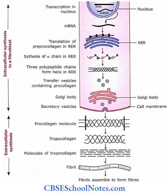
Intracellular Synthesis
- With the help of mRNA, amino acids are arranged sequentially to form alpha polypeptide chains.
- These chains are then transferred to the lumen of the rough endoplasmic reticulum.
- The polypeptide chains undergo the following modifications in the rough endoplasmic reticulum:
- Proline and lysine amino acids of the chain are converted to hydroxyproline and hydroxylysine.
- Hydroxylysine combines with sugar groups.
- Three polypeptide chains now form a helix (triple helix). However, both the terminal ends of these chains remain uncoiled.
- This molecule is now called procollagen. [Vitamin C is necessary for the formation of procollagen. Therefore, the deficiency of vitamin C leads to non-healing of wounds, and the formation of bone is impaired.]
- These procollagen molecules now move from the rough endoplasmic reticulum to the Golgi complex. There occurs the packaging of soluble procollagen in secretory vesicles.
- From here these molecules are secreted in the extracellular space through secretory vesicles.
Extracellular Synthesis
- Various enzymes act on the terminal uncoiled portion of procollagen molecules and cleave it from the rest of the coiled portion. These enzymes (procollagen peptidase) are secreted by fibroblasts themselves.
- After cleavage of the procollagen molecule, the remaining molecule is now called a collagen molecule or tropocollagen molecule.
- The tropocollagen molecules aggregate in an orderly manner to form the collagen fibrils.
- The collagen fibrils assemble to form microscopically visible collagen fiber.
Extracellular Synthesis Remember
Collagen is not only synthesized by fibroblasts but also by many other cells of the body, i.e., chondroblasts, osteoblasts, epithelial cells resting on the basement membrane, mesenchymal cells, perineurial cells, cementoblasts, odontoblasts, and smooth muscle cells.
Elastic Fibres
The diameter of elastic fibers is 0. 1 to 0.2 pm. They run singly and branch to form a network in loose areolar tissue but are present in bundles in ligamentum flava and ligamentum nuchae.
- Chemically, elastic fibers consist of a protein called elastin. The fiber is surrounded by a glycoprotein called fibrillin. Fibrillin plays an important role in organizing elastin into fibers.
- Absence of fibrillin results in the formation of elastin sheets or lamellae, instead of elastic fibers, as found in blood vessels.
- Elastic fibers are strong and can be stretched up to 150% of their relaxed length. Their elasticity is due to the protein elastin. These fibers are stained dark brown with orcein and Verhoeffs methods.
- They are synthesized by fibroblasts and smooth muscle cells of blood vessels. Elastic fibers are mainly found in skin, blood vessels, and lung tissue.
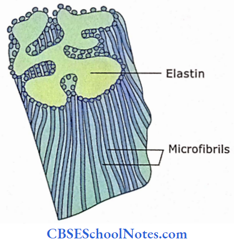
Elastic Fibres Remember
- In relaxed conditions, the molecules of elastin are coiled and cross-linked to each other to form a network. The stretching leads to the uncoiling of these molecules leading to an increase in the length of the elastic ligament or membrane or lamella, etc.
- When the stretching force is withdrawn, the elastin molecules return to a relaxed condition (coiled condition).
![]()
Elastic Fibres Clinical Applications
Marfan Syndrome
- It is a disorder, which is due to the abnormal development of elastic fibers. Marfan’s syndrome is inherited as autosomal dominant and is due to a mutation of the fibrillin gene(FBN1).
- Tissues rich in elastic fibers, i.e., large arteries, I periosteum (a membrane covering the bone), and the ligaments that suspend the lens of the eye are weakened.
- This results in blurred vision due to displacement of the lens. Long bones become abnormally large and there occurs the weakening of the wall of the aorta. The weakened aorta may suddenly rupture leading to sudden death.
Comparison of the properties of connective tissue fibers
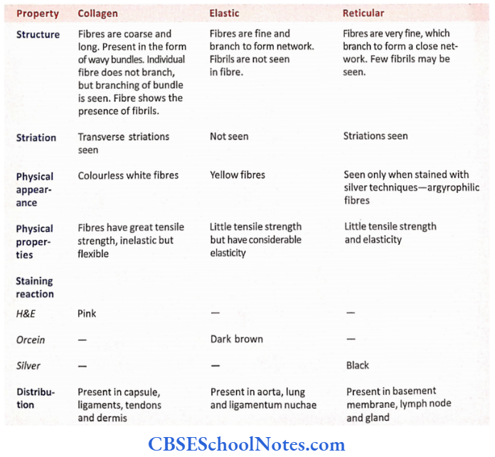
Reticular Fibres
- These are fine delicate strands (20-40 nm in diameter), that form a supportive network for many tissues.
- They do not run in bundles.
- Chemically, they consist of collagen (type 3) and have a coating of glycoprotein.
- They can be stained black by the silver impregnation method. Because of their affinity to silver salts, these fibers are called argvrophilic. These fibers are also PAS-positive.
- These fibers are also synthesized by fibroblasts. The other cells, which produce reticular fibers are reticular cells in lymphatic tissues, Schwann cells in endoneurium, and smooth muscle cells in blood vessels and the intestine.
- Reticular fibers provide support and strength and form a supporting framework around fat cells, nerve fibers, and smooth muscles. They also form the framework of the spleen, lymph nodes, bone marrow, liver, and glands.
- These fibers also help to form the basement membrane.
Reticular Fibres Remember
Reticular fibers are arranged in the form of a network (meshwork), which is necessary to provide support to glandular and epithelioid tissues.
3. Cells of Connective Tissue
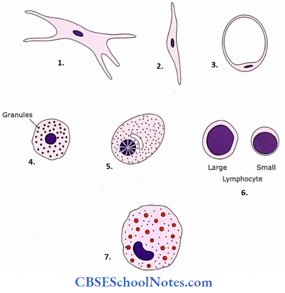
The cells of connective tissue are of two different types: fixed cells and free cells. The fixed cells are long-lived. These include fibroblasts, myofibroblasts, pericytes, fat cells, mast cells, and pigment cells.
The free cells arc short-lived wandering cells that are continually replaced by cells of the blood. These cells include eosinophils, neutrophils, monocytes, lymphocytes, mast cells, and plasma cells.
Fibroblasts
- They are the most numerous connective tissue cells and are present in almost all types of connective tissues.
- Fibroblasts are large, fiat, spindle-shaped cells that have many branching processes.
- Fibroblasts are responsible for the synthesis of extracted lularmatrix (secretion of ground substance and all types of connective tissue fibers).
- Inactive fibroblasts are called fibrocytes. Active fibroblasts have a large quantity of basophilic cytoplasm and euchromatic nuclei.
- They become very active during wound repair and synthesize collagen fibers. Here, they lie parallel to the long axis of the fibers.
Fibroblasts Remember
Fibroblasts are the most numerous connective tissue cells and are involved in the synthesis of all types of connective tissue fibers and ground substances.
- Active fibroblast
- Inactive fibrocyte
- Fat cell
- Mast cell
- Plasma cell
- Lymphocyte and
- Macrophage.
Myofibroblast
The myofibroblast is a cell showing features of both fibroblast and smooth muscle cells. In appearance, these cells resemble fibroblasts but contain actin and myosin filaments in large amounts.
- The myofibroblasts possess contractile properties and behave like smooth muscle cells. However, a myofibroblast differs from a smooth muscle cell in that it is not surrounded by basal lamina.
- Their activity is responsible for wound closure after tissue injury due to the contraction of the wound. They are also found in periodontal ligaments probably helping in tooth eruption.
Pericytes
Pericytes surround the endothelial cells of capillaries and venules. They are surrounded by their basal lamina, which fuses with the basal lamina of endothelium.
Pericytes show features of both endothelial and smooth muscle cells. They contain actin and myosin filaments and, thus are involved in contraction.
Fat Cells
Fat cells (adipose cells) synthesize and store large quantities of lipids. A fat cell is spherical. The nucleus is flattened and displaced to one side.
- The lipids occupy almost the whole of the cell, pushing the cytoplasm as a thin rim around it. The cell may occur singly as in loose areolar tissue or they may occur in groups as in adipose tissue.
- Fat cells are stained orange with Sudan 3 stain. In H and E, stains fat is dissolved, so cells appear empty (un-stained). Their appearance is like a signet ring. Fat cells are fully differentiated and do not undergo cell division.
Pigment Cells
Pigment cells are capable of synthesizing pigment melanin which is a dark brown pigment. These cells are called melanocytes.
These cells are found in the skin, iris, some areas of the brain, and the choroid. These cells are star-shaped with branching processes. In the skin, they protect the tissue from the harmful effects of ultraviolet rays.
Macrophages (histiocytes)
Macrophages develop from monocytes of blood. Macroph ages are capable of eating bacteria and cellular debris by the process of phagocytosis. They have irregular shapes with short branching projections.
- They have an indented nucleus. Macrophages are of two types: fixed macrophages and wandering macrophages. When the need arises macrophages may fuse to form giant cells (Langhans cells).
- The Langhans cells are very large and may contain up to 100 nuclei. They can engulf large foreign bodies.
Mast Cells
Mast cells are found mostly close to the blood vessels in connective tissue. These cells are small and oval. The nucleus is centrally placed and the cytoplasm contains many granules.
- These cytoplasmic granules take purple to red color when stained with toluidine blue stain, while the nucleus is stained blue. This kind of color reaction is called metachromasia.
- These granules contain heparin which acts as an anticoagulant of blood. Mast cells also release histamine that in turn causes contraction of smooth muscles (mainly of bronchioles) and dilates small blood vessels as part of allergic reaction.
- Leukotricnc C released by mast cells causes bronchospasm. Mast cells also produce TNF-a (tumor necrosis factor-a), interleukins, growth factors, and prostaglandins.
- The release of chemicals from mast cells promotes allergic reactions (For Example., immediate hypersensitivity reactions or anaphylactic shock).
Mast Cells Remember
Mast cells contain numerous granules in their cytoplasm These granules contain heparin, histamine, prostaglandin din, and many other chemicals involved in the inflammatory process.
Plasma Cells
Plasma cells arise from B-lymphocytes. They synthesize antibodies against antigens. Thus these cells are found more in connective tissue at the time of infection. Plasma cells are ovoid.
- The cytoplasm is basophilic due to the presence of abundant RER. There is a clear area of cytoplasm near the nucleus for the Golgi complex. The nucleus is round and eccentrically placed.
- The chromatin pattern is unique, giving it a cartwheel appearance. All the above features indicate that plasma cells are actively involved in protein (antibody) synthesis. The toxic antigen may be neutralized when it combines with the respective antibody.
Plasma Cells Remember
Plasma cells originate from B lymphocytes and synthesize antibodies.
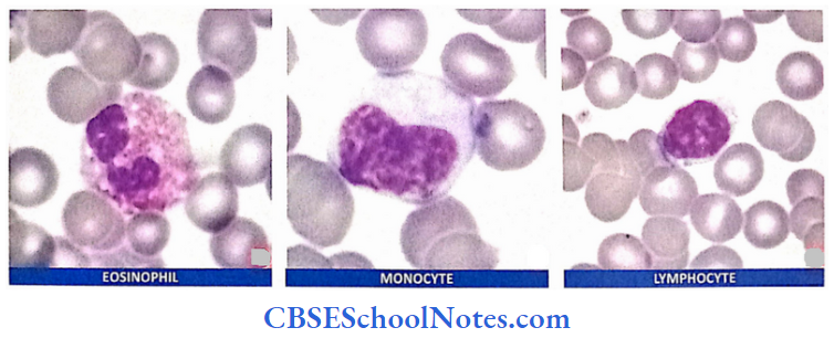
White Blood Cells
Three different types of white blood cells are seen in connective tissues, i.e., lymphocytes, monocytes, and eosinophils. Lymphocytes play an important role in the defense of the body against microorganisms (bacteria etc.).
Monocytes act as macrophages, while eosinophils play an important role in allergic reactions. Thus all these cells function towards the defense of the body.
Plasma Cells Clinical Applications
Mast Cells and Anaphylactic Shock
Some persons when exposed to an antigen (substances foreign to the body, For Example. ( bee venom after a bee sting, some food, or certain drugs) becomes oversensitive to the antigen.
- This means that they produce antibodies as allergic reactions against antigens. These antibodies remain attached to the surface of mast cells.
- When they are exposed to the same antigen (bee venom, food, or drug) for the second time the antigen-antibody reaction takes place on the surface of the mast cells.
- This triggers the release of the contents of mast cell granules (i.e., heparin, histamine, and other chemicals) within a few minutes after the bee sting or taking the drug.
- Histamine causes contraction of smooth muscles (mainly of the bronchiole leading to wheezing and difficulty in breathing); the dilatation of blood vessels leading to the swelling of the face and sudden fall in blood pressure.
- This condition is called an anaphylactic shock. Anaphylactic shock is a serious condition and if not treated urgently may lead to death.
Functions Of Connective Tissue
The following main functions can be attributed to the connective tissue:
- Support: Connective tissue provides support to the epithelium, For Example., lamina propria.
- Strength: It provides tensile strength to those areas that are subjected to mechanical stress, For Example., the dermis of the skin, ligament retinaculum, etc.
- Storage: Fat cells store fat while ground substances store water, ions, and inorganic materials.
- Transport: Water, ions, and inorganic materials are transported from blood to the various tissues of the body through a connective tissue matrix.
- Packing: Connective tissues act as packing material as they fill spaces, For Example., loose connective tissue and adipose tissue. Connective tissue also forms capsules around organs.
- Repair: Connective tissue helps in wound healing.
- Defense: The cells of the connective tissue (plasma cells, lymphocytes, macrophages, monocytes, and eosinophils) function toward the defense of the body.