
Circulatory System
The circulatory system consists of the heart and blood vessels.
- The heart is a muscular pump, which facilitates the circulation of blood through all tissues and organs of the body by its rhythmic concentration.
- Blood vessels form a closed system of tubes that carry blood away from the heart to the tissues ofthe body and then return it to the heart.
- Blood vessels consist of arteries capillaries and veins.
- Arteries are blood vessels that carry blood from the heart to calories
- The capillaries are thin-walled blood vessels arranged in an .work within the tissue. The exchange of substances between blood and tissue (distribution of oxygen and nutrients to the tissues and collection of carbon dioxide and other waste products from them) takes place through the walls of capillaries.
- The veins return blood to the heart
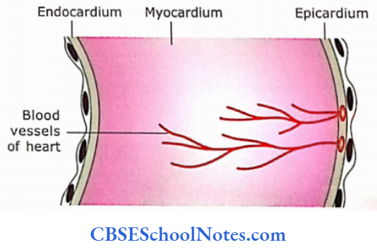
The heart and blood vessels are all lined on the inner surface of the heart face by endothelial cells (simple squamous cells). For the histological structure of heart and the structure of the myocardium (heart muscles) .
The three layers of the heart (epicardium, myocardium and endocardium) are homologous to three layers of blood vessels (tunica intima, tunica media and tunica adventitia, see below
Circulatory system Remember:
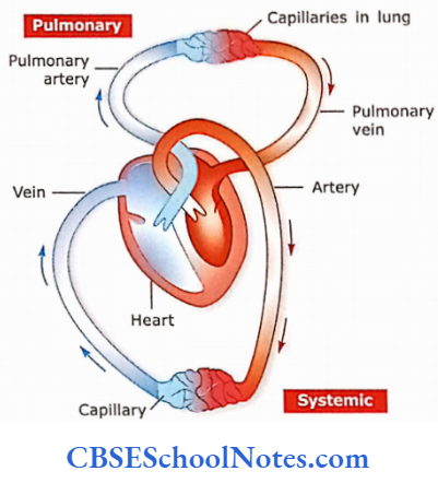
The circulatory system consists of the heart and blood vessels, hence sometimes also known as the cardiovascular system. It consists of two kinds of circulation, i.e., pulmonary circulation and systemic circulation. Pulmonary circulation carries blood to the lungs, while systemic circula¬ tion carries blood to various tissues of the body
General Structure Of Blood Vessels
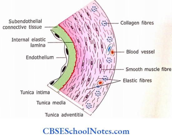
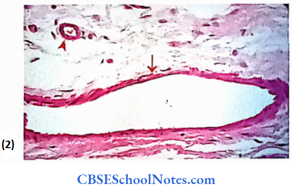
Three layers of tissue constitute the wall of a typical blood vessels
- Tunica intima: It consists of a single layer of squamous endothelial cells and underlying sub-endothelial connective tissue
- Tunica media: It consists of smooth muscle cells arranged concentrically around the lumen of the vessels
- Tunica adventitia: It mostly consists of connective tissue arranged parallel to the long-axis lumen of the vessel
Blood vessels Remember:
The tunica adventitia of blood vessels, which Itself consists of connective tissue, blonds with the surrounding connective tissue. The histological structure of various kinds of blood vessels is described below in detail.
Arteries
As stated earlier, arteries are vessels, which conduct blood from the heart to capillaries. Arteries branch repeatedly between the largest arteries to the capillary plexus. Because of this repeated branching, the total cross-sectional area of the vascular system increases to 800 times that of the aorta. With an increase in the branching, there is a gradual decrease in the rate of blood flow. This slow flow provides ample time for the exchange of substances through capillaries.
General Structure of Arteries
The wall of every artery passes following three characteristic layers
- Tunica intima: It is the innermost layer. It consists of the following four components
- The endothelium is made up of simple squamous epithelium that faces the lumen and comes in contact with blood
- Basal lamina
- Subendothelial connective tissue is made up of cate connective tissue.
- Internal elastic lamina is made up of a layer of elastic material.
- Tunica media: The tunica media Is the Intermediate layer surrounding the tunica Intlma, ft Is usually the thickest layer and consists of elastic fibres and smooth muscle cells In varying proportions depending on the size of the artery see below).
- The smooth muscle cells and elastic fibres are arranged circularly or helically).
- The tunica media Is separated from tunica adventitia by external elastic lamina composed of elastic fibres.
- The Internal elastic lamina Is fenestrated permitting the diffusion of substance Into deeper regions of the arterial v/aJJ to nourish the cells,
- Tunica adventitia (external):
- This Is the outer coat of an artery and is composed mainly of connective tissue elements (predominantly of collagen fibres and connec¬ tissue cells).
- The collagen fibres in adventitia run longitudinally
Arteries Clinical Application
Question 1. What are the functions of endothelial cells?
Answer:
Besides providing the smooth surface in the lumen of blood vessels for free flow of blood, endothelial cells also perform
The following important functions:
- Secretion: They secrete type II, IV and V collagen, lamin, endothelin (a vasoconstrictor substance), nitric oxide and von Willebrand factor. The Von Willebrand factor facilitates the coagulation of platelets during clot formation.
- Site for storage of enzymes: They possess many membrane-bound enzymes. The angiotensin-converting enzyme converts angiotensin I to angiotensin II for the regulation of blood pressure. Endothelial cells also bind lipoprotein lipase, which breaks the lipoproteins. Enzymes of endothelial cells inactivate serotonin, bradykinin, prostaglandins, thrombin and noradrenaline.
Arteries Of Remember:
Endothelial cells perform many functions, which are necessary for the functional and structural integrity of vessel wall itself
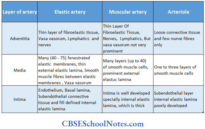
Arteries Of Classification
Arteries are classified into three types
- Elastic (conducting) arteries
- Muscular (distributing) arteries
- Arterioles
This classification is based on the diameter of arteries, thickness of the wall and dominant component of their tunica media (elastic material or muscle fibres). There is always a gradual change seen from large-size elastic arteries to muscular arteries to arterioles.
1. Elastic Arteries:
These are also called conducting vessels as their main function is to conduct the blood from the heart to the muscular arteries. Examples of these arteries are large-size arteries like the aorta, pulmonary trunk and their main branches,i.e., brachiocephalic, common carotid, subclavian and common iliac.
They are called as elastic arteries because tunica media in these arteries is predominantly made up of elastic fibres , Usually the diameter ofan elastic artery is more than 1 cm.
- TunicaIntima:
- Endothelium: It is a simple squamous epithelium resting on basal lamina. These flat cells are elongated and oriented with their long axis to the direction of blood flow.
- Subendothelial layer: It consists of connective tissue. This layer contains collagen and elastic fibres, smooth muscle cells, fibroblasts and macrophages.
- The internal elastic membrane: In large-size elastic arteries, this layer is not visible because it becomes difficult to differentiate it from many elastic layers present in tunica media
- Tunica Media:
- It is the thickest of three layers of elastic arteries. Tunica media contains a high proportion of elastic fibres in the form of sheets or lamellae, which are arranged as concentric fenestrated sheets.
- In between elastic lamellae are layers of smooth muscle cells, type collagenous fibres and ground substance. Compared to elastic layers, smooth muscle lay¬ ers are thin and circularly oriented.
- There are about 50 lamellae of elastic material in the human aorta. In histological sections, internal and external elastic laminae are not differentiated from other elastic lamellae of tunica media. Smooth muscle cells of media are considered to produce elastin and collagen.
- Tunica Adventitia:
- This layer is made up of a connective tissue layer. It is rela¬ tively thin and contains longitudinally running collagen fibres. There is also a loose network of elastic fibres.
- It contains fibroblasts, macrophages and mast cells. As the media is very thick, the diffusion of metabolites from the lumen of large-sized arteries is inadequate.
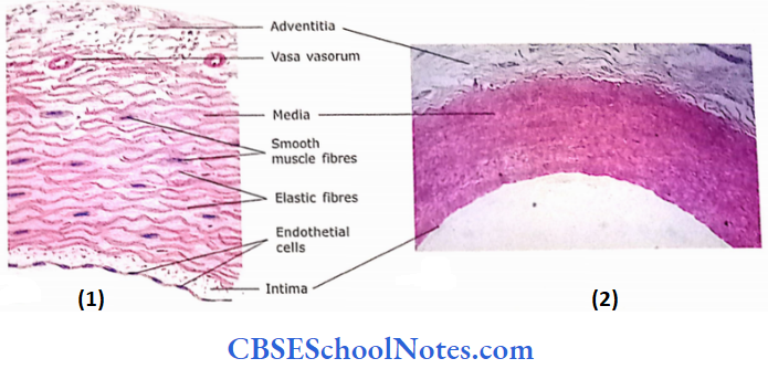
- Hence, tunica adventitia contains blood vessels (vasa vasorum), which supply blood to adventitia and outer media, while inner media and intima are supplied by blood flowing through the lumen of the artery.
- Adventitia also contains nerve bundles and lymph vessels.

Elastic Arteries Remember:
Tunica media of elastic arteries is predominantly composed of concentric layers of fenestrated elastic membranes. Smooth muscle cells are less abundant in this type of conducting arteries
Elastic Arteries Functions:
- Elastic arteries conduct the blood from the heart to medium size (muscular) arteries.
- Their elastic recoil is responsible for the continuous flow of blood through arteries
Elastic Recoil:
The large arteries (elastic arteries) nearest the heart are subjected to the greatest contractile forces and therefore possess greaterproportion of elastic tissue in tunica media. As the blood is pumped from the heart into large arteries, their elastic walls distend, accommodating the surge of blood
The distention of the elastic wall accumulates the potential energy. During the diastole (relaxation of ventricles), the distended arteries come back to their original size because of elastic recoil.
This elastic recoil converts the potential energy (stored energy) in the vessel into the kinetic energy of the blood. The blood continues to move through the arteries even during diastole because recoil acts as an additional force that helps the blood to flow continuously
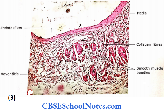
2. Muscular Arteries:
These are also known as medium-sized arteries. Usually, the diameter of the lumen of the muscular artery is 2-10 mm. As these arteries regulate the flow of blood to an organ or tissue these are also called distributing arteries (For example, brachial, femoral, ulnar, radial, renal, etc).
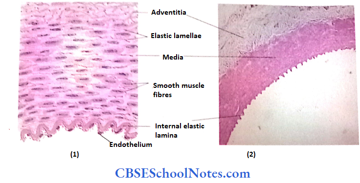

With the gradual change of elastic arteries to muscular arteries, the elastic material decreases and smooth muscle cell becomes the main constituent of tunica media. Muscular arteries are of smaller diameter than the elastic arteries. The internal elas¬tic lamina (membrane) is visible
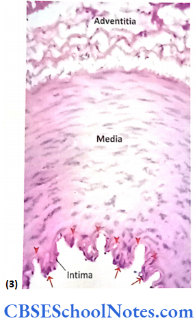
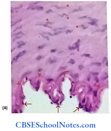
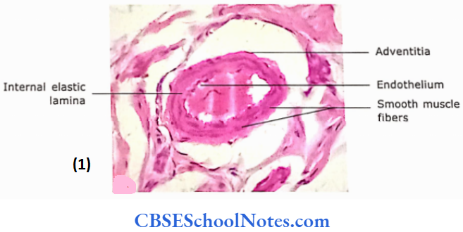
- TunicaIntima: The layers of endothelial cells and basal lamina are same as in large elastic arteries. The subendothelial connective tissue is much less or even absent. In the histological section, the internal elastic lamina is prominent and seen as a wavy structure due to the contraction of smooth muscle in tunica media(after death due to rigormortis). The internal elastic lamina is clearly visible only in muscular arteries
- Tunica Media: Smooth muscle cells are predominantly present in the form of circumferentially or spirally oriented layers. The 75% mass of media is formed by smooth muscle cells and associated reticular fibres. Media also contains type collag¬enous fibres and relatively little elastic material. Each smooth muscle cell is surrounded by its basal lamina. Gap junctions are present between adjacent muscle cells for coordinated contraction
- Tunica Adventitia: It is made up of connective tissue. Although it is thicker than the tunica adventitia of elastic arteries, both are almost similar histologically.
Muscular Arteries Remember:
Tunica media of muscular arteries is the thickest layer of the vessel wall and predominantly consists of helically arranged smooth muscle cells
Muscular Arteries Functions:
- The smooth muscle of the tunica media of the muscular artery can alter the size of its lumen by contraction or relaxation.
- In this way, muscular arteries are capable of regulating the How of blood as per cent of regions supplied by them.
- The small size muscular lyrics are of 1- 2 mm in diameter having 8-10 layers of smooth muscle cells in the tunica media
Some Important Differences Between Elastic and MuscularArteries:
- The diameter of the lumen of the elastic artery is much more compared to the thickness of its wall. On the other hand, the thickness of the wall of the muscular artery is rela¬tively more compared to the diameter of its lumen.
- The internal and external elastic laminae are visible in the muscular arteries, while they are will-defined in the elastic artery because the media predominantly contains elastic laminae, thus internal and external elastic laminae are difficult to distinguish.
- The tunica adventitia of the elastic artery is relatively thin compared to the muscular artery
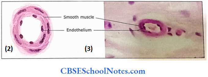
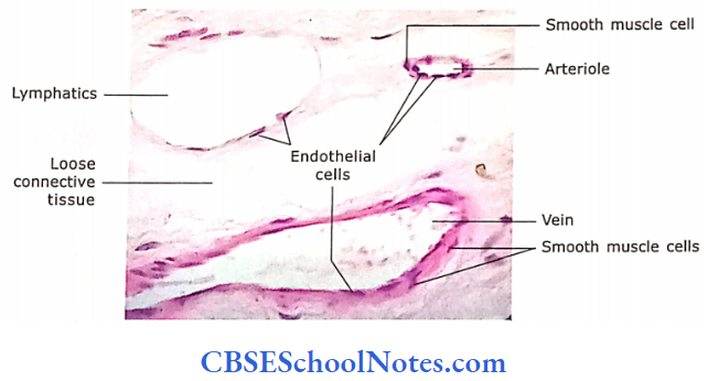
3. Arterioles:
Small arteries having a size less than 100 μm are classified as arterioles. They terminate into the capillary network. The precapillary terminal arterioles may have a diameter as less as 12 μm noon.
Comparison between various types of arteries:
- TunicaIntima: The tunica intima is formed by endothelial cells resting on the basal lamina, which in turn is supported by thin layer of subendothelial connective tissue. There is an absence of internal and external elastic. lamina in most of the arterioles
- Tunica media: It consists of a thin layer of circularly arranged smooth muscle cells. In large-size arterioles, there may be 2 or 3 layers of smooth muscle cells while in terminal arterioles there is only one layer of smooth muscle cells. In terminal arteries, these smooth muscle cells may act as precapillary 1 2 t sphincter, which regulates the flow of blood through the capillary network depending on the metabolic needs ofthe tissue
- Tunica Adventitia: The tunica adventitia is thin and well-defined and consists of collagen fibres and occasional fibroblasts. Sometimes in very small arteriole adventitia is not visible
Arterioles Remember:
Although the arterioles are less than 0.1 mm (100 millimicrons) they consist of all three vascular coats
Arterioles Functions:
- Arterioles regulate the blood flow through capillaries.
- The change in the diameter of arterioles (vasoconstriction or dilatation) can also significantly alter blood pressure.
- Arterioles that supply the blood to the capillaries are called as metarterioles, the smooth muscle layer is not continuous but individual muscle cells are placed at intervals. Their contraction controls the blood flow into the capillary bed.
Metarterioles Remember:
Smooth muscles of metarterioles may act as a precapillary sphincter, which controls the blood flow in the capillary bed (i.e., dilation increases the blood flow, while constriction reduces it). This vascular resistance also controls the systemic arterial blood pressure
Arteriosclerosis Clinical Applications
Arteriosclerosis:
- It is an age-related change of arteries and is usually seen after middle age.
- There is generalized slow thickening of the intima of arteries.
- This is due to an increase in collagen and the accumulation of lipids in the intima.
- This leads to the diffuse narrowing of the lumen of blood vessels.
Atherosclerosis:
- It is a pathological condition. Here, there is a patchy accumulation of lipid, fibrous tissue and macrophages in tunica intima.
- The elevated plaque of the intima leads to the narrowing of the arterial lumen. Formation of blood [clots on the plaque may occlude the lumen.
- When this occurs in the coronary artery it may lead to myocardial infarction (heart attack) and when it occurs in the brain it may lead to cerebral thrombosis (stroke)
Hypertension or High Blood Pressure:
- When the systolic blood pressure exceeds more than 140 mmHg and the diastolic more than 90 mmHg for a prolonged period, then the condition is known as high blood pressure or hypertension.
- From middle age onwards, this condition results mainly due to increased vascular resistance. This increased resistance is due to the narrowing of the lumen of small arteries and arterioles.
- This narrowing may be secondary to the increase in the amount of smooth muscle in the wall or due to the active contraction of the smooth muscle.
- The amount of smooth muscle increases because of the multiplication of preexisting smooth muscle cells.
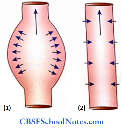
Aneurysm:
- It is a sac-like dilatation of the wall of an artery. It occurs due to weakness in the wall of the artery resulting from the replacement of elastic fibres by collagen fibres.
- The disease is usually seen in the aorta and carotid vessels in old age where it may be associated with syphilis, Marfan syndrome, Ehlers-Danlos syndrome (both syndromes are associated with connective tissue disorders) and atherosclerosis.
- If not detected and repaired early, death may result due to rupture leading to rapid loss of massive blood.
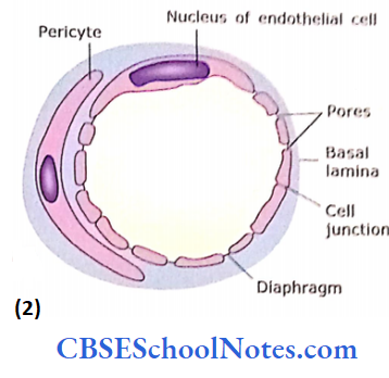
Capillaries
Capillaries are thin-walled endothelial-lined microscopic vessels that connect arterioles and venules. They form an extensive network of vessels almost in every tissue of the body. The diameter of the capillary is between 5-10 μm and the length is approximately around 50 μm.
As the diameter of erythrocytes is about 7 μm, they pass through the lumen of the capillary, one at a time. While they are passing through the narrow lumen (5-8 μm) there occurs deformation in their shape. The surface area of all capillaries of the human body is roughly 1,000 square meters. The total length of capillaries in the body is about 40,000 miles
Capillaries Remember:
The diameter of capillaries is sometimes smaller than the diameter of an RBC, so red blood cells range themselves in a file and also fold on themselves to pass through the capillary.
Microcirculation:
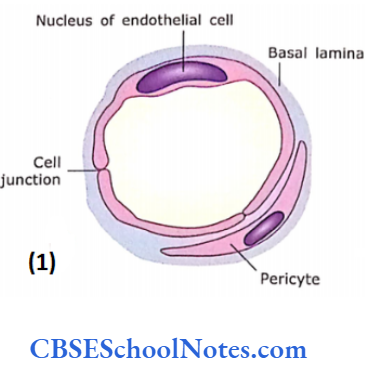
The flow of blood through a capillary is called microcirculation. The capillaries consist of a single layer of extremely thin endothelial cells with a basal lamina supported by a loose network of reticular fibres. The lumen of the capillary may be lined by only one cell or by portions of two or even three cells. These endothelial cells are elongated in the direction of blood flow. The margins of endothelial cells are joined with tight junctions (zonulae occludens).
Sometimes, discontinuities in the cell junction may be seen through which fluid or leucocytes may pass. The thin margin of cells may also overlap. In some capillaries, pericytes are found in association with endothelium and embedded within the basal lamina of endothelium.
Pericytes possess long oval euchromatic nuclei and several peripheral processes that clasp the capillary wall. Pericytes are unspecialized cells capable of differentiating into fibroblasts or smooth muscle cells as per the need
There is no tunica media or adventitial layers in capillaries:
Capillaries are of three different types
- Continuous capillaries
- Fenestrated capillaries
- Sinusoids
Layers in capillaries Remember:
Capillaries consist of a single layer of endothelial cells resting on basal lamina. Tunica media and tunica adventitia both are absent in capillaries
The speed of flow of blood through capillaries is very slow. Thus, enough time becomes available for the exchange between blood and tissue
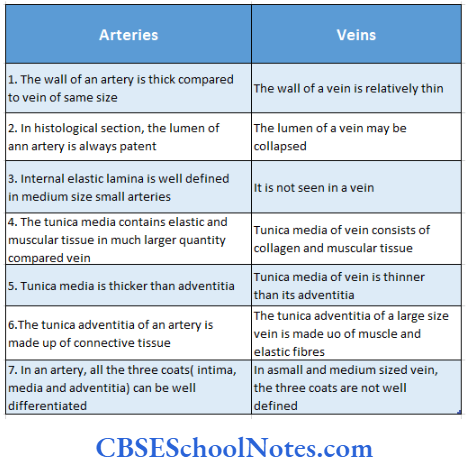
1. Continuous Capillaries:
In the continuous type of capillaries, the plasma membrane of endothelial cells forms a continuous tube. The peripheral portions of the endothelial cells are very thin. Endothelial cells are held together by tight junctions. The basement membrane surrounds the endothelium.
The main feature of the continuous capillary is the presence of pinocytic vesicles that indicate the mode of transepithelial transport (through the cytoplasm of epithelial cells). These types of capillaries are found in tissues like muscle, brain, connective tissues, skin and lung. Sometimes this kind of capillary is also called somatic capillary
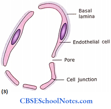
2. Fenestrated Capillaries:
In the fenestrated type of capillaries, the endothelial cells have many circular pores ranging from 50 to 100 nm in diameter. Apore is usually closed by thin diaphragm, which is thinner than plasma membrane and resembles in appear¬ ance thin basal lamina. The diffusion of substances takes place through the diaphragm.
This type of capillary is found in the pancreas, endocrine glands, intestinal villi, choroid plexus and ciliary processes of the eye. This kind of capillary is also called as visceral capillary. The capillaries of renal glomeruli have pores, which are not closed by the diaphragm. However, they have unusually thick basal lamina.
3. Sinusoids:
Sinusoids are special kinds of capillaries that are large (about 30-40 pm in diameter) and irregular in shape. Because of this, the flow of blood is sluggish which allows sufficient time for the exchange of substances between blood and tissue fluid.
Large pores or slits are seen in endothelial cells, which are large pores or slits are seen in endothelial cells, which are not closed by diaphragms. Blood and tissue fluid can easily diffuse through these pores.
A basal lamina may or may not be found over these endothelial discontinuities(slit or pores) thus increasing the exchange between blood and tissue. Sinusoids may also contain specialized lining cells that are adapted to the special function of tissue, i.e. sinusoids in the liver contain phagocytic cells (Kupffer’s cells) which remove bacteria and other foreign particles from the blood. The sinusoidal capillaries or sinusoids are found in the liver, bone marrow, spleen, anterior pituitary gland, parathyroid and adrenal medulla.
Capillaries Functions
- The exchange of gases, fluid and molecules between blood and tissue takes place through the walls of capillaries.
- In the continuous capillaries, the mechanism of exchange across the capillary wall is due to transcytosis (through cytoplasm) while in fenestrated capillaries the exchange takes place through diffusion of substances across the diaphragm, which is 100 times more rapid than across continuous capillaries.
- When the need arises endothelin is secreted by capillary endothelium and reaches the muscle cells of blood vessels where it helps in the contraction of smooth muscle leading to an increase in blood pressure. Prostacyclin, a potent vasodilator, is also released by capillaries. NO and O2 tension are other vasodilating agents
Differences between the three types of capillaries:

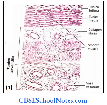
Veins
Veins are classified as large, medium and small-sized veins. Small size veins arc also known as venules. which are further classified as post-capillary and muscular venules. Though the large and medium-sized veins have the same three layers (i.e., tunica intima, media and adventitia) as seen in large and medium-sized arteries, these layers are not well defined in veins.
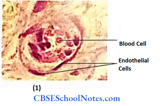
The large and medium veins and corresponding arteries usually travel together and thus can be compared in histological sections. For comparison of arteries and veins
Differences between artery and vein:
Veins Remember:
Veins are classified based on their size (diameter) and the thickness of their wall
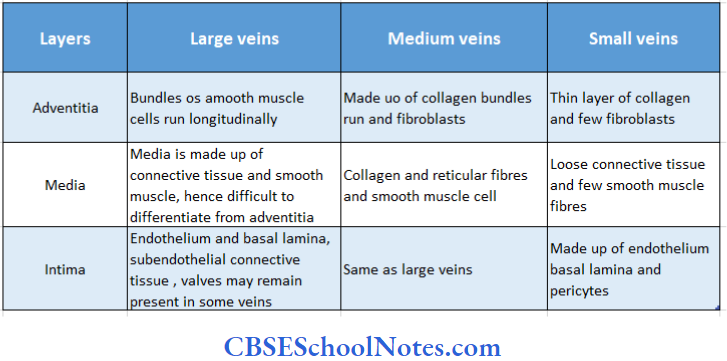
1. Large veins
Large veins have the following three layers
- TunicaIntima: It consists of endothelial cells resting on the basal lamina, itself is supported by a small amount ofsubcndothclial con¬nective tissue and a few smooth muscle cells
- Tunica Media: It consists of smooth muscle cells, collagenous fibres and fibroblasts. Smooth muscle cells are usually arranged longitudinally and circularly. Tunica media in veins is thin compared to that of arteries. As smooth muscles are present both in intima and media, it becomes difficult to distinguish the boundary between the two
- Tunica Adventitia: This layer is always thicker than media and contains smooth muscle cells, which arc oricntcii longitudinally. Adventitia also contains bundles of collagen elastic fibres and fibroblasts. The thin-walled large veins are protected from stretching (because of movements of the diaphragm) by longitudinally oriented smooth muscles and elastic fibres

2. Medium Veins
Medium-sized veins will show all three layers as described for large-size veins. However, it becomes gradually difficult to distinguish all three layers with decreasing si/e of medium-sized veins. The tunica intima consists of little or no subendothelial connective tissue. Media consists of a few layers of smooth muscle and associated collagen and elas¬ tic fibres. The adventitia is typically thicker than media and consists of collagen and elastic fibres
As stated earlier, veins are accompanied by arteries of corresponding size and it would be easier to identify first a medium size artery, which would help to identify the medium vein lying close to it in a histological section.
3. Small Veins or Venules
Small size veins are also known as venules that are of two different types, i.e., postcapillary venules and muscular venules.
Two to three capillaries converge to form postcapillary venules, which are the smallest veins (10-0.5 νm). These are lined by endothelium, basal lamina and pericytes. Pericytes are branching cells, which may be outside the basal lamina or sometimes enclosed by basal lamina. The postcapillary venules possess special permeability (diffusion of fluid and white blood cells into surrounding tissue and absorption from tissue into venules)
Comparison between various types of veins:
Veins Clinical Application
Varicose Veins:
Varicose veins are abnormally enlarged tortuous veins seen in the legs of an old person. The varicose veins result due to the failure of valves, which help the blood flow in the antigravity direction. Sometimes, varicose veins may also be caused due to loss of muscle tone and degeneration of vessel walls.
These veins may also result from prolonged standing as in case of the washerman remaining standing while washing the clothes. Two other sites where varicose veins are common are the oesophagus and anal canal where it is known as haemorrhoids (or piles)
The larger venules are called muscular venules because of the presence of one or two layers of smooth muscle cells outside the tunica intima. These muscle layers constitute tunica media that is absent in postcapillary venules. The tunica adventitia is thicker than the tunica media
Blood Supply And Nerve Supply To Blood Vessels
1. Blood supply to blood vessels:
“Vasa vasorum” is the name given to the small arteries and veins that enter the vessel wall and branch profusely in tunica adventitia and

Outer part of tunica media of size vessels. They provide nutrition and remove wastes from the outer part of the vessels as the inner part of the wall is directly supplied by nutrients from the lumen of the vessel
2. Nerve supply of vessels:
The sympathetic part ofthe autonomic nerves is vasomotor to the smooth muscle cells of the blood vessels. Their stimulation leads to the release of norepinephrine, which causes the constriction ofthe smooth muscle cells of tunica media (vasoconstriction). Arteries that supply skeletal muscles also receive parasympathetic (cholinergic) nerves, which are responsible for vasodilation.