
Cerebral Hemispheres
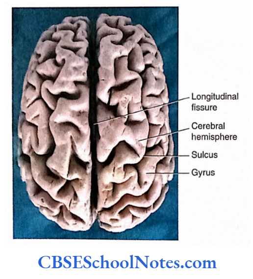
The cerebrum is the largest part of the brain that occupies the anterior and middle cranial fossae of the skull. It consists of two cerebral hemispheres which are partially separated from each other by a longitudinal fissure.
At the bottom of the longitudinal fissure, the right and left cerebral hemispheres are connected by a great commissure of white fibers known as the corpus callosum.
Each cerebral hemisphere is covered by a layer of grey matter, known as the cerebral cortex. It shows complicated folds known as gyri and grooves between gyri are called sulci.
The formation of gyri and sulci is an attempt to increase the surface area of the cerebral cortex which has to be accommodated into the cranial cavity.
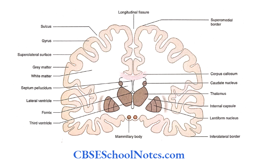
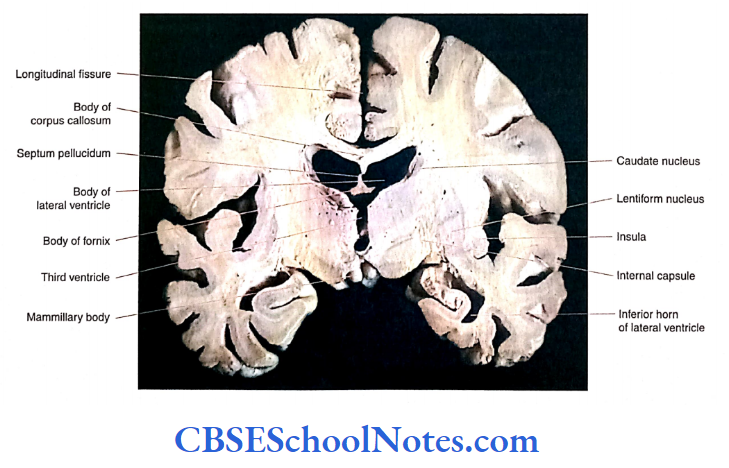
Deep into the cortex, the central core of each cerebral hemisphere is formed by white matter.
Within the white matter of the cerebral hemisphere, large masses of grey matter are present which are called basal nuclei (mainly caudate and lentiform nuclei).
Read and Learn More Neuroanatomy
Each cerebral hemisphere has an extensive cavity known as the lateral ventricle.
External Features Of The Cerebral Hemisphere
Each cerebral hemisphere presents with three surfaces, three borders, and three poles. These borders, surfaces, and poles can be easily identified with the help of figures showing the lateral aspect, medial aspect, and inferior aspect of the cerebral hemisphere.
A coronal section passing through the hemisphere will also help in identifying these features.
Surfaces
Each cerebral hemisphere consists of superolateral, medial, and inferior surfaces.
Superolateral surface: The superolateral surface is a large and convex surface that lies between superomedial and inferolateral borders. The convexity faces superolaterally.
Medial surface: The medial surface is seen in the midsagittal section of the brain. It lies between the superomedial and the inferomedial borders.
Inferior surface: The inferior surface is divided into the orbital surface and tentorial surface by the stem of the lateral sulcus.
The orbital surface is formed by the frontal lobe while the tentorial surface is formed by the inferior surfaces of the temporal and occipital lobes.
Borders
Each cerebral hemisphere consists of superomedial, inferolateral, superciliary, and inferomedial borders.
Superomedial border: The superomedial border is the upper border of the cerebral hemisphere and extends between the frontal and occipital poles. It separates a large superolateral surface from the medial surface.
Inferolateral border: The inferolateral border separates the superolateral surface from the tentorial surface and extends between the temporal and occipital poles.
Superciliary border: The superciliary border separates the superolateral surface from the orbital surface of the frontal lobe. It extends between the frontal pole and the stem of the lateral sulcus.
Inferomedial border: The inferomedial border is subdivided into medial orbital and medial occipital borders. The medial orbital border separates the orbital surface from the medial surface while the medial occipital border separates the tentorial surface from the medial surface.
Poles
Students are requested to identify the frontal, occipital, and temporal poles in the cerebral hemisphere with the help of
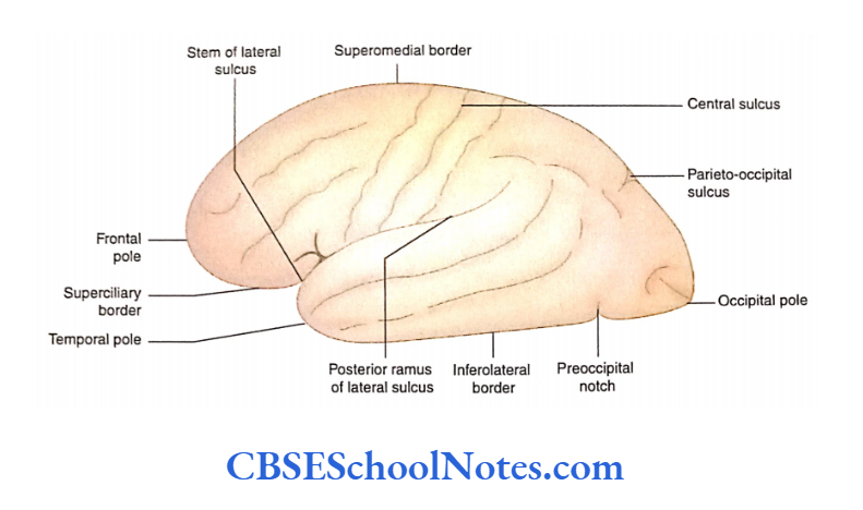
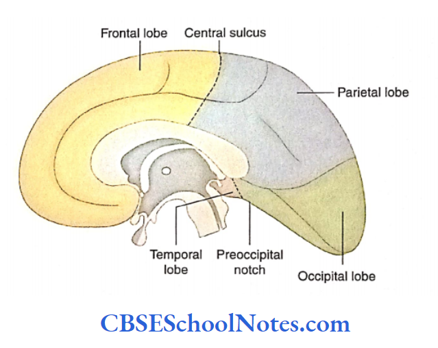
Important Sulci on the Superolateral Surface of the Cerebral Hemisphere. The cerebral hemisphere is divided into four lobes with the help of major sulci on the superolateral surface.
These important sulci are central sulcus, posterior ramus of lateral sulcus, parieto-occipital sulcus and pre-occipital notch. Identify these important sulci on the superolateral surface.
Lobes Of The Cerebral Hemisphere
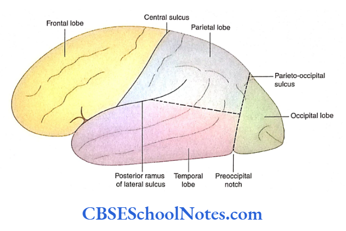
With the help of four major landmark sulci and two imaginary lines, the cerebral hemisphere is divided into four lobes.
These are the frontal, parietal, temporal, and occipital lobes on the superolateral surface of the hemisphere. Various lobes on the superolateral surface are determined as follows:
The frontal lobe is bounded by the central sulcus (behind) and by the posterior ramus of the lateral sulcus (below).
The parietal lobe lies behind the central sulcus. Posteriorly, it is bounded by the first imaginary line and below by the posterior ramus of the lateral sulcus and the second imaginary line.
The temporal lobe is situated below the posterior ramus of the lateral sulcus and the second imaginary line. Posteriorly, this lobe is limited till the first imaginary line.
The occipital lobe lies behind the first imaginary line. The boundaries of the frontal, parietal, temporal, and occipital lobes on the medial and inferior surfaces of the cerebral hemisphere are shown respectively.
Sulci and Gyri of the Cerebral Hemisphere
Except for the major sulci and gyri mentioned in the preceding text, individual variation may be seen in the presence or absence of many other gyri and sulci.
A brief account of the major sulci and gyri on various surfaces of the cerebral hemisphere is as follows.
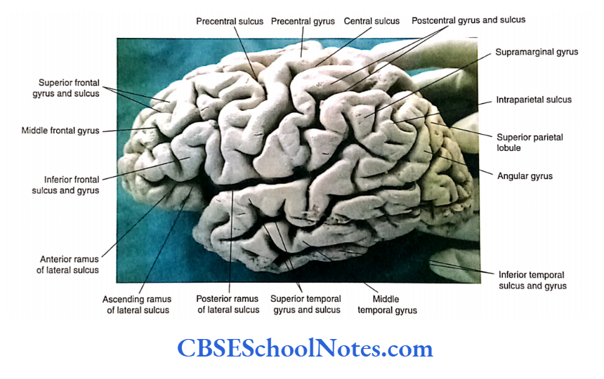
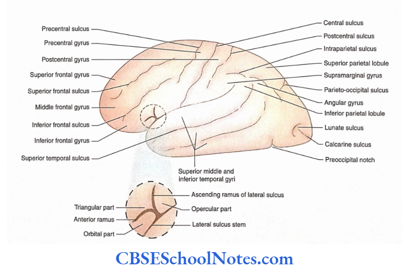
Superolateral Surface
The superolateral surface of the cerebral hemisphere presents important sulci and gyri in each of the lobes. Students should learn these sulci and gyri with the help of

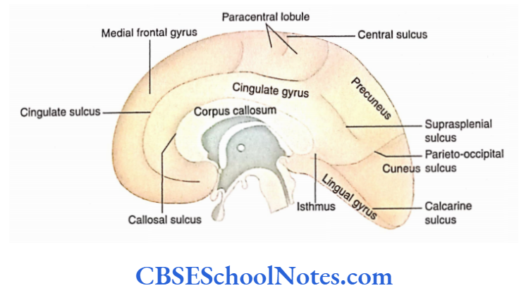
Medial Surface
The medial surface shows the following major sulci callosal, cingulate, parieto-occipital, supraspinal, and calcarine. These sulci are located around the corpus callosum.
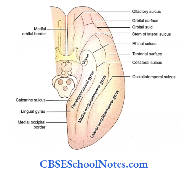
Inferior Surface
The inferior surface of the cerebral hemisphere is divided into two surfaces by the stem of the lateral sulcus: Orbital and Tentorial. The orbital surface lies anterior to the stem of the lateral sulcus while the tentorial surface lies posterior to it.


Cerebral Hemispheres Summary
- The cerebrum is situated on the diencephalon and brainstem. It is the largest part of the brain. It consists of two cerebral hemispheres.
- Each cerebral hemisphere is covered by a layer of grey matter, known as the cerebral cortex.
- The cerebral hemisphere shows the presence ofsulci and gyri. The formation ofsulci and gyri is an attempt to increase the surface area of the cerebral cortex.
- Each cerebral hemisphere consists of three surfaces (superolateral, medial, and inferior), three borders (superomedial, inferomedial, and inferolateral), and three poles (frontal, parietal, and occipital).
Each cerebral hemisphere consists of four lobes:
- Frontal,
- Parietal, Temporal and Occipital.
- Each lobe of the cerebral hemisphere shows many sulci and gyri on various surfaces of the cerebral hemisphere.
Cerebral Hemispheres Multiple Choice Questions
Question 1. Which of the following statements about the cerebral hemisphere is false?
- It is present in the anterior and middle cranial fossae
- The cerebral cortex shows complicated foldings called gyri
- Deep in the cerebral cortex, the white matter contains large masses of grey matter called caudate and lentiform nuclei
- Each cerebral hemisphere contains a cavity known as the third ventricle
- The right and left cerebral hemispheres are connected by corpus callosum
Answer: 4. The right and left cerebral hemispheres are connected by the corpus callosum
Question 2. Which of the following statements about the cerebral hemisphere is/are true?
- It shows three surfaces—superolateral, medial, and inferior
- It shows three borders—superomedial, inferomedial, and inferolateral
- It shows three poles—frontal, occipital, and temporal
- All of the above
Answer: 4. All of the above
Question 3. The cerebral hemisphere is divided into the following lobes
- Frontal
- Orbital
- Parietal
- Temporal
- Occipital
Answer: 2. Orbital
Question 4. The lentiform nucleus is situated deep in the insular cortex
- It is a hidden part of the cerebral cortex
- It consists of long and short gyri
- The lentiform nucleus is situated deep in the insular cortex
- The insula is involved in motor and sensory activities of the autonomic nervous system
- All of the above
Answer: 5. All of the above
Question 5. The following major sulci are present on the medial surface of the cerebral hemisphere except for?
- Callosal
- Cingulate
- Parieto-occipital
- Interparietal
- Calcarine
Answer: 4. Interparietal
Question 6. Which of the following major sulci are present on the inferior surface of the cerebral hemisphere?
- Orbital
- Olfactory
- Collateral
- Occipito-temporal
- All of the above
Answer: 5. All of the above
Question 7. Which of the following statement(s) about the parahippocampal gyrus is/are true?
- It lies between the medial occipital border and the collateral sulcus
- It is continuous posteriorly with the lingual gyrus
- The anterior end of the parahippocampal gyrus shows a hook-like projection called an uncus
- All of the above
Answer: 2. It is continuous posteriorly with the lingual gyrus