
Cerebellum
- The cerebellum is a part of the hindbrain. It lies behind the pons and the upper medulla. The fourth ventricle separates the cerebellum from the pons and the medulla.
- The cerebellum is concerned with motor activities without any conscious awareness. The functions of the cerebellum include maintenance of posture and balance, control of muscle tone and coordination of activities of various muscle groups.
External Features Of Cerebellum
The cerebellum consists of two cerebellar hemispheres and a central part called vermis. The vermis unites the two cerebellar hemispheres.
Surfaces of Cerebellum
- The cerebellum presents superior and inferior surfaces. The superior surface is flattened and presents a superior vermis in the midline.
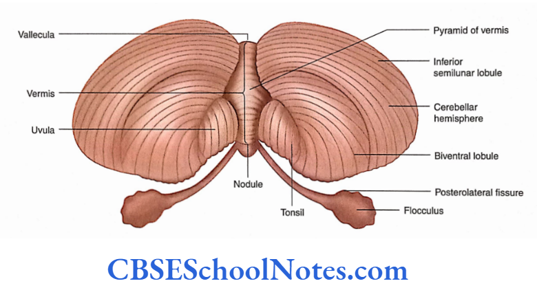
- The inferior surface of the cerebellum is convex and shows a deep depression between the two hemispheres known as vallecula. The inferior vermis lies on the floor of the vallecula.
Read and Learn More Neuroanatomy
Fissures and Lobes of Cerebellum
The cerebellum is divided into lobes with the help of two deep fissures:
- Primary fissure and
- Posterolateral fissure.
Posterolateral fissure.
The posterolateral fissure separates the posterior lobe of the cerebellum from the flocculonodular lobe. The flocculonodular lobe is present on the inferior surface of the cerebellum. The above two fissures divide the cerebellum into three lobes.
Horizontal Fissure
The horizontal fissure is important as it demarcates the superior cerebellar surface from the inferior one.
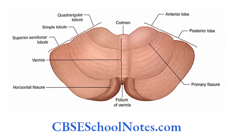
Other Fissures and Lobules
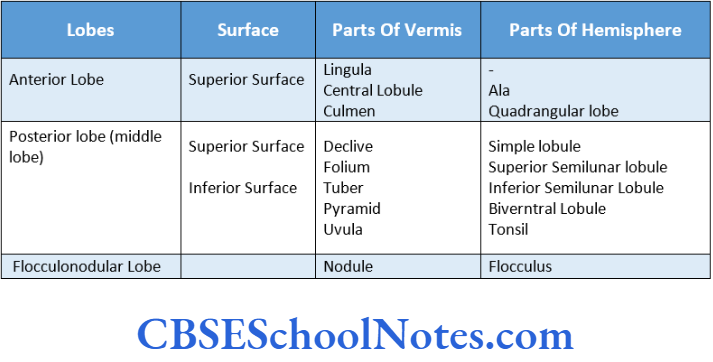
The anterior and posterior lobes are further subdivided into many lobules with the help of other fissures. The names of these lobules are provided
Cerebellar Tonsils
- The cerebellar tonsils are small lobules located on the inferior surface of the cerebellum.
- The cerebellar tonsils are important structures clinically because, in cases of raised intracranial pressure, the cerebellar tonsils may herniate into the foramen magnum.
- The protruded tonsil may block the circulation of the cerebrospinal fluid between the subarachnoid space in the cranial cavity and the subarachnoid space around the spinal cord.
- The herniated tonsils may also compress the vital centres of the brainstem; if severe, they may result in the death of an individual.
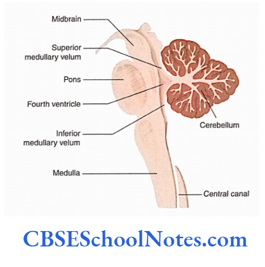
Relation of the Cerebellum To The Fourth Ventricle
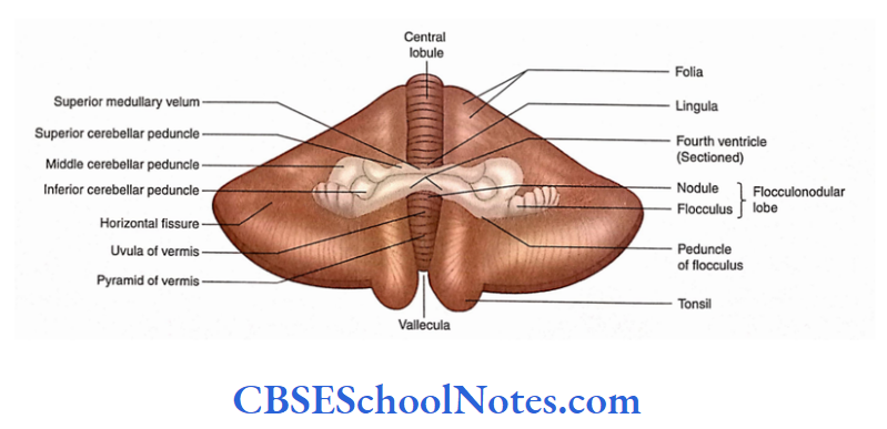
The relation of the cerebellum to the brainstem is shown in the midsagittal section. The fourth ventricle lies between the pons and the upper medulla anteriorly and the cerebellum posteriorly. The roof of the fourth ventricle trick- is formed above by the superior medullary velum.
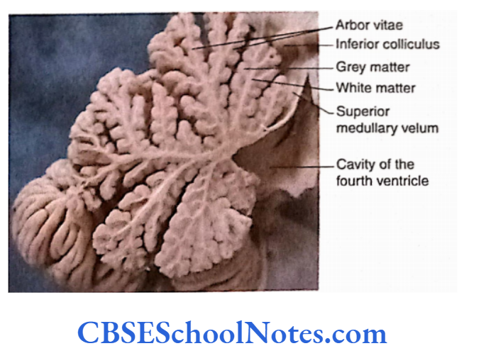
Cerebellar Cortex
- The surface of the cerebellum is covered by grey matter, called the cerebellar cortex. The cortex is marked by a series of slender, parallel ridges called folia.
- These ridges are narrow leaf-like bands which are arranged transversely on the surface of the cerebellar cortex.
- The midsagittal section of the hindbrain also demonstrates the central core of white matter which shows complex tree-like branchings. This arrangement is known as ‘arbour vitae’. This branching pattern is covered by grey matter (cerebellar cortex.
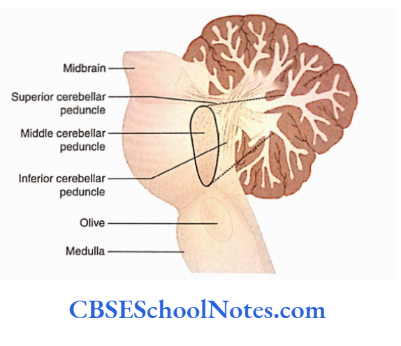
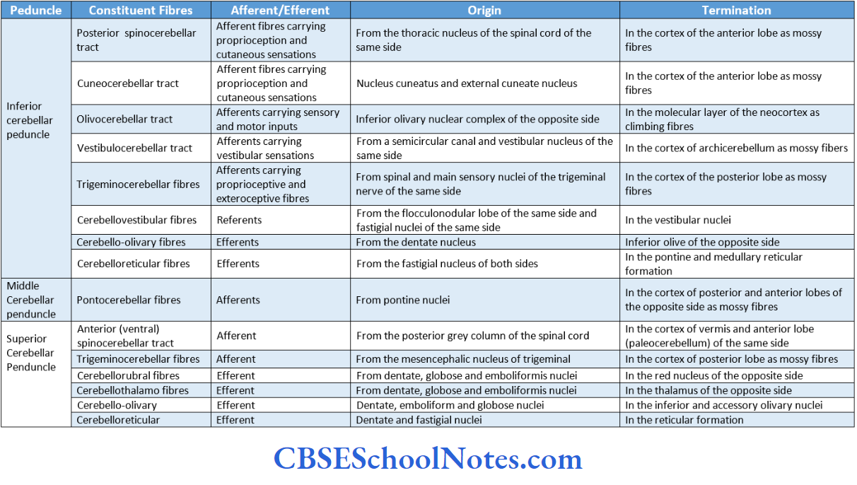
Cerebellar Peduncles
The cerebellum is attached to the brainstem by three pairs of cerebellar peduncles. The superior cerebella peduncles connect it to the midbrain, the middle connects it to the pons And their inferior peduncle connects it to the spinal cord.
The afferent and efferent fibres enter and leave the cerebellum through this peduncle.
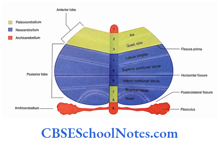
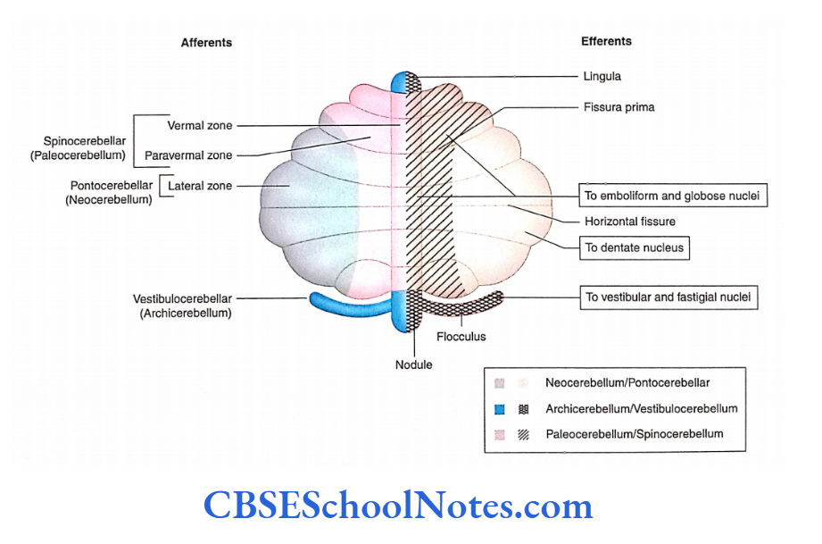
Phylogenetic Classification Of Cerebellum
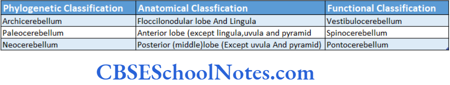
Based on evolution and function, the cerebellum can be divided into three parts; Archicerebellum paleocerebellum and neocerebellum
Archicerebelium
- The archicerebellum is phylogenetically the oldest part of the cerebellum. Anatomically, it is represented by the flocculonodular lobe and the lingula of the anterior lobe.
- Functionally, the archicerebellum is related to the vestibular system and its main function is to maintain posture and balance.
Paleocerebellum
- Paleocerebellum consists of the anterior lobe (except lingual), uvula and pyramid of the posterior lobe.
- As this part of the cerebellum is functionally related to the spinal cord, it is also known as the spinocerebellum. Paleocerebellum regulates the muscle tone.
Neocerebellum
- The neocerebellum consists of the posterior lobe (except the uvula and pyramid). It is functionally related to the cerebral cortex. It receives inputs from the contralateral cerebra cortex via corticopontocerebellar pathways.
- This part of the cerebellum is also known as pontocerebellum and is concerned with the coordination of the voluntary motor function.
Functional Division Of Cerebellum
- The functional divisions of the cerebellum are vestibulocerebellum, spinocerebellum and pontocerebellum.
- The vestibulocerebellum consists of the flocculonodular lobe and receives inputs from the vestibular nerve and nuclei.
- The spinocerebellum consists of a major part of the vertical and paranormal zones.
- The pontocerebellum consists of the lateral zone of the cerebellar hemisphere.
- To a certain extent, these functional divisions of the cerebellum correspond to phylogenetic divisions (i.e. archicerebellum, paleocerebellum and neocerebellum)
- The functional division, functional anatomy and phylogenetic classification of cerebellum are correlated.
Internal Structure Of Cerebellum
The interior of the cerebellum is composed ofthe following: A thin layer of grey matter, the cerebellar cortex. The cerebellar cortex is folded into folia (gyri).
Deep into the cerebellar cortex, the core of the cerebellum is formed by white matter.
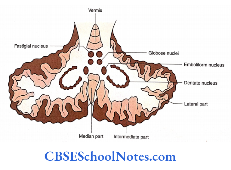
Deep within the cerebellar white matter, four pairs of nuclei are present.
Cerebellar Cortex
The histological structure of the grey matter of the cerebellar cortex is uniform throughout the cerebellum.
The cortex is composed of three layers:
- Molecular,
- Purkinje and
- Granular layers.
Deep to the granular layer, the cerebellar cortex lies in contact with the white matter.
The afferent fibres to the cerebellar cortex are of two different types:
- Climbing and Mossy fibres while efferents are axons of Purkinje cells. The efferent fibres terminate in the cerebellar nuclei.
- Students should learn the microscopic structure of the cerebral cortex from a textbook of histology
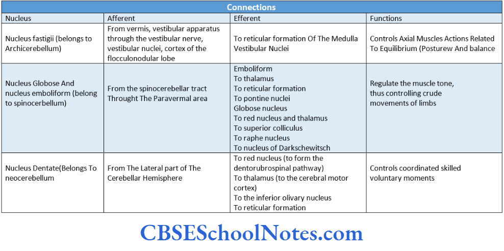
Cerebellar Nuclei
Four pairs of cerebellar nuclei are present deep within the cerebellar white matter.
These nuclei consist of most of the neurons, which give origin to the efferent fibres of the cerebellum. From the medial to the lateral side, they are named as follows:
- Fastigial
- Globose
- Emboliform
- Dentate
The connections and functions of these nuclei.
White Matter of the Cerebellum
A large body of white matter is present in the centre of each cerebellar hemisphere. Intracerebellar nuclei are present within the white matter of each cerebellar hemisphere.
The dentate nucleus occupies a large area on the lateral part of white matter.
The white matter of the cerebellum consists of two types of fibres: Intrinsic and Extrinsic.
1. Intrinsic fibres: The axons of the Purkinje cells, which originate in the cerebellar cortex and terminate in the intracerebellar nuclei are the intrinsic fibres of the cerebellum.
Extrinsic fibres: The extrinsic fibres connect the cerebellum with other parts of the central nervous system—brain and spinal cord.
These fibres, afferents and efferents, enter or exit the cerebellum through the cerebellar peduncles.
The cerebellar peduncles connect the cerebellum with the medulla oblongata, pons and midbrain. The fibres of three peduncles.
Functions Of Cerebellum
1. The cerebellum controls the muscular contraction efficiently, in an automatic manner and at an unconscious level.
2. The main function of the cerebellum is the coordination of motor activities so that the voluntary movements are smooth, balanced and accurate.
This is achieved by:
Maintenance of posture and balance with the help of the vestibulocerebellum, control of muscle tone with the help of the spinocerebellum and coordination and integration of various muscle groups, required for a given motor act, with the help of pontocerebellum.
3. The cerebellum helps in learning motor skills, with gradual training.
The lesions of the cerebellum are classified into the following categories:
- Lesion of vermis (spinocerebellum)
- Lesion of the flocculonodular lobe (archicerebellar syndrome)
- Lesion of the lateral hemisphere (neocerebellar syndrome)
- Lesion of Vermis
Patients show the following disorders:
Ataxia: It is defined as an uncoordinated sequence of movement. The patient stands with the legs spread apart walks on a wide base (wadding gait) and sways from side to side or backwards. Therefore, he takes the help of a wall while walking.
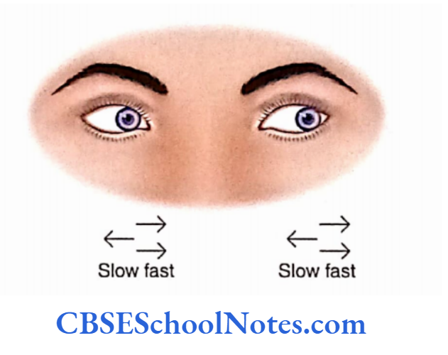
Cerebellar nystagmus: It is defined as conjugate involuntary oscillatory movements of the eyes. Nystagmus occurs due to the interruption of connections of the vermis with the ocular motor nuclei.
Speech disorder: This occurs due to a lack of coordination among muscles of speech (i.e. asynergy of speech muscles). Archicerebellum Syndrome Archicerebellum syndrome is due to a lesion of the flocculonodular lobe and uvula.
This produces truncal stance and gait ataxia. It also produces cerebellar nystagmus, vertigo and vomiting.
Neocerebeliar Syndrome
Neocerebeliar syndrome is due to a lesion of afferent pathways, cortex, intracerebellar nuclei of the cerebellar hemisphere or efferent pathways (superior cerebellar peduncle).
This syndrome shows the following signs:
Ataxia: The movements are not smooth but intermittent and jerky.
Dysmetria: It is defined as the inability to measure the distance correctly for reaching the intended target.
When the patient tries to touch an object, the finger overshoots the mark or deviates from it (described as past pointing), for example finger-nose test.
Asynergy: In the performance of fine movements, there is a lack of coordination among different muscle groups. This leads to a succession of mechanical or puppet-like movements.
Hypotonia: This results in muscle weakness and rapid fatigability. This is due to loss of deep cerebellar nuclei leading to hypotonia of peripheral muscles.
Intentional tremor: When a purposeful movement (finger-nose test) is attempted, then an involuntary, rhythmical wavering movement of the hand occurs.
Speech disorder: This is due to asynergy involving muscles used in speech. Speech becomes thick and monotonous.
Nystagmus: It is present if the vermis is also involved


Cerebellum Summary
- The cerebellum is part of the hindbrain. It lies behind the pons and the medulla in the posterior cranial fossa.
- It consists of two large lateral masses (cerebellar hemispheres) and a central part called vermis.
- The cerebellum is divided into many lobes (i.e. anterior, posterior and flocculonodular) with the help of primary fissure and posterolateral fissure. The posterior (middle) lobe is the largest and is present on both the surfaces of the cerebellum (i.e. superior and inferior surfaces).
- The surface of the cerebellum is covered by grey matter called the cerebral cortex. The grey matter is arranged in parallel ridges called folia.
- Based on evolution, the cerebellum can be divided into three parts: archicerebellum, paleocerebellum and neocerebellum.
- This classification of cerebellum roughly correlates with the functional classification of cerebellum, i.e. vestibulocerebellum, spinocerebellum and pontocerebellum.
- The archicerebellum is represented by the flocculonodular lobe and lingual. It is the oldest part of the cerebellum.
- Paleocerebellum consists of the anterior lobe, uvula and pyramid of the posterior lobe. The neocerebellum consists of the posterior lobe (except the uvula and pyramid). It is the most recent in development.
- The cerebellar cortex (grey matter) is folded into many folia (gyri). The histological structure of the cortex is uniform throughout the cerebellum. The cortex is composed of three layers: the molecular layer, the Purkinje layer and the granular layer.
- Deep in the cerebellar cortex is the presence of white matter. Four pairs of cerebellar nuclei (fastigial, globose, emboliform and dentate) are present deep within the cerebellar white matter.
- The white matter of the cerebellum consists of extrinsic and intrinsic fibres. The intrinsic fibres are mostly axons of Purkinje cells which originate in the cerebellar cortex and terminate in the intracerebellar nuclei.
- The extrinsic fibres connect the cerebellum with other parts of the central nervous system (brain and spinal cord).
- The extrinsic fibres are afferents and efferents. These fibres pass out or come into the cerebellum through three cerebellar peduncles on each side.
- The inferior, middle and superior cerebellar peduncles connect the cerebellum with the medulla oblongata, pons and midbrain, respectively.
- For the constituent fibres of various peduncles.
- The main functions of the cerebellum can be summarised as follows:
- Maintenance of posture and balance with the help of vestibulocerebellum
- Control of muscle tone with the help of spinocerebellum
- Coordination and integration of various muscle groups (required for a given motor act) are achieved with the help of pontocerebellum.
Multiple Choice Questions
Question 1. Which of the following statements about the cerebellum is false?
- It occupies the posterior cranial fossa
- It lies below the tentorial cerebella
- It lies behind the pons and medulla
- It is part of the brainstem
- It is mainly concerned with the maintenance of posture and balance
Answer: 4. It is mainly concerned with the maintenance of posture and balance
Question 2. All the statements regarding the cerebellum are correct except?
- It consists of two lobes (hemispheres) and a vermis
- It consists of superior and inferior surfaces
- There are two deep fissures—primary and posterolateral
- The posterolateral separates the anterior lobe of the cerebellum from the posterior lobe
- The flocculonodular lobe constitutes a very small part of the cerebellum
Answer: 4. The flocculonodular lobe constitutes a very small part of the cerebellum
Question 3. Which of the following statements about the cerebellar peduncle is true?
- The superior cerebellar peduncle connects it to the midbrain
- The middle peduncle connects it to the pons
- The inferior peduncle connects it to the medulla
- All of the above
Answer: 4. All of the above
Question 4. Which of the following facts regarding the input (afferent) fibres of the cerebellum is false?
- Afferent fibres are of two types—climbing and mossy
- Climbing fibres originate from the inferior olivary complex
- Mossy fibres originate in vestibular nuclei, pontine nuclei and spinal cord
- Mossy fibres ascend to the molecular layer of the cerebellar cortex and form synaptic contacts with the dendritic tree of Purkinje cells
Answer: 4. Mossy fibres ascend to the molecular layer of cerebellar cortex and form synaptic contacts with the dendritic tree of Purkinje cells
Question 5. The following nuclei are present in each cerebellar hemisphere except
- Fastigial
- Vestibular
- Globose
- Emboliformis
- Dentate
Answer: 2. Vestibular
Question 6. The following afferent fibres (tracts) are present in the inferior cerebellar peduncle except
- Posterior spinocerebellar
- Olivocerebellar
- Vestibulocerebellar
- Anterior spinocerebellar
- Trigeminocerebellar
Answer: 4. Trigeminocerebellar
Question 7. The following are the functional divisions of the cerebellum except
- Vestibulocerebellum
- Spinocerebellum
- Pontocerebellum
- Olivocerebellum
Answer: 4. Olivocerebellum
Question 5. The following are the functions of the cerebellum except
- Maintenance of posture and balance
- Coordination of exteroceptive sensory impulses
- Control of muscle tone
- Coordination of various muscle groups required for a given motor act
Answer: 2. Coordination of exteroceptive sensory impulses
Question 6. Most of the efferents of the cerebellum are axons of
- Cerebellar nuclei
- Golgi cells
- Basket cells
- Purkinje cells
Answer: 4. Purkinje cells
Question 10. The branches of the following arteries are responsible for the supply of blood:
- Vertebral artery
- Vertebral and basilar arteries
- Basilar artery
- Internal carotid artery
- Vertebral and internal carotid arteries
Answer: 2. Vertebral and basilar arteries
Question 11. Most of the afferent fibres to the cerebellum make synaptic contact with
- Cerebellar nuclei
- Golgi cells
- Granule cells
- Purkinje cells
- Basket cells
Answer: 2. Granule cells