
Central Nervous System
The central nervous system (CNS) consists of the brain and the spinal cord, which are made up of nervous tissues (nerve cell bodies, axons and dendrites, neuroglial cells, and blood vessels.
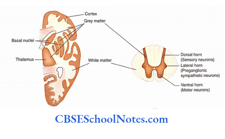
The surface of the brain is covered with a thin layer of grey matter.
This grey matter is called the cortex. However, many areas of grey matter are also present deep within the white matter of the brain. These areas are called ‘nuclei’.
In the spinal cord, grey matter is situated centrally and is ‘H’ shaped. The grey matter is surrounded by the white matter.
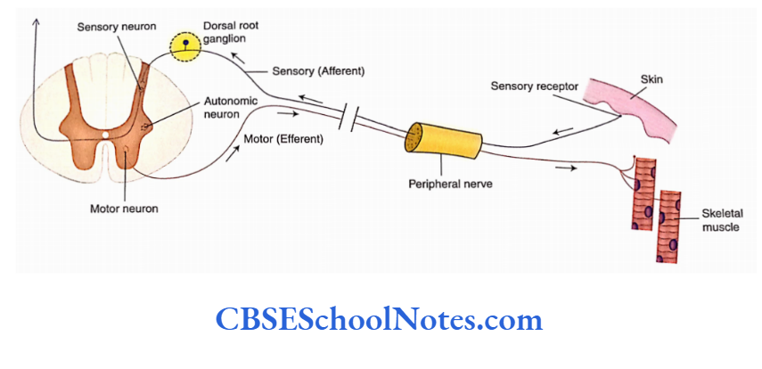
The grey matter shows two ventral (anterior) grey columns (horns) that contain motor neurons.
The dorsal grey columns (horns) contain sensory neurons. In the thoracic and upper lumbar regions, the grey matter also exhibits a lateral horn containing preganglionic autonomic neurons.
Read and Learn More Neuroanatomy
The white matter of the spinal cord is the collection of nerve fiber bundles, which are known as tracts. There are some ascending and descending tracts.
Central Nervous System Function
Both the brain and the spinal cord are surrounded by three membranes called meninges, namely the pia mater, arachnoid mater, and dura mater. Pia mater is
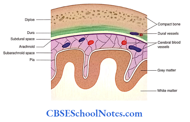
A thin delicate membrane, which intimately covers the outer surface of the brain and the spinal cord.
The space between the pia and the arachnoid mater is filled with cerebrospinal fluid (CSF). Blood vessels also run in subarachnoid space.
Central Nervous System Function
The dura mater is the outermost membrane that is very tough. It is fused with the bones of the cranial cavity (brain box).
Brain
The brain lies in the cranial cavity and is continuous with the spinal cord just below the level of the foramen magnum.
The brain, in the ascending order, is divided into three principal parts:
- Hindbrain
- Midbrain
- Forebrain
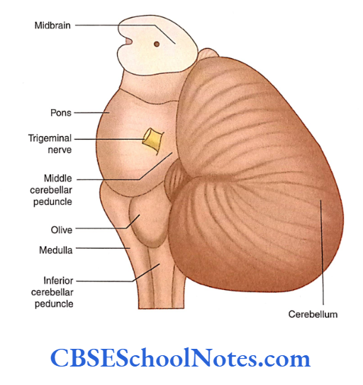
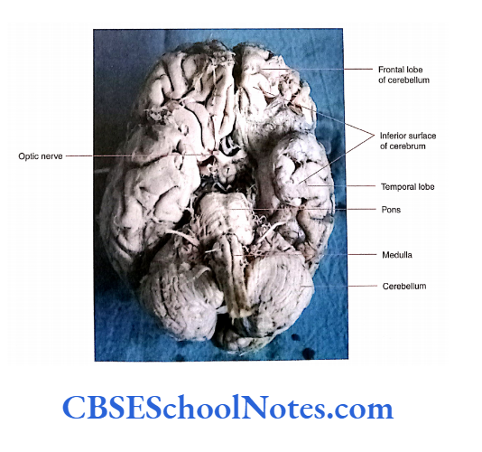
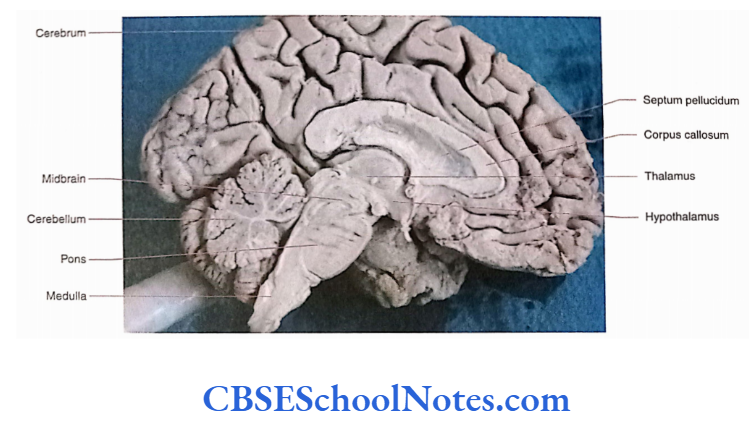
The medulla, pons, and midbrain are called brainstems. The midbrain is a small part, which connects the forebrain to the hindbrain. Posterior to the brainstem lies the cerebellum. The forebrain lies superior to the brainstem.
Hindbrain
The hindbrain consists of the medulla oblongata, pons, and cerebellum.
Medulla Oblongata
- Medulla oblongata is also known as medulla. It is conical in shape and continuous below the spinal cord. (just below the foramen magnum) and above with the pons.
- On the ventral aspect, there is the presence of an anterior median fissure, which divides it into right and left halves.
- On either side of the fissure is a ridge called a pyramid. Posterolateral to the pyramid is an oval swelling, known as the olive.
- Posterolateral to olives are inferior cerebellar peduncles, which contain the white fibers and connect the medulla to the cerebellum.
- The lower part of the medulla contains the central canal, which is continuous below the central canal of the spinal cord.
- The central canal opens above in the fourth ventricle, which is situated posterior to the upper part of the medulla and pons.
Pons
- Pons lies above the medulla and below the midbrain. On the ventral aspect, it forms a bulge that is convex from side to side and above downwards.
- On each side, the pons are continuous with the middle cerebral peduncle, which extends posteriorly into the cerebellum.
Central Nervous System Function
Cerebellum
- The cerebellum overlaps the posterior surfaces of the brainstem, i.e. midbrain, pons, and medulla.
- It lies in posterior cranial fossa inferior to a horizontal fold of dura matter (tentorium cerebelli).
- The surface of the cerebellum is covered by grey matter which shows a large number of closely set transverse fissures.
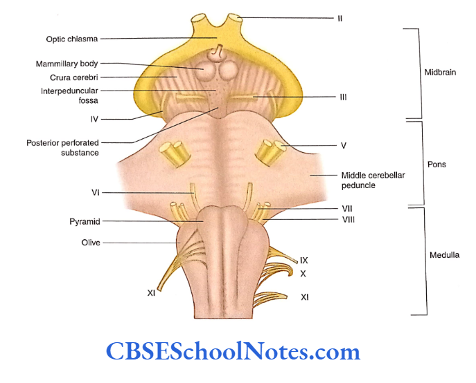
Midbrain
- The midbrain is the short segment of the brainstem situated above the pons.
- It joins the forebrain to the hindbrain. On the ventral aspect, it exhibits two broad white bundles of nerve fibers, the crura cerebri.
- These crura cerebri form the posterolateral boundary of a fossa called the interpeduncular fossa containing a posterior perforated substance.
- The posterior surface of the midbrain is deeply situated and overlapped by the cerebellum. It consists of four small swellings, called the colliculi (two superior and two inferior
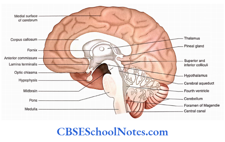
colliculi). - The midbrain is traversed by a narrow canal, the cerebral aqueduct, which is connected superiorly with the third ventricle and below with the fourth ventricle.
Forebrain
The forebrain consists of the diencephalon and cerebrum.
Diencephalon (Thalamus and Hypothalamus)
- The diencephalon is deeply situated in the forebrain. Only the inferior surface of the hypothalamus is visible in the roof of the interpeduncular fossa.
- The inferior surface of the hypothalamus is formed by the tuber cinereum and mammillary bodies.
- The thalamus and hypothalamus are seen on the lateral surface of the third ventricle in the median sagittal section of the brain.
- The thalamus is situated dorsal to the hypothalamus. The third ventricular cavity is situated between the right and the left diencephalons.
Cerebrum
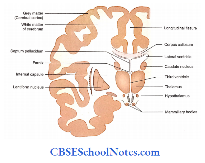
- The cerebrum is the largest part of the brain. The cerebrum consists of two cerebral hemispheres which are separated from each other by a midline fissure, called the longitudinal fissure.
- At the bottom of this fissure, there is the presence of the corpus callosum. The corpus callosum is a thick band of nerve fibers that connects the corresponding areas of two cerebral hemispheres (right and left).
- Each cerebral hemisphere is covered by grey matter (cortex). The surface of each hemisphere shows extensive folding to increase the surface area of the cortex.
- Thus, the cortex presents a large number of grooves (sulci) and ridges (gyri).
- Deep to the grey matter of the cortex, each hemisphere has a central core of the white matter.
- Many large collections of grey matter are present within the subcortical white matter and are referred to as basal nuclei (e.g. caudate and lentiform nuclei.
- Each cerebral hemisphere contains a large cavity, called the lateral ventricle. Each cerebral hemisphere has three surfaces: superolateral, inferior, and medial.
Ventricular System of the Brain
- The brain has large cavity-like spaces that are referred to as ventricles. The ventricles are named lateral ventricles, third ventricles, and fourth ventricles.
- The lateral ventricle is a large cavity situated in each cerebral hemisphere.
- Both the lateral ventricular cavities communicate with a median narrow cavity (third ventricle), which is situated between the right and the left diencephalons.
- The third ventricle communicates below with the fourth ventricle through the cerebral aqueduct of the midbrain.
- The fourth ventricle is continuous below with the central canal situated in the lower medulla and spinal cord.
- The ventricular system is lined with epithelium known as ependyma and at places it contains choroid plexuses which secrete CSF.
Spinal Cord
- The spinal cord is about a 45-cm long, cylindrical structure situated within the vertebral canal of the vertebral column. In adults, it extends from the medulla to the superior border of the second lumbar vertebrae.
- Within the vertebral canal, the cord is well protected by three meninges (pia, arachnoid, and dura) and CSF.
- Meninges covering the spinal cord are continuous above with the meninges covering the brain.
- The spinal cord shows two enlargements—cervical and lumbar—which give origin to cervical and lumbosacral.
Peripheral Nervous System
- The peripheral nervous system (PNS) is that part of the nervous system that is present outside the CNS (brain and spinal cord).
- This part of the nervous system is responsible for carrying the impulses to and away from the CNS.
- The PNS consists of nerve fibers and cell bodies located outside the CNS.
- The collection of cell bodies outside the CNS is called a ganglion, for example, a collection of neurons in the dorsal root ganglion, ganglion of cranial nerve, and sympathetic and parasympathetic ganglia.
Peripheral nerves take their origin from the CNS (brain and spinal cord) and are classified into cranial and spinal nerves. Ganglia are associated with cranial and spinal nerves.
- Cranial nerves
- Spinal nerves
- Ganglia
A typical spinal nerve is attached to the spinal cord through ventral nerve roots (containing motor axons or fibers) and dorsal nerve roots (containing sensory fibers).
The dorsal root contains a ganglion (dorsal root ganglion) in which cell bodies of sensory neurons are situated.
Cranial Nerves
- The 12 pairs of nerves that arise from the brain are called cranial nerves.
- All the cranial nerves come out from the cranial cavity through the foramina present at the base of the skull (cranium).
- Cranial nerves innervate the head and neck and to a lesser extent the thoracic and abdominal viscera.
- Out of 12 cranial nerves, the first two (olfactory and optic) are attached to the forebrain while the remaining 10 are attached to the brainstem.
- Some cranial nerves also carry autonomic (parasympathetic) fibers.
Ganglia
Ganglia are a collection of neurons outside the CNS and are a part of the PNS as they are present in association with cranial and spinal nerves. They are of two types: Sensory and Autonomic.
Some Important Terms
The following terms will be frequently used during discussion in the book.
Grey and White Matter
A section of the fresh brain or spinal cord shows some areas as white and others as grey.
- White matter is the aggregation of myelinated processes (fibers). As the myelin is white, the collection of fibers also looks white.
- On the other hand, grey matter of the nervous system is the collection of nerve cell bodies, dendrites, unmyelinated axons starting from or ending on the nerve cells (forming a synaptic junction), and neuroglial cells.
- The grey matter looks greyish as there is no myelin in these areas. Blood vessels are present in both grey and white matter.
- The grey and white matter of the brain and spinal cord.
Cortex and Nucleus
- The thin layer of grey matter on the surface of the cerebrum and cerebellum is called the cortex. Deep in the cortex is the presence of white matter.
- A nucleus is a mass of grey matter embedded within the white matter inside the CNS (brain and spinal cord).
- The nucleus consists of an aggregation of nerve cell bodies within the CNS, which share a common function, for example, the thalamus and caudate nucleus.
Fibers in a Peripheral Nerve
A peripheral nerve is a mixed nerve containing both afferent (sensory) and efferent (motor) fibers.
Afferent Fibres
The fibers that carry sensory impulses from various parts of the body (skin and deeper structures) to the CNS are called afferent fibers (sensory nerve fibers).
Efferent Fibres
The fibers which carry impulses from the CNS to various parts of the body are called efferent fibers. Many of these fibers are connected to muscles and are called motor nerve fibers.
Summary
- The CNS consists of the brain and the spinal cord.
- The brain consists of the forebrain, midbrain, and hindbrain.
- Both the brain and spinal cord are made up of grey and white matter.
The CNS is surrounded by three meninges:
- Pia,
- Arachnoid and
- Dura mater.
- The hindbrain consists of the medulla, pons, and cerebellum. The forebrain consists of the diencephalon (thalamus and hypothalamus) and cerebrum. The midbrain joins the forebrain with the hindbrain.
- Each cerebral hemisphere presents three surfaces: Superolateral,
- Medial and
- Inferior.
- Each surface is covered by cortex which shows a large number of sulci and gyri.
- Each hemisphere has a central core of white matter. Many large collections of grey matter are present within the white matter and are referred to as nuclei.
- The brain contains many large cavities called ventricles, i.e. lateral ventricles, third ventricles, and fourth ventricles.
- The PNS consists of 1 2 pairs of cranial and 31 pairs of spinal nerves.
- Sensory ganglia are a collection of nerves in association with cranial and spinal nerves.
- Autonomic ganglia are classified into sympathetic and parasympathetic.
- Sympathetic ganglia are located in the sympathetic trunk while parasympathetic ganglia are located close to the organ they supply.
Central And Peripheral Nervous Systems Multiple Choice Questions
Question 1. Which of the following statements about the spinal cord is true?
- The ventral grey horn contains motor neurons
- The dorsal grey horn contains sensory neurons
- Lateral grey horn contains preganglionic autonomic
neurons - All of the above
Answer: 4. All of the above
Question 2. Which of the following statements about the grey matter of the nervous system is true?
- It contains nerve cell bodies
- It contains neuroglial cells
- There is no myelin in grey matter
- It contains blood vessels
- All of the above
Answer: 5. All of the above
Question 3. Which of the following ganglia is a collection of nerve cell bodies?
- Spinal root ganglion
- Sympathetic ganglion
- Parasympathetic ganglion
- All of the above
Answer: 5. All of the above
Question 4. Which of the following statements is false?
- Nucleus is the mass of grey matter in the spinal cord
- The nucleus is the mass of grey matter in the brain
- The nucleus is a thin layer of grey matter on the surface of the cerebellum
- The thalamus is an example of nucleus
Answer: 3. Nucleus is a thin layer of grey matter on the surface of the cerebellum
Question 5. The collection of white matter in the CNS is known as
- Tract
- Funiculus
- Lemniscus
- All of the above
Answer: 4. All of the above
Question 6. Which are not the afferent fibers?
- They carry the sensory impulse
- They carry the motor impulse
- They carry both motor and sensory impulses
- They carry impulses from the brain to various parts of the body
Answer: 2. They carry the motor impulse
Question 7. Hindbrain consists of
- Medulla, pons, and cerebellum
- Midbrain, pons, and medulla
- The midbrain, pons, medulla, and cerebellum
- Midbrain, pons, and cerebellum
Answer: 1. Medulla, pons, and cerebellum
Question 8. Which of the following statements is true?
- Pons is connected to the cerebellum through middle cerebellar peduncles
- The midbrain is connected to the cerebellum through superior cerebellar peduncles
- The medulla is connected to the cerebellum through inferior cerebellar peduncles
- All of the above
Answer: 4. All of the above
Question 9. Diencephalon consists of
- Telencephalon
- Thalamus
- Hypothalamus
- Corpus striatum
Answer: 2. Thalamus
Question 10. The peripheral nerve consists of
- Cranial nerves
- Spinal nerves
- Nerve fibers and cell bodies outside the CNS
- All of the above
Answer: 4. All of the above
Question 11. The ganglia are ofthe following type:
- Sensory ganglia of cranial nerves
- Autonomic ganglia
- Dorsal root ganglia of spinal nerves
- Ventral root ganglia of spinal nerves
Answer: 1. Sensory ganglia of cranial nerves.