
Cartilage
we have seen that cartilage is classified as a specialized connective tissue. It consists of all the three components of a connective tissue, i.e., ground substance, fibres and cells.
Cartilage differs from other connective tissues by the presence of substances in the intercellular matrix that give firmness to it.
The firm pliable matrix is capable of resisting mechanical stresses. Three types of cartilage are found in the body:
- Hyaline cartilage
- Elastic cartilage
- Fibrocartilage
Although all the above cartilages consist of cells (chondrocytes), ground substance and fibres, they differ from each other because of the type and amount of fibres present in them.
- Cartilage is not supplied with the blood vessels(it is avascular). However, it is usually surrounded by a membrane called as perichondrium, which is rich in blood vessels.
- The chondrocytes receive nutritive substances from blood vessels in the perichondrium by diffusion through the ground substance. P
- Erichondrium is present in most of the hyaline cartilage, and elastic cartilage but absent in fibro cartilage
Cartilage Remember
Cartilage is a special kind of connective tissue consisting 1 of chondrocyte cells, connective tissue fibres and specialized ground substances. The firm pliable matrix is capable of resisting mechanical stresses.
Hyaline Cartilage
Hyaline Cartilage Location
It is the most abundant type of cartilage in the human body. This
cartilage is found in the fetal skeleton, ends of adult bones(articular cartilage), nose, costal cartilage, trachea, bronchi and larynx.
Hyaline Cartilage Description
It consists of a homogeneous, transparent and amorphous intercellular matrix. The matrix consists of collagen fibres and ground substance.
Throughout the matrix, cartilage cells (chondrocytes) are present in small spaces called lacunae. Hyaline cartilage is surrounded by perichondrium.
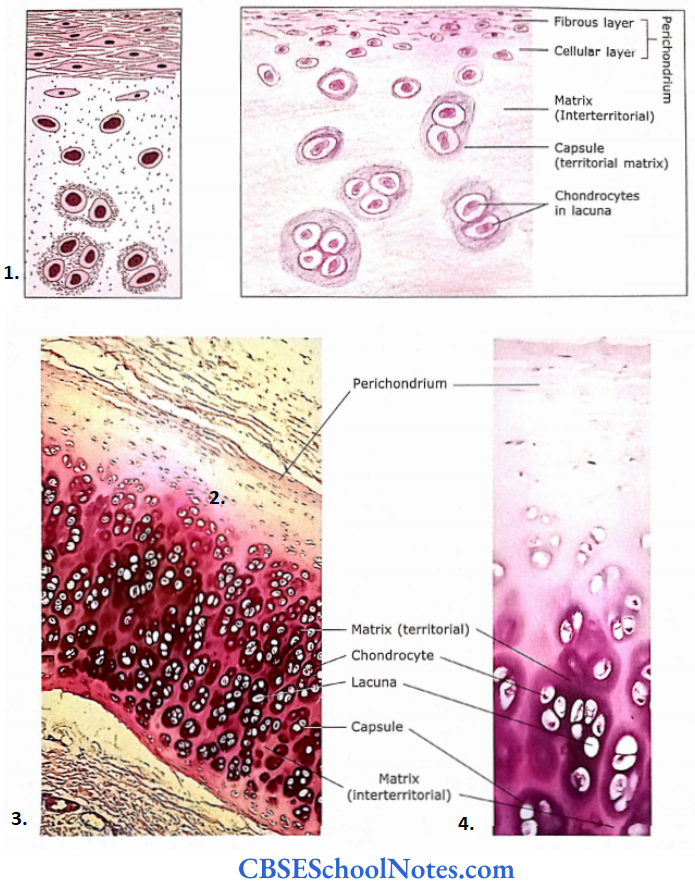
- Schematic diagram showing features of hyaline cartilage
- As seen under a microscope.
- Photomicrograph of hyaline cartilage perichondrium in the upper part chondrocytes are present in lacunae
- Photomicrograph of hyaline cartilage perichondrium in the lower part chondrocytes are present in lacunae
1. Fibres
The ground substance of hyaline cartilage contains fine type II collagen fibres that are about 15-40 nm in diameter. The thin fibres do not show cross striations and form a three-dimensional network in the ground substance.
- The thicker fibres show cross-striations and are arranged in a direction that can resist the strain to which cartilage is subjected. Collagen fibres provide stability and strength to the cartilage.
- These fibres constitute about 40% of the dry weight of cartilage. These fibres are not seen in the histological section because they have the same refractive index as that of ground substance.
- Some other types of collagen fibres(types 6,9,10 and 11) are also present in this cartilage in small amounts.
2. Ground Substance
It is a featureless(homogeneous) gel-like substance that stains blue with a basic dye(haematoxylin). The main constituent of ground substance is sulphated proteoglycans (aggrecan).
- The basophilia of the matrix is due to the presence of chondroitin and keratan sulfate which are acidic. The highest concentration of these substances is around the lacuna as it is newly formed.
- This dark blue staining around the lacuna is called a capsule or territorial matrix. As we go away from the lacuna the concentration of sulphated proteoglycans becomes less and less.
- Thus the matrix does not show that intense blue staining that is seen in a capsule. This matrix is called an inter-territorial matrix.
Hyaline Cartilage Remember
The matrix of hyaline cartilage consists of type 2 collagen fibres and sulfated proteoglycans, glycoproteins and water.
The matrix is homogenous as fibres are not seen in the histological section because they have the same refractive index as that of ground substance.
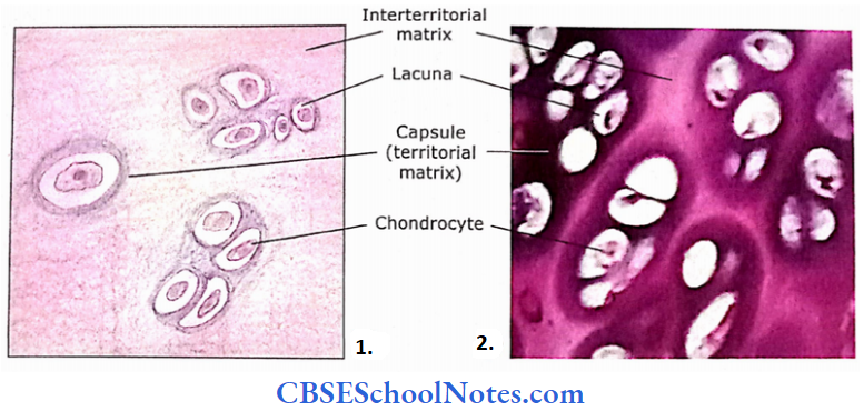
- A portion of hyaline cartilage showing lacunae containing shrunken chondrocytes.
- Photomicrograph of a portion of hyaline cartilage
Hyaline Cartilage Further Details
Chemical Composition of Ground Substance
The principal constituent of ground substance is proteoglycan which is present in high concentration and is responsible for the firmness of matrix.
- The proteoglycan molecule consists of a core protein from which many glycosaminoglycan molecules radiate in a bottlebrush configuration. There are two types of glycosaminoglycans in the proteoglycans of hyaline cartilage.
- These are chondroitin sulphate and keratan sulphate. About 80-100 molecules of proteoglycans are joined to a long molecule of hyaluronic acid to form large hyaluronate proteoglycan aggregates.
- These aggregates are then bound to the thin collagen fibres present in the matrix. The glycosaminoglycan side chains form electrostatic bonds with the collagen.
Thus, the ground substance and collagen form a cross-linked molecular framework that resists tensile forces.
- The ground substance also contains other types of proteoglycans that do not form aggregates. It also contains various other types of glycoproteins.
- The ground substance also can collect large amounts of water molecules.
- Because of a high degree of hydration hyaline cartilage solutes can diffuse easily through the matrix and it acts as an effective weight-bearing cartilage (For Example., articular cartilage).
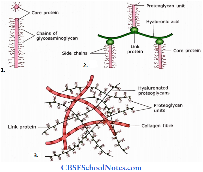
- A proteoglycan subunit is formed with a core protein to which about 100 glycosaminoglycan units(chains) are joined in bottlebrush configuration.
- About 80-100 molecules of proteoglycans are joined to a long molecule of hyaluronic acid with the help of a link protein.
- These hyaluronate proteoglycan aggregates bound to thin collagen fibres present in the matrix.
3. Cells
Only one type of cell(chondrocyte) is seen in the cartilage. These cells occupy lacunae in the matrix. In H and E preparation, these cells are usually shrunken and the gap is seen between the cell and the margin of a lacuna.
- However, in elec tron microscopy cell appears elliptical and occupies a complete lacuna. Chondrocytes are responsible for the synthesis of collagen fibres and ground substances.
- In young cartilage, chondrocytes show mitotic cell division and give rise to daughter cells. These newly formed chondrocytes produce fibres and ground substances around themselves.
- Groups of daughter cells from one chondro cytes remain in close relationship and form cell nests. As newly divided chondrocytes produce a matrix, this surrounds them, and the chondrocytes move away from each other.
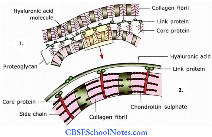
- The hyaluronate proteoglycan aggregates bound to two thin collagen fibrils.
- Enlarged view of area enclosed in a rectangle. The chondroitin sulphate side chains of proteoglycan form an electrostatic bond with the collagen.
Perichondrium
Hyaline cartilage, on its free surface, is always covered with a fibrovascular membrane called perichondrium. The perichondrium is absent in the cartilage where it forms.
- The free surface as in joint cavity articular cartilage) and where cartilage makes direct contact with bone (i.e., costal cartilage making direct contact with rib; epiphyseal cartilage making contact with metaphyses and epiphyses in a developing bone).
- Perichondrium consists of two layers, i.e., an outer fibrous layer(made up of dense irregular fibrous connective tissue) and an inner cellular layer(made up predominantly of cells which may convert to chondrocytes when the cartilage is growing.
- In adult cartilage, only a fibrous layer is present. Although cartilage itself is avascular, the fibrous layer of the perichondrium has blood capillaries.
- These capillaries provide nutrition to cartilage cells by diffusion through the matrix. The hyaline cartilage displays both appositional and interstitial growth.
In the case of appositional growth, there is the addition of new cartilage at its surface, while in the case of interstitial growth new cartilage is formed within the existing cartilage by division and differentiation of chondrocytes.
Perichondrium Functions
- Articular cartilage provides cushioning and a smooth surface for movements at joints.
- Although it is flexible, it provides support because of its firmness (For Example., as in costal cartilage).
- Firmness of cartilage keeps the lumen of the trachea and bronchi patent.
- Most long bones of the fetus, to begin with, are cartilage models. Later this cartilaginous model is replaced by bone.
Elastic Cartilage
Elastic Cartilage Location
The elastic cartilage is present in the pinna of the ear, epiglottis, tips of arytenoids, corniculate and cuneiform cartilages of the larynx, external auditory meatus and auditory tube.
Elastic Cartilage Description
This cartilage is highly elastic. It looks yellow in the fresh state and is hence sometimes called yellow elastic cartilage. The elastic cartilage also consists of ground substance fibres, cells and perichondrium.
Fibres
Elastic cartilage contains a meshwork of branching and anastomosing elastic fibres that give it a yellow appearance. The elastic fibres are more heavily concentrated in the centre of cartilage than near the perichondrium.
This cartilage also contains a few type 2 collagen fibres. Elastic fibres are not seen in H and E stains but are visualized by special staining methods for elastic fibres(orcein stain).
Ground Substance
As in hyaline cartilage, it also contains proteoglycans.
Cells
- Chondrocytes are present in lacunae. These cells are bigger than cells present in hyaline cartilage and are present singly or in groups of two.
- Cells are closely placed as the intercellular ground substance is much less than in hyaline cartilage.
- Although the matrix of hyaline cartilage calcifies in old age, the matrix of elastic cartilage does not calcify with age.
Perichondrium
It is the same as described in hyaline cartilage.
Elastic Cartilage Remember
Elastic cartilage is differentiated from hyaline cartilage as it contains elastic fibres in the matrix as well as in the perichondrium.
Elastic Cartilage Functions
It provides shape and support to the organ, with elasticity.
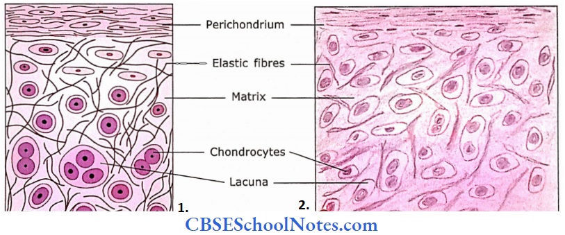
- Schematic diagram of elastic cartilage.
- under microscope.
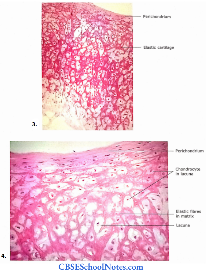
- Low power view of elastic cartilage from epiglottis.
- Photomicrograph of elastic cartilage
Fibrocartilage
It is also known as fibrocartilage it contains bundles of thick collagen fibres. The histological structure of fibro-cartilage resembles dense regular connective tissue. One may confuse it with the section of a tendon. However, it can be identified because chondrocytes are seen between collagen bundles.
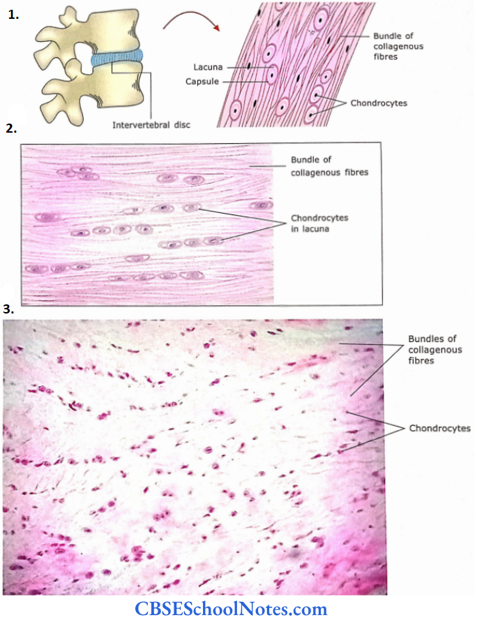
- Schematic diagram of fibrocartilage.
- Microscope.
- Section of fibrocartilage from intervertebral disc.
Fibrocartilage Location
Fibrocartilage is found in intervertebral discs (annulus fibrosus), pubic symphysis and manubriostemal joint.
- The menisci of the knee joint and articular disc of tempo roman tabular and sternoclavicular joints are also fibrocartilage nous. Similarly, the glenoidal and acetabular labrum is also made up of fibrocartilage.
- Sometimes, this cartilage is seen at the site of insertion of tendon in bone.
Fibrocartilage Description
Fibres
In fibrocartilage, all the collagen fibres are of type 1 and type 2 varieties. The proportion of type 1 and type 2 fibres varies in different types of fibrocartilage. In the intervertebral disc, both type 1 and type 2 are in equal proportion.
Ground Substance
A minimal amount of basophilic ground substance is seen around the chondrocyte.
cells
Very few chondrocytes are seen, which are oriented between large collagenous fibre bundles. Chondrocytes are either present in a row or scattered singly between bundles of fibres.
Perichondrium is absent in fibrocartilage.
Fibrocartilage Functions
Fibrocartilage is capable ofresisting compressive and shear forces, i.e., deformation, For Example., intervertebral disc.
Fibrocartilage Remember
Fibrocartilage consists of abundant type 1 collagen fibres and minimal ground substance. The perichondrium is absent in this type of cartilage.
Fibrocartilage Clinical Applications
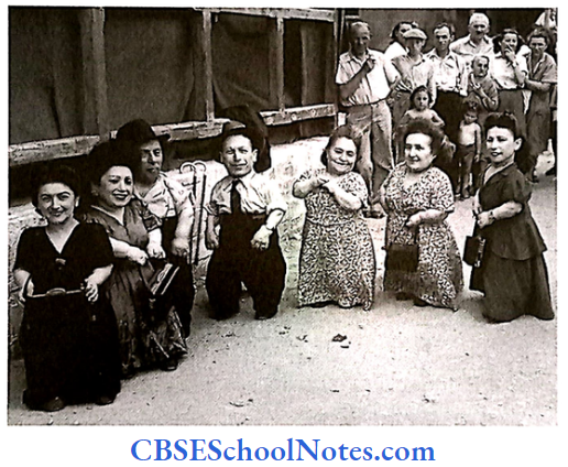
- Pseudoachondroplasia
- It is due to a defect of cartilage oligomeric matrix protein (COMP) in the joints and is characterized by a more typical development of the head and face.
- In this condition, there occurs the reduced proliferation of cartilage cells in the epiphyseal plate of long bones. This causes shortening of limbs, resulting in short stature.
- However, the head and face are normal. The achondroplasia is inherited as an autosomal dominant disease. Shown in the accompanying photograph are seven pseudochondroplastic members of the Ovitz family.
- A family of Romanian Jews who toured Eastern Europe as a musical troupe before World War 2 (their taller siblings working backstage), survived imprisonment at Auschwitz, and the family immigrated to Israel. They were photographed on arrival in Haifa in 1949.
- Osteoarthritis
- Osteoarthritis is a disease of old age. In this condition, the articular cartilage of interphalangeal joints of the hand, hip joint and knee joint progressively become thinner and ultimately break.
- This exposes the bone beneath the cartilage. It is a highly painful condition and leads to a walking disability.
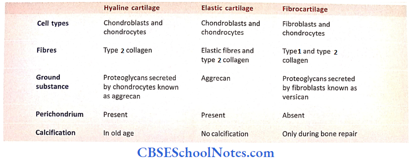
Comparison Between different types of cartilages
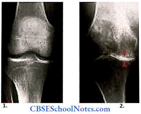
- Healthy diseased
- Knee joints.