
Brainstem Midbrain
- The midbrain is the uppermost part of the brainstem, connecting the pons and the cerebellum with the forebrain.
- It measures about 2 cm in length. The midbrain is traversed by a cerebral aqueduct, which connects the third ventricle to the fourth ventricle.
- The midbrain contains nuclei of origin for cranial nerves III (oculomotor) and IV (trochlear).
Brainstem Midbrain External Features
The midbrain presents ventral, dorsal, and right and left lateral surfaces.
Ventral Surface
The ventral surface shows the presence of two crura cerebri.
The crura cerebri (cerebral peduncles) are rope-like thick bundles of white fibres.
The two crura cerebri diverge from each other when traced from pons towards the cerebral hemispheres.
They form the lateral boundaries of the fossa called the interpeduncular fossa. The interpeduncular fossa contains the posterior perforated substance and mammillary bodies.
Read and Learn More Neuroanatomy
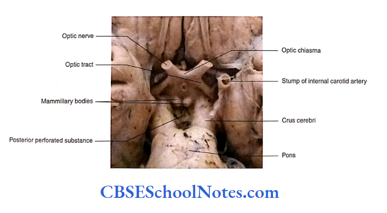
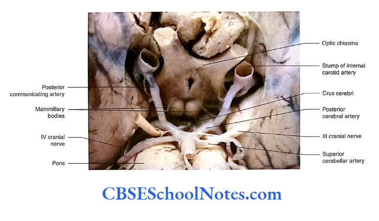
The 3 cranial nerve (oculomotor) is seen to arise from the medial aspect of the crus cerebri. The 5 cranial nerve (trochlear) comes to the ventral side from the lateral aspect of the midbrain.
Each crus cerebri is crossed transversely from above downwards by the optic tract (as it passes backwards towards the lateral geniculate body), posterior cerebral artery and superior cerebellar artery.
Dorsal Surface
- The dorsal surface ofthe midbrain presents four rounded eminences called colliculi or corpora quadrigemina
- The superior pair of eminences is called the superior colliculi and the inferior pair is referred to as the inferior colliculi.
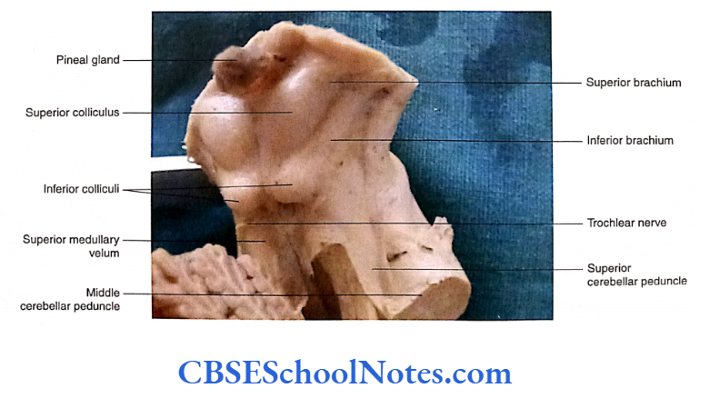
- The 4 cranial nerves (trochlear) take origin just below the inferior colliculi.
- After taking origin, the pair of trochlear nerves run forwards across the lateral aspect of the midbrain to appear on its ventral aspect where they lie between the superior cerebellar and posterior cerebral arteries
Lateral Surface
- On each lateral surface ofthe midbrain, two thick bands of white fibres are seen. These are known as brachium colliculi.
- There is a brachium for each ofthe superior and inferior colliculi.
- The brachium of the superior colliculus passes upwards, forwards and laterally to the lateral geniculate body and optic tract.
- The brachium of the inferior colliculus, on each side, connects the inferior colliculus to the medial geniculate body.
Brainstem Midbrain Internal Structure
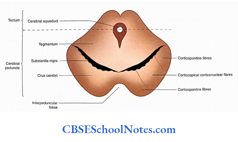
To understand the internal structure of the midbrain we will have to look at its transverse section The most prominent structure seen in this section is the cerebral aqueduct.
This duct is surrounded by central grey matter.
A transverse line drawn through the cerebral aqueduct divides the midbrain into two parts:
- The part lying behind the transverse line is called the tectum
- The part lying ventral to this line is made up of right and left cerebral peduncles.
Tectum
- The tectum is the dorsal part of the midbrain. Its structure is different at the upper and lower levels of midbrain.
- At the upper level, the tectum consists of a pair of superior colliculi and at the lower level, it has a pair of inferior colliculi.
Cerebral Peduncle
Each of the cerebral peduncles, from the ventral to the dorsal side, is made up of three parts. These are as follows:
- Crus cerebri (also called basis peduncle)
- Substantia nigra
- Tegmentum
The structure of crus cerebri and substantia nigra remains the same throughout the length of the midbrain. However, the structure ofthe tegmentum is different at the Upper and lower levels of midbrain.
Crus Cerebri
The crus cerebri is semilunar in section and consists of a dense mass of descending white fibres.
The crus cerebri is continuous above the internal capsule and below the basilar part of the pons.
The fibres in the crus cerebri consist of the following: corticopontine fibres, corticospinal fibres and corticonuclear fibres
The corticospinal and corticonuclear fibres occupy the intermediate three-fifths of the crus cerebri while the corticopontine fibres occupy the latter one-fifth and medial one-fifth of the crus cerebri.
Corticospinal fibres are motor fibres originating in the precentral gyrus of the cerebral cortex. These fibres pass through the basilar part of the pons to enter the pyramid of the medulla.
Corticonuclear fibres also arise from the precentral gyrus and terminate in the motor nuclei of cranial nerves of the opposite and same side.
Corticopontine fibres terminate by forming synapses with the pontine nuclei located in the pons.
The axons of pontine nuclei then pass to the cerebellar hemisphere of the opposite side through the middle cerebella.
Substantia Nigra
The substantia nigra is a band of dark grey matter which is present dorsal to crus cerebri and ventral to tegmentum.
It appears dark because of the presence of pigmented nerve cells. These nerve cells contain the melanin pigment, and dopamine is synthesised in the substantia nigra.
Tegmentum
- The tegmentum is the region of the midbrain that lies between the substantia nigra and tectum.
- It consists of grey and white matter. The tegmentum of the midbrain is an upward continuation of the tegmentum of the pons.
- As the structure of crus cerebri and substantia nigra remains the same throughout the midbrain, we shall learn about the structure of tegmentum and tectum at lower and upper levels (i.e. at the level of inferior and superior colliculi)
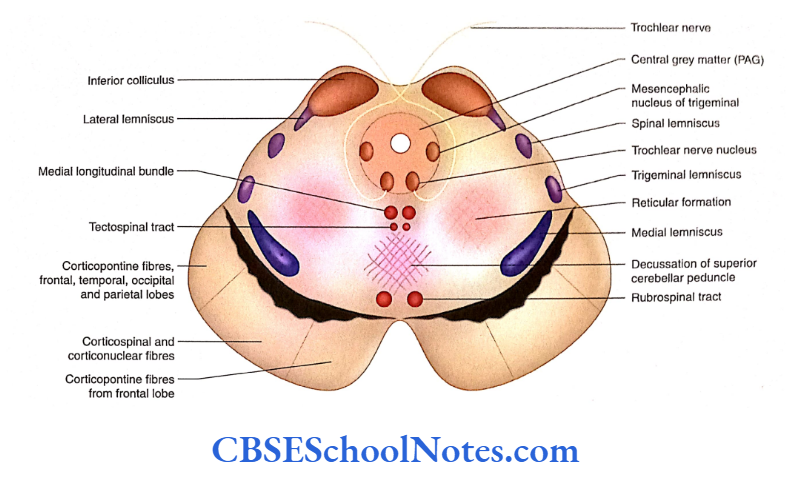
- Transverse Section of Midbrain at the Lower Level [Level of Inferior Colliculus)
- This level corresponds to the level of the inferior colliculus
Tegmentum
The arrangement of grey and white matter in the tegmentum at the level of inferior colliculus is described in the following text.
Arrangement of Grey Matter
The grey matter present in this section is identified as follows:
1. Periaqueductal grey matter: The grey matter surrounding the cerebral aqueduct is known as periaqueductal grey (central grey) matter. At this level, it contains trochlear (motor nuclei) and mesencephalic (sensory) nuclei.
Trochlear nerve nucleus: The trochlear nudeus is situated in the ventral periaqueductal grey matter near the midline.
This nucleus is situated immediately posterior to the medial longitudinal bundle (MLB). The fibres of the trochlear nerve run dorsally and decussate before coming out on the dorsal surface of the midbrain.
The mesencephalic nucleus of the trigeminal nerve: The mesencephalic nucleus lies in the lateral part of the central grey matter.
The neurons of this nucleus receive proprioceptive impulses from the muscles of mastication, facial muscles, ocular muscles and teeth.
2. Nudeus of reticular formation: This consists of many small nuclei located in the area of the midbrain reticular formation.
Arrangement of White Matter
The tegmentum of the midbrain contains all the white bundles as seen in the tegmentum of the upper pons.
The white fibre bundles that are seen in this region are given in the following text:
Decussation of superior cerebellar peduncles: This is seen in the midline ventral to the central grey matter. After decussation, the fibres form ascending and descending tracts.
Arrangement of various lemnisci: The medial, trigeminal, spinal and lateral lemnisci are arranged dorsomedial to substantia nigra, in order from medial to lateral side. The lateral lemniscus ends in the inferior colliculus.
The other white fibres present near the midline of the tegmentum are MLB, tectospinal tract, decussation of the superior cerebellar peduncle (described in the preceding text) and rubrospinal tract, in order from the dorsal to the ventral side preceding text) and rubrospinal tract, in order from the dorsal to the ventral side.
Tectum
The tectum is the dorsal part of the midbrain, and the transverse section of the midbrain at the lower level passes through the inferior colliculus.
Inferior colliculus: The inferior colliculus is a large nucleus of the auditory pathway (acts as a relay station) and is associated with auditory and audiovisual reflexes
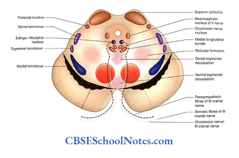
Transverse Section of Midbrain at the Upper Level (Level of Superior Colliculus)
Tegmentum
The characteristics of tegmentum as seen in the transverse section of the midbrain at the level of superior colliculus are as follows
Arrangement of Grey Matter
The grey matter depicts the following important nuclei:
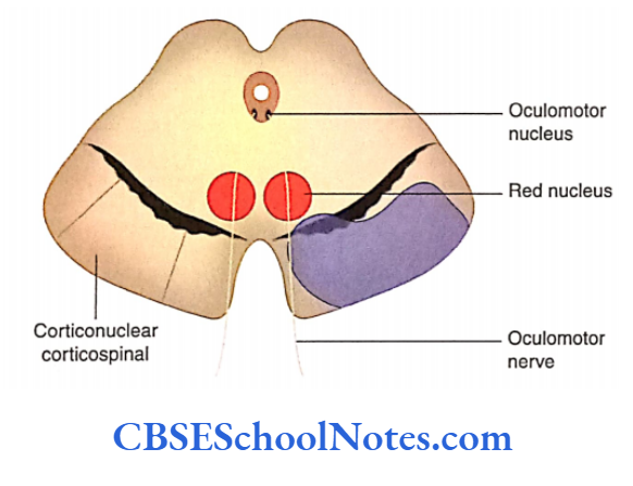
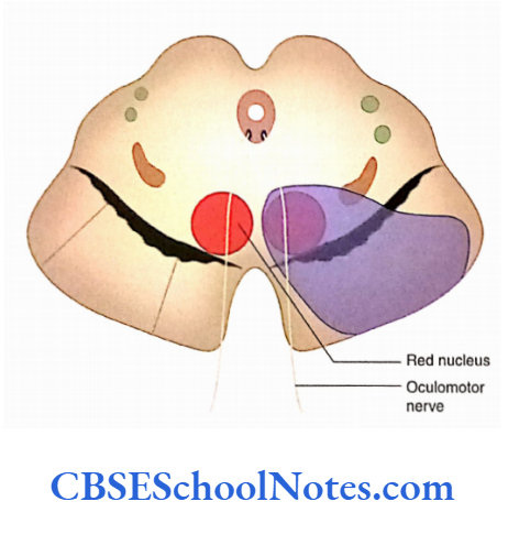
Oculomotor nerve nucleus: It is situated ventral to periaqueductal grey matter close to midline. The axons of the oculomotor nucleus pass ventrally, traversing the red nucleus, and come out as an oculomotor nerve in the interpeduncular fossa.
Edinger-Westphal nucleus: This is a visceral motor nucleus which is situated dorsal to the oculomotor nuclear complex.
This nucleus gives rise to preganglionic parasympathetic fibres which travel with the oculomotor nerve to relay in the ciliary ganglion. The postganglionic fibres supply the ciliary muscle and the constrictor papillae muscle of the iris.
Mesencephalic nucleus: This nucleus maintains the same position as in the lower level (in the lateral part of central grey matter).
Red nucleus: This is the most prominent collection of neurons in the tegmentum. It is an oval column of cells extending in the upper half of the midbrain. It lies dorsal to substantia nigra and close to midline.
The afferent fibres reach the red nucleus from the cerebellum (through the superior cerebellar peduncle), cerebrum, basal ganglia, superior colliculus, hypothalamus, substantia nigra and spinal cord.
The efferent fibres from the red nucleus cross in the ventral tegmental decussation and then go to the spinal cord (rubrospinal tract), cranial nerve motor nuclei 3, 4, 5, 6, 7 (supranuclear), olivary nucleus, reticular formation (rubroreticular), substantia nigra, cerebral cortex and thalamus.
The red nucleus is considered an important motor nucleus of the extrapyramidal system (maintaining posture and muscle tone).
Arrangement of White Matter
The following groups of white fibres, in the form of tracts and lemnisci, are seen:
Lemnisci: Medial, trigeminal and spinal lemnisci occupy the ventral part of the tegmentum, in order from the medial to the lateral side. The lateral lemniscus is not found at this level as it has already terminated in the inferior colliculus.
Medial longitudinal bundle: The MLB is placed just in front of the oculomotor nucleus, in the paramedian position.
Dorsal tegmental decussation—tectospinal and tectobulbar tracts: This decussation is present in front of the MLB at this level. The fibres of the tectospinal tract arise from the superior colliculi and decussate in the median plane.
After decussation, the fibres descend as the tectospinal tract which is present in front ofthe MLB, in the paramedian position.
Some fibres of this tract end in the motor cranial nerve nuclei (3, 4 and 6) inside the brainstem. This tract is known as the retrobulbar tract and is considered a pathway for reflex movements of the eye in response to visual stimuli.
Ventral tegmental decussation and rubrospinal tract: The fibres of the rubrospinal tract arise from the lower part of the red nucleus and soon cross to the opposite side.
The crossing of fibres lies in the ventral part of the tegmentum and is known as ventral tegmental decussation.
After decussation, these fibres descend through the brainstem as a rubrospinal tract. The fibres of the rubrospinal tract terminate on the anterior horn cells of spinal grey matter.
Tectum
The tectum is the dorsal part of the midbrain which, at this level, consists of the superior colliculus and pretectal nucleus.
- Superior colliculus: The superior colliculus has a complex structure. It is made up of seven alternating layers of white and grey matter. It receives fibres from the retina and other sources and sends fibres to the brainstem and spinal cord through the tectobulbar and tectospinal tracts. It is concerned with retlex movements of the head and neck in response to visual stimulus.
- Pretectal nucleus: The pretectal nucleus lies lateral to the superior colliculus from where it extends upwards till the level of posterior commissure. The pretectal nuclei receive fibres from both retinae ami visual cortex through the optic tract and superior brachium. The pretectal nuclei send efferents (axons) to the Edinger-Westphal nucleus on the same and opposite side.
Brainstem Midbrain Summary
The midbrain is the uppermost part of the brainstem, connecting the pons and the cerebellum with the forebrain. It contains nuclei of origin for cranial nerves 3 and 4.
External features
- The midbrain presents ventral, dorsal and lateral surfaces. The ventral surface shows the presence of two crura cerebri which enclose posterior perforated substance and mammillary bodies. The 3 cranial nerves arise from the medial aspect of the cerebri.
- The dorsal surface of the midbrain presents a superior and an inferior pair of colliculi.
- The 4 cranial nerves (trochlear) originate from either side of the midline, just below the inferior colliculi.
The lateral surface of the midbrain shows two thick bands of white fibres:
- Superior and
- Inferior brachium.
Internal structure
On the section of a midbrain, a transverse line drawn through the cerebral aqueduct divides the midbrain into two parts:
- Cerebral peduncle and Tectum.
- Cerebral peduncle
The cerebral peduncle is further divided, in the ventrodorsal direction, into crus cerebri, substantial nigra and tegmentum. The structure of crus cerebri and substantia nigra remains the same throughout the length of the midbrain.
However, the structure of the tegmentum and tectum varies at the upper and lower levels of the midbrain.
- Crus cerebri: It consists of a dense mass of descending white fibres, that is, corticospinal, corticonuclear and corticopontine.
- Substantia nigra: It is a band of dark grey matter containing melanin and iron pigments in nerve cells. It is present just posterior to the crus cerebri. It synthesises the neurotransmitter dopamine.
- Tegmentum: It consists of grey and white matter and is situated between substantia nigra and tectum.
At the level of the inferior colliculus, the following features are seen in the tegmentum:
The central grey matter contains trochlear and mesencephalic nuclei. Trochlear nerves come out of the dorsal aspect of the midbrain. The reticular formation is present ventrolateral to central grey matter.
Decussation of superior cerebellar peduncles is seen in the midline ventral to central grey matter.
The arrangement of various lemnisci (ML, TL, SL and LL) is dorsomedial to substantia nigra.
The medial longitudinal bundle, tectospinal tract, decussation of the superior cerebellar peduncle and rubrospinal tract are seen in the paramedian position.
At the level of the superior colliculus, the following features are seen in the tegmentum:
- The oculomotor nerve nucleus is situated ventral to periaqueductal grey in the midline. The Edinger-Westphal nucleus is dorsal to the oculomotor nucleus.
- The red nucleus is situated dorsal to substantia nigra close to midline. It is a motor nucleus of the extrapyramidal system. The mesencephalic nucleus maintains the same position as in the lower level.
- The medial, trigeminal and spinal lemnisci are located in the ventral part of the tegmentum, in order from the medial to the lateral side.
- The medial longitudinal bundle is situated ventral to the oculomotor nucleus in the paramedian position.
- Dorsal and ventral tegmental decussation is located in the midline of the tegmentum.
Tectum
Three different collections of grey matter are found in tectum:
- Superior colliculus,
- Inferior colliculus and
- Pretectal nucleus.
Superior colliculus: The superior colliculus is a flattened mass formed by seven alternating layers of white and grey matter.
Inferior colliculus: It is a large nucleus of the auditory pathway and is associated with auditory and audiovisual reflexes.
Pretectal nucleus: The pretectal nucleus is situated superolateral to the superior colliculus.
It is a small mass of diffuse neurons. Stimulation of one pretectal nucleus leads to constriction of the pupil in both eyes because it innervates the Edinger-Westphal nuclei of both sides.
Brainstem Midbrain Multiple-Choice Questions
Question 1. The following structures cross crus cerebri from above downwards except
- Optic tract
- Posterior cerebral artery
- Superior cerebral artery
- Trochlear nerve
Answer: 4. Trochlear nerve
Question 2. The cranial nerve which takes origin from the dorsal aspect of the brainstem is
- Oculomotor nerve
- Trochlear nerve
- Accessory nerve
- Vagus
Answer: 2. Trochlear nerve
Question 3. Which of the following statements is false?
- Inferior colliculi are centres for auditory pathways and auditory reflexes
- The brachium of the inferior colliculus connects the inferior colliculus to the medial geniculate body
- The brachium conveys auditory fibres to the medial geniculate body
- From the medial geniculate body, fibres travel in optic radiation
Answer: 4. From the medial geniculate body, fibres travel in the optic radiation
Question 4. In a cross-section, each cerebral peduncle, from the ventral to the dorsal side, is made up of the following except
- Crus cerebri
- Substantia nigra
- Tegmentum
- Tectum
Answer: 4. Tectum
Question 5. Which of the following statements about crus cerebri is false?
- Fibres in crus cerebri are corticospinal, corticonuclear and corticopontine
- Corticospinal and corticonuclear fibres occupy about medial three-fifths of crus cerebri
- The lateral one-fifth is occupied by corticopontine fibres descending from the temporal, occipital and parietal lobes
- Corticopontine fibres belong to the extrapyramidal system
Answer: 2. Corticospinal and corticonuclear fibres occupy about medial three-fifths of crus cerebri
Question 6. Which of the following statements about substantia nigra is false?
- It is a band of dark grey matter
- It extends throughout the length of the midbrain
- It is connected mainly to the corpus striatum
- Dopamine synthesised by it reaches the corpus striatum
- None of the above
Answer: 5. None of the above
Question 7. Which of the following statements about substantia nigra is true?
- It synthesises dopamine and substance P
- It also synthesises GABA, 5-HT and ENK
- In Parkinsonism, the synthesis and transport of dopamine are defective
- In Huntington’s disease, the production of GABA is reduced
- All of the above
Answer: 5. All of the above
Question 8. The tegmentum of the midbrain at the level of inferior colliculus contains the following nuclei except
- Periaqueductal grey
- Trochlear nucleus
- Edinger-Westphal nucleus
- Mesencephalic nucleus
- The dorsal nucleus of Raphe
Answer: 3. Mesencephalic nucleus
Question 9. Which of the following structures of white matter is present near the midline of the tegmentum at the level of inferior colliculus?
- Medial longitudinal bundle
- Tectospinal tract
- Decussation of the superior cerebellar peduncle
- Rubrospinal tract
- All of the above
Answer: 5. All of the above
Question 10. Which of the following nuclei is absent in the tegmentum of the midbrain at the level of superior colliculus?
- Oculomotor nerve nucleus
- Edinger-Westphal nucleus
- Superior salivatory nucleus
- Red nucleus
- Mesencephalic nucleus
Answer: 3. Superior salivatory nucleus
Question 11. Which ofthe following lemniscus is absent in the tegmentum of the midbrain at the level of the superior colliculus?
- Medial lemniscus
- Trigeminal lemniscus
- Spinal lemniscus
- Lateral lemniscus
Answer: 4. Lateral lemniscus
Question 12. Which of the following statements about the inferior colliculus is false?
- It receives afferent from lateral lemniscus
- The lateral lemniscus brings proprioceptive impulses to the inferior colliculus
- It sends efferents to the medial geniculate body
- The medial geniculate body in turn sends fibres to the auditory cortex in the temporal lobe.
Answer: 2. The lateral lemniscus brings proprioceptive impulses to the inferior colliculus.