
Ventricles Of The Brain
- Ventricles of the brain are cavities that are present inside the brain. They are lined with ependyma and filled with cerebrospinal fluid (CSF).
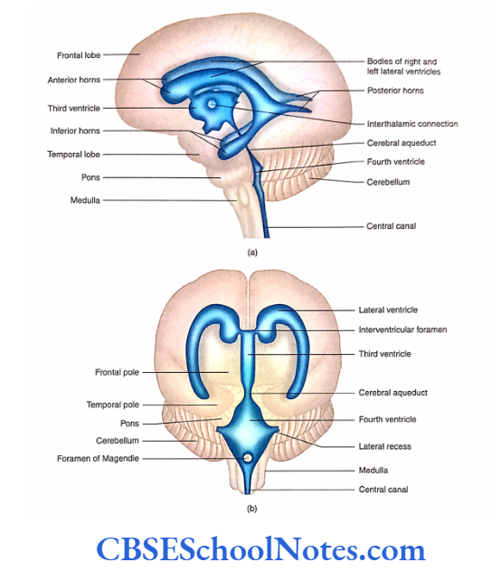
- The ventricles are four in number and are named lateral ventricles (a pair) third and fourth.
- These CSFs flow from the lateral ventricles to the third ventricle and from the third to the fourth ventricle.
- The fourth ventricle is continuous with the subarachnoid space through openings in its roof (refer to Chapter 23 for circulation of CSF).
- The choroid plexus, which is responsible for the production of CSF, is present in all the cavities (ventricles).
Lateral Ventricle
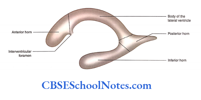
- Each cerebral hemisphere has a cavity called the lateral ventricle, which consists of the anterior horn, body, posterior horn, and inferior horn.
- The central part or the body of the lateral ventricle is situated in the parietal lobe.
- The central part of the right and left lateral ventricles lies close to the median plane separated from each other by septum pellucidum.
- The anterior horn extends into the frontal lobe, the posterior horn extends into the occipital lobe and the inferior horn extends into the temporal lobe.
- Each lateral ventricle communicates with the third ventricle through the interventricular foramen.
- Central part
- Anterior horn
- Posterior horn
- Inferior horn
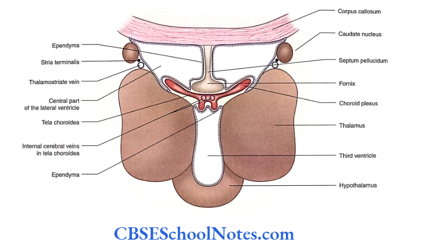
Central Part
Read and Learn More Neuroanatomy
The central part of the lateral ventricle extends J anteroposteriorly between the interventricular foramen and the splenium of the corpus callosum. The central part becomes continuous anteriorly with the anterior horn while posteriorly with the posterior and inferior horns. Moreover, the central part of the ventricle is triangular in cross-section and thus has three walls.
- Roof: It is formed by the undersurface of the corpus callosum.
- Floor: It is formed from the lateral to the medial side by the caudate nucleus, thalamostriate vein and stria terminalis, upper surface of the thalamus, choroid plexus and fornix.
- Medial wall: It is formed by the septum pellucidum.
Most medially, there is a slit-like space between the upper surface of the thalamus and fornix. ‘Ibis space is called a choroid fissure through which the choroid plexus invaginates the lateral ventricle.
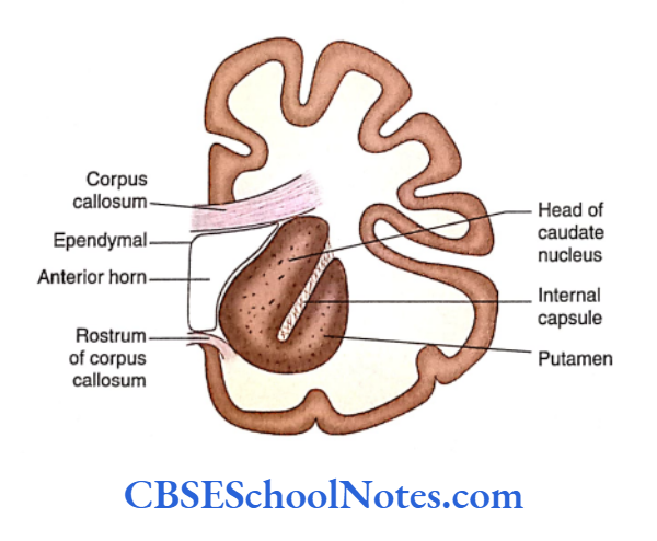
Anterior Horn
The anterior horn is present anterior to the interventricular foramen and extends forwards, laterally and downwards in the frontal lobe of the brain. It is triangular in section and has
- Roof: Undersurface of corpus callosum
- Floor: Upper surface of the rostrum of the corpus callosum
- Medial wall: Septum pellucidum
- Lateral wall: Head of the caudate nucleus
- Anterior wall: Genu of corpus callosum.
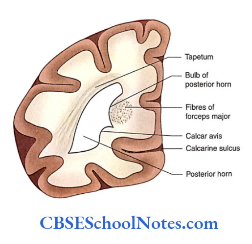
Posterior Horn
The posterior horn extends backwards and medially into the occipital lobe
Roof and lateral wall: Tapetum of corpus callosum
Medial wall: Formed by two elevations, i.e. bulb of the posterior horn and calcar avis.
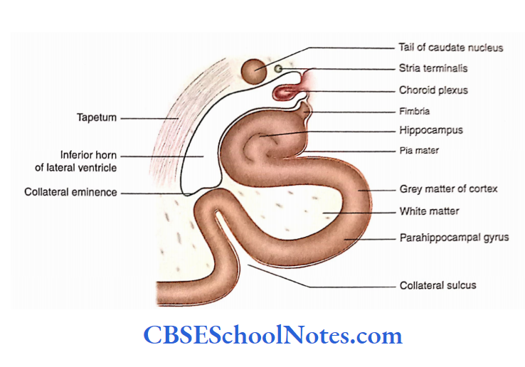
Inferior Horn
The inferior horn begins at the posterior end of the body ofthe lateral ventricle at the level ofthe splenium of the corpus callosum.
The inferior horn at first passes backwards and laterally and then passes downwards behind the thalamus into the temporal lobe.
In the temporal lobe, it takes a turn forward and ends in uncus. The inferior horn has a roof and a floor.
Roof: Above by stria terminalis and tail of caudate nucleus; below and laterally by tapetum. The amygdaloid nucleus lies in its anteriormost part of the roof.
Floor: Medially, it is formed by the hippocampus and laterally by collateral eminence.
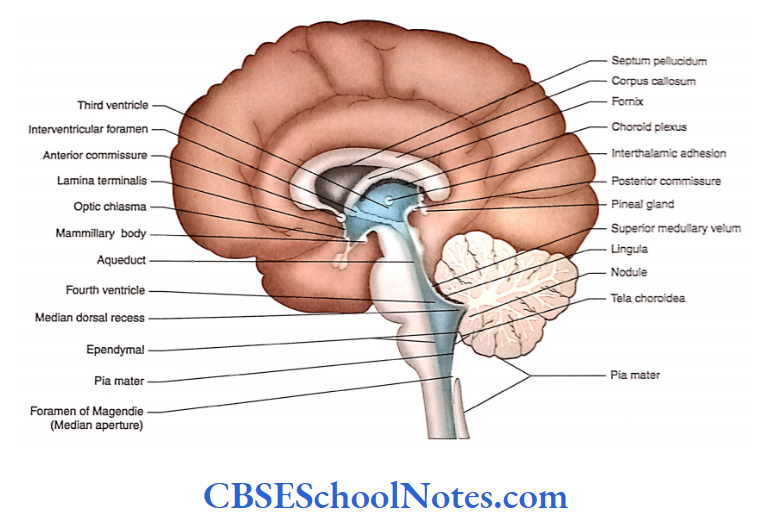
Third Ventricle
The third ventricle is a slit-like narrow space of diencephalon. It is situated between two thalami.
The cavity of the third ventricle communicates with the right and left lateral ventricles through the interventricular foramen. Posteriorly, the cavity communicates with the fourth ventricle through the cerebral aqueduct.
As the cavity is situated in the midline, it has two lateral walls. It also consists of an anterior wall, posterior wall, roof and floor.
Anterior Wall
The anterior wall is formed by the lamina terminalis, anterior commissure and anterior column of the fornix.
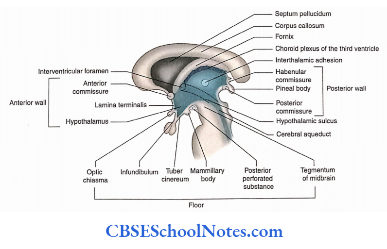
Lateral Wall
The lateral wall is large and is divided by the hypothalamic sulcus into an upper thalamic part and a lower hypothalamic part.
Two thalamines are usually connected by interthalamic adhesion.
Posterior Wall
From above downwards, it is formed by supraspinal recess, habenular commissure, pineal recess in the stalk of the pineal gland and posterior commissure.
Roof
The roof is formed by the ependyma stretching between two thalamis. From the roof two choroid plexuses protrude, one on either side of the median plane.
Floor
The floor is formed by the following structures as traced anteroposteriorly: optic chiasma, infundibulum, tuber cinereum, mammillary bodies, posterior perforated substance and tegmentum of midbrain Two recesses are seen in the floor.
These are the optic recess (above) and the optic chiasma) and infundibular recess (above the infundibular stalk).
Fourth Ventricle
The fourth ventricle is found in the hindbrain. Above, it communicates through the cerebral aqueduct with the third ventricle and below it communicates with the central canal of the closed part of the medulla oblongata.
The fourth ventricle lies in front of the cerebellum and behind the lower part of the pons and upper part medulla.
The cerebellum forms the roof of the fourth ventricle and its floor is formed by the pons and upper part of the medulla. The cavity of the fourth ventricle consists of a floor, roof
and lateral wall.
Floor
In the anatomical position, the floor of the fourth ventricle is the anterior wall of the ventricle. The shape of the floor is diamond shaped and hence sometimes called rhomboid fossa.
The floor is divided into upper and lower triangular parts by bundles of transversely running fibres (striaemedullaris)
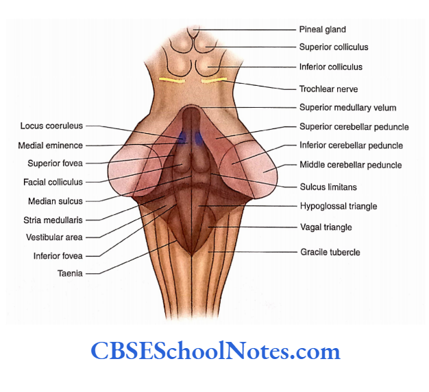
The upper triangular part is formed by the posterior surface of pons while the lower triangular part is formed by the posterior surface of the upper medulla.
Right and Left Halves
The floor of the ventricle is also divided into right and left halves by a median sulcus. On either side of the median sulcus, a longitudinal elevation is known as the median
or medial eminence.
Upper Part
- Immediately above the striae medullaris, on either side, the medial eminence shows a slight swelling known as facial colliculus. This swelling is produced due to fibres of facial nerve looping around the abducent nerve nucleus.
- At the upper level of the facial colliculus, there is a triangular depression—the superior fovea.
- Just above the superior fovea, the sulcus limitans ends in an area which is bluish and is called locus coeruleus.
- The bluish colouration is due to the presence of the melanin pigment present in the noradrenergic neurons of the nucleus coeruleus.
Lower Part
- The lower part of the floor, below the striae medullaris, shows the presence of two small triangles on either side of the median sulcus.
- These triangles are known as hypoglossal and vagal triangles.
- The vagal triangle is situated lateral to the hypoglossal triangle. The dorsal nucleus of the vagus and solitary nuclei lie deep in the vagal triangle.
- The hypoglossal nerve nucleus lies deep in the hypoglossal triangle.
- The lower part of the sulcus limitans close to the apex of the vagal triangle presents a depression—the inferior fovea.
Roof
The roof of the fourth ventricle is tent-shaped and projects posteriorly towards the cerebellum.
Upper Part
The upper part of the roof is formed on each side by superior cerebellar peduncles. The interval between these peduncles is bridged by a thin sheet of white matter—the superior medullary velum.
Lower Part
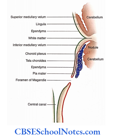
The lower part of the roof is formed, in small part, by the inferior medullary velum and in large part by tela choroidea.
- The right and left inferior medullary vela are present on each side of the nodule of the cerebellum. The inferior medullary velum is made up of a thin sheet of white matter and merges posteriorly in the white matter of the cerebellum.
- The inner surface of the inferior medullary velum is covered by ependyma while its outer surface is covered by pia mater.
- At the free margin of the inferior medullary velum, the ependyma and pia mater come close to each other and form the tela choroidea of the fourth ventricle. Thus, the lowest part of the roof of the fourth ventricle is membranous.
- Laterally, on either side, this membrane fuses with the inferior cerebellar peduncle.
- The lower part of this membrane presents a large median aperture (foramen of Magendie) through which the fourth ventricle communicates with the cerebellomedullary cistern.
- The roof also presents two lateral apertures through which the ventricle communicates with the subarachnoid space.
Lateral Walls
The upper part of the fourth ventricle is bounded laterally by the right and left superior cerebellar peduncles.
The lower part is bounded laterally by the right and left inferior cerebellar peduncles and gracile and cuneate tubercles.
Cavity of the Fourth Ventricle
The cavity of the fourth ventricle is somewhat diamond-shaped and lined by ependyma. It has superior, inferior and two lateral angles. The cavity communicates above with the third ventricle through the cerebral aqueduct.
Openings
- The fourth ventricle communicates below (at its inferior angle) with the central canal of the medulla oblongata.
- It has three openings in the roof—one median and two lateral through which it communicates with the subarachnoid space.
- Two lateral openings (foramina of Luschka), one on each side, lie in the lateral angle of the ventricle between the inferior cerebellar peduncle and the flocculus.
- Through this opening, the CSF escapes into the subarachnoid space. The choroid plexus of the fourth ventricle also protrudes through this opening.
Recesses
The cavity has many pouch-like protrusions which are known as recesses. It has a pair of lateral recesses, a single median dorsal recess and a pair of lateral dorsal recesses.
Summary
- Ventricles are CSF-filled cavities of the brain. These cavities are lined with ependyma.
- Each cerebral hemisphere contains a large C-shaped cavity known as a lateral ventricle.
Each lateral ventricle consists of a body and three horns:
- Anterior, Posterior and Inferior.
- The anterior horn extends into the frontal lobe, the posterior horn in the occipital lobe and the inferior horn in the temporal lobe.
- The third ventricle is situated in the midline and has two lateral walls, anterior wall, roof, floor and posterior wall. The cavity of the third ventricle communicates with the right and left lateral ventricles through the interventricular foramen. Below, it also communicates with the fourth ventricle through the cerebral aqueduct.
- The fourth ventricle is located between the brainstem and the cerebellum. It communicates below with the central canal.
- The floor of the fourth ventricle is diamond-shaped; hence, sometimes it is called rhomboid fossa. The fourth ventricle has three openings in the roof—one median and two lateral.
Multiple Choice Questions
Question 1. Following are parts of the lateral ventricle except
- The central part of the body
- Anterior horn
- Posterior horn
- Superior horn
- Inferior horn
Answer: 4. Superior horn
Question 2. Which of the following facts regarding the central part of the lateral ventricle is false?
- It extends between the interventricular foramen and the splenium
- It has three walls—roof, floor and medial wall
- The medial wall is formed by septum pellucidum
- The floor is formed by the thalamus and caudate nucleus
- None of the above
Answer: 5. None of the above
Question 3. Which of the following facts regarding the third ventricle is false?
- It is a slit-like narrow space of diencephalon
- It communicates with the lateral ventricle through the interventricular foramen.
- It communicates with the fourth ventricle through the central canal
- It has two lateral walls
Answer: 3. It communicates with the fourth ventricle through the central canal
Question 4. Which of the following structures is not seen in the floor of the fourth ventricle?
- Stria terminalis
- Median sulcus
- Median eminence
- Sulcus limitans
- Vestibular area
Answer: 1. Stria terminalis
Question 5. Which of the following structures are present in the roof of the fourth ventricle?
- Superior cerebellar peduncles
- Superior medullary velum
- Inferior medullary velum
- Tela choroidea
- All of the above
Answer: 5. All of the above