
Bones Of The Vertebral Column
The vertebral column is present in the central region of the body. It is the main constituent of the axial skeleton.
The column is also called as spine or backbone. The vertebral column extends from the base of the skull to the tip of the coccyx.
The vertebral column is composed of a series of many irregular bones called vertebrae. There are 33 vertebrae in the column, which are connected by intervertebral joints.
The important intervertebral joint is made up of a fibrocartilagenous intervertebral disc that binds the bodies of two adjacent vertebrae.
The length of the vertebral column is about 70 centimeters in an adult male. About l/4th length of the column is formed by intervertebral discs.
The adult vertebral column is divided into 5 different regions:
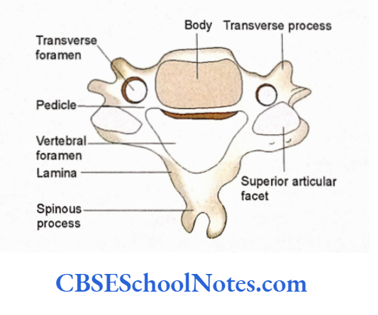
The Cervical Region: This part of the column is present in the neck and consists of seven cervical vertebrae.
The Thoracic Region: It is present in the thorax and consists of 12 thoracic vertebrae.
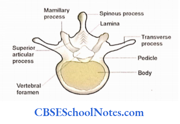
The Lumbar Region: It is present in the abdominal part and is made up of 5 lumbar vertebrae.
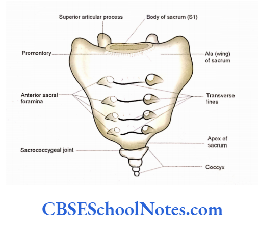
The Sacral Region: It is present in the pelvic region and consists of five fused sacral vertebrae. These five fused vertebrae are considered as a single bone, the sacrum.
The Coccygeal Region: It is present at the lower end of the column. It is usually a single bone, which is formed by the fusion of four coccygeal vertebrae.
When you shall view the articulated vertebral column from the front (anterior aspect) you shall notice the progressive increase in the width of the vertebral bodies from above downwards (from C2 to L5). This is because, at each vertebral segment, some more load is added to the column.
Read and Learn More Human Osteology Notes
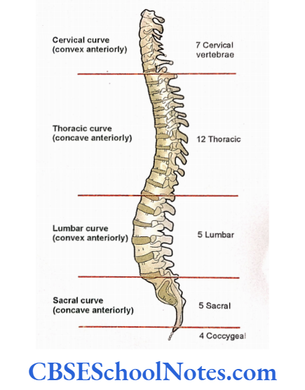
Curves of the Vertebral Column
When viewed from the lateral side, the vertebral column of an adult shows four curvatures, i.e., cervical, thoracic, lumbar, and sacral.
- The cervical and lumbar curves are convex anteriorly, while the thoracic and sacral curves are concave anteriorly.
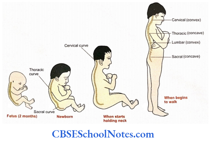
- The thoracic and sacral curvatures are called primary curvatures. They are present at the time of birth. These curvatures are formed mainly due to the shapes of vertebrae.
- The cervical and lumbar curvatures are called secondary curvatures because they develop after birth. Cervical curvature develops after the child starts holding the head on the neck. The lumbar curvature develops after an infant assumes an upright posture and begins to walk. Thus secondary curvatures develop due to the posture.
- In adults, cervical and lumbar intervertebral discs are thicker anteriorly thus contributing to anterior convexity. The posterior aspect of the column is formed by laminae, spinous processes, and articular facets.
- The adjacent spinous process and laminae are inter¬ connected with the help of ligaments, while facets form joints.
Functions of the Vertebral Column
- The vertebral column acts as a rigid but flexible column.
- It transmits the weight of the body.
- It supports the head.
- The vertebral column protects the spinal cord and part of the spinal nerves.
- It gives attachments to the ribs and muscles of the back.
Structure And Functions Of A Typical Vertebra
In the following paragraphs, only the generalized description of vertebrae is given.
Vertebral Body The detailed description of cervical vertebrae is given in Chapter 7, thoracic vertebrae in Chapter 5, and lumbar, sacral, and coccyx in Chapter 6.
Though the vertebrae of different regions of the vertebral column vary in size, shape, and other characteristics many basic features are common in these vertebrae.
A vertebra from the mid-thoracic region is best suited to study the basic features of a typical vertebra.
The following description of a typical vertebra is based on the features of a mid-thoracic vertebra.
A typical vertebra consists of:
- A vertebral body.
- A vertebral (neural) arch with seven processes.

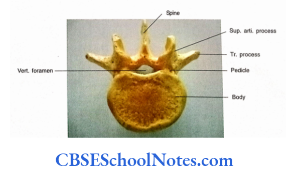
Vertebral Body
- Hold a typical thoracic vertebra in your hand and note the following:
- The body of a vertebra is situated anteriorly. It is somewhat cylindrical.
- The cylindrical body is rounded from side to side. Its superior and inferior surfaces are flat.
- Thus the body has six surfaces (anterior, posterior, superior, inferior, and two lateral).
- The anterior, lateral, and posterior surfaces contain minute foramina for the vessels.
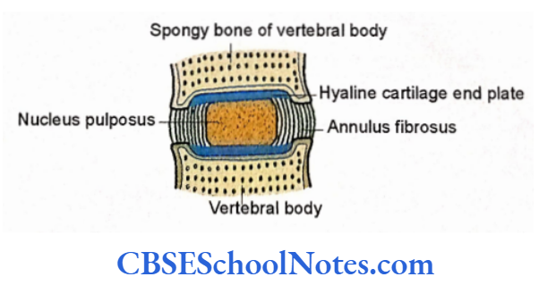
- The body is made up of spongy bone and covered by a thin layer of compact bone.
- However, its superior and inferior surfaces are not covered by compact bone but by a thin layer of hyaline cartilage in living persons. The upper and lower surfaces give attachments to the intervertebral discs.
Vertebral Arch
The vertebral arch is situated posterior to the body of the vertebra. It consists of a pair of pedicles and laminae. Seven processes arise from the vertebral arch of a typical vertebra.
Pedicles.
- The pedicles are short, stout bars that are attached to the posterolateral aspects of the body.
- They are attached close to the superior border of the body.
- Pedicles project posteriorly and somewhat laterally from the body to unite with the laminae.
- If we look at the lateral aspect of a vertebra there is the presence of an inferior vertebral notch just below the pedicle.
- This notch is bounded anteriorly by the body superiorly by the pedicle and posteriorly by the inferior articular process.
- The superior vertebral notch is situated above The pedicle. It is much shallower as compared to the inferior vertebral notch.
Laminae
- These are flat vertical plates of the bone that join in the midline to form the posterior portion of the vertebral arch.
- They extend backward and medially from the pedicles.
- Posteriorly, the lamina of the right and left sides fuse in the midline to form a spinous process.
- The body, pedicles, and laminae of a vertebra together enclose a foramen called vertebral foramen.
- Collectively the vertebral foramina of the successive vertebrae form the vertebral canal that transmits the spinal cord.
Transverse Processes
The transverse process extends laterally on each side from the point where the lamina and pedicle join each other.
Articular Processes
There are two superior and two inferior articular processes. They also arise from the junction of pedicle and lamina.
Each particular process bears a smooth articular facet. In the thoracic vertebrae, the superior articular facets face posterolaterally, while the inferior ones face anteromedially.
Spinous Process
It projects posteroinferiorly in the midline from the junction of two laminae
Functions of the Various Components of a Vertebra
- The bodies and intervertebral discs are involved in the transmission of the load of the trunk to the lower limbs.
- The intervertebral discs are shock absorbers and also permit various movements between two successive bodies.
- Articular processes (and facet joints) allow and guide the movements between adjacent vertebrae.
- Facet joints are also involved in the transmission of load. The magnitude of the load transmitted by facet joints varies in various regions of the column, i.e., cervical and lumbar facets are highly loaded while thoracic is least loaded.
- Lamina is also involved in the transmission of load as it passes from superior to inferior articular facet joints.
- In the thoracic region, where the column is concave anteriorly, the load passes from the vertebral arch (lamina) to the body.
- While, in the lumbar region, where the column is concave posteriorly, load passes from the body to the vertebral arch. The transmission of load between the body and the vertebral arch is through the pedicles.
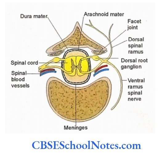
- The spinal cord and its meninges are well protected in the vertebral foramen (canal).
- The transverse and spinous processes give attachment to muscles and act as levers for various movements of the vertebral column.
- Transverse processes in the thoracic region are also involved in the transmission of load from the ribs to the laminae.
Articulation between Two Successive Typical Vertebrae
The articulations between two successive thoracic vertebrae are. Adjacent vertebrae are connected at three intervertebral joints, i.e., one median joint between bodies and two joints between the articular processes of successive vertebrae.
The two adjacent vertebral bodies are joined by the intervertebral disc, which is made up of fibrocartilage. Each disc consists of annulus fibrosus (outer fibrous part) and nucleus pulposus (inner soft part).
The two superior articular processes of a vertebra articulate with the two inferior articular processes of the vertebra situated above it.
Similarly, two inferior articular processes of the vertebra articulate with the two superior articular processes of the vertebra situated below it.
The joints between articular processes are synovial and are also known as facet joints.
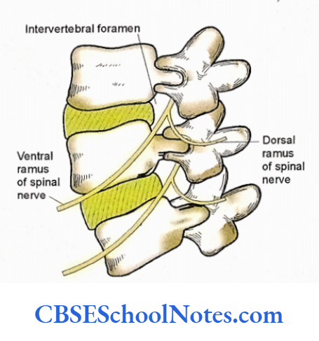
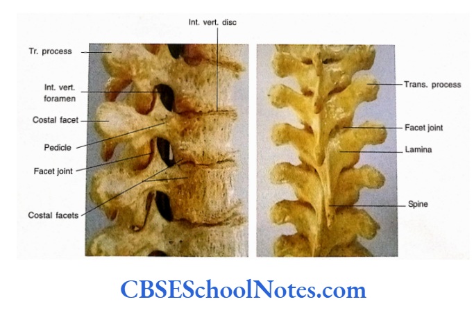
The vertebral foramina of the successive vertebrae forms a continuous vertebral canal, that contains the spinal cord and its meninges. The superior and inferior vertebral notches of the adjacent vertebrae join to form the intervertebral foramen through which passes the spinal nerves and vessels.
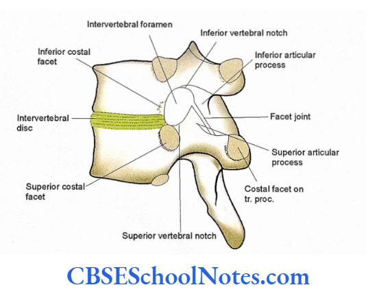
The boundaries of intervertebral foramen are formed anteriorly by the body of the upper vertebra, the intervertebral disc, and a small part of the body of the lower vertebra.
The upper and lower boundaries are formed by pedicles of the upper and lower vertebrae respectively. The posterior boundary is formed by the lamina of the upper vertebra and facet joint
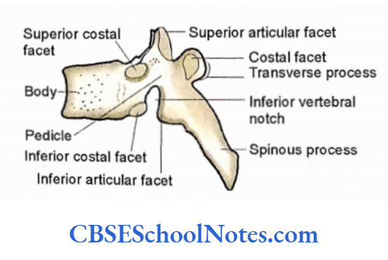
The articular facets are present on the body and transverse processes of the thoracic vertebra. These are called costal facets.
The costal facets of adjacent bodies articulate with the head of the rib. The facet on the transverse process articulates with the tubercle of the rib.
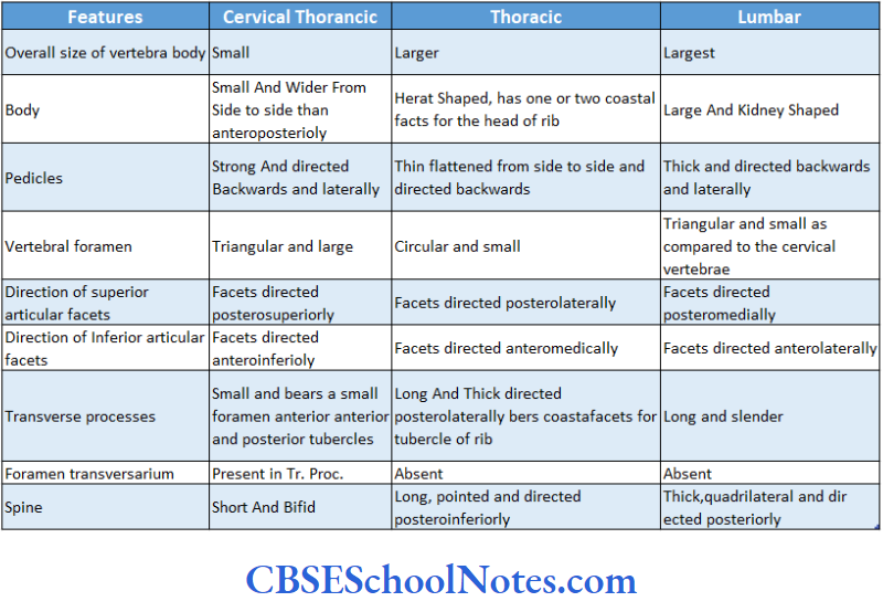
Principle Distinguishing Features of the Vertebrae of the Various Regions of the Vertebral Column Besides the features mentioned, the following features will help students to distinguish cervical, thoracic, and lumbar vertebrae from one another:
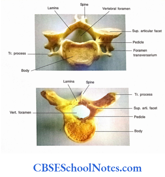
A cervical vertebra can be easily identified because of the presence of a foramen in its transverse process. This foramen is called as foramen transversarium.
A thoracic vertebra is recognized by the presence of articular facets on the body and transverse processes. These facets are called costal facets, which articulate with the ribs.
A lumbar vertebra is recognized because it has a large kidney-shaped body. There is the absence of foramen transversarum in the transverse processes. There is also the absence of costal facets on the body and transverse processes.
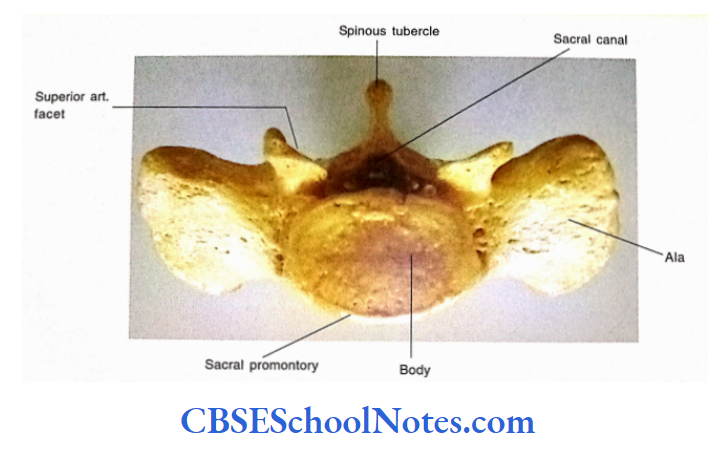
The sacrum is easily identified because of its shape. As it is formed by the fusion of five sacral vertebrae, it is a single, curved, and triangular bone.
The coccyx is formed by the fusion of four coccygeal vertebrae. Coccyx is identified by its small size and fused nature.
Movements Occurring in the Vertebral Column
As the sacral and coccygeal vertebrae are fused, no movements are possible between these vertebrae.
The cervical, thoracic, and lumbar vertebrae are not fused hence, are mobile.
Two adjacent vertebrae are joined with each other at three intervertebral joints, i.e., by intervertebral disc (between two adjacent bodies) and by two synovial joints (between articular. processes).
The movements between two adjacent vertebrae are slight. But when movements between series of vertebrae are added, the column shows considerable flexibility.
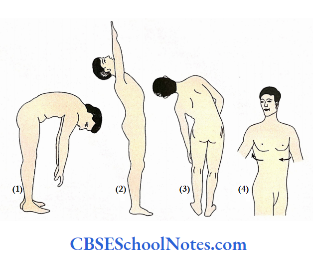
The following movements are possible in the vertebral column:
- Flexion: Forward bending.
- Extension: Backward bending.
- Lateral flexion: Side bending.
- Rotation: Twisting.
In different regions, i.e., cervical and lumbar regions are more mobile as compared to thoracic. Almost all types of movements are possible in the cervical region.
Rotation movement is the main movement of the thoracic region, but this movement is not possible in the lumbar region.
Particular Features of a Typical Vertebra
There are many ligaments, which connect adjoining vertebrae. With the help of these ligaments and intervertebral joints, a flexible but rigid column is formed.
Attachments of Ligaments
The anterior longitudinal ligament is attached to the anterior surfaces of the bodies of successive vertebrae. It is a continuous ligament, which extends from the base of the skull to the sacrum.
The posterior longitudinal ligament is also continuous and attached to the posterior surface of the bodies of vertebrae.
The transverse processes of the adjacent vertebrae are connected by intertransverse ligaments.
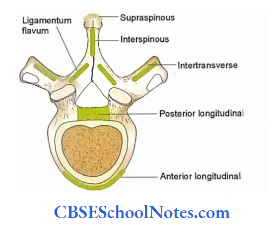
- The Ligamentum flava connects the laminae of adjacent vertebrae.
- Similarly, the spinous processes of adjacent vertebrae are connected by interspinous ligaments.
- In the cervical region, the tips of the spines are connected by the elastic ligament, called as ligamentum nuchae.
- The supraspinous ligaments are attached to the tips of spinous processes of vertebrae between the 7th cervical vertebra to the sacrum.
Attachments of Muscles
- Various muscles are attached to the various aspects of vertebrae in different vertebral regions. The attachments of these muscles are described along with the bones of that region.
Clinical importance
Abnormal Curvatures
Following abnormal curvature may be present in the vertebral column:
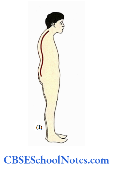
Kyphosis
This is due to the abnormal increase in the thoracic curvature (increase in the thoracic concavity anteriorly). It is usually seen in old age due to osteoporosis. The osteoporosis leads to the erosion of the anterior part of one or more vertebrae, leading to an increase in concavity.
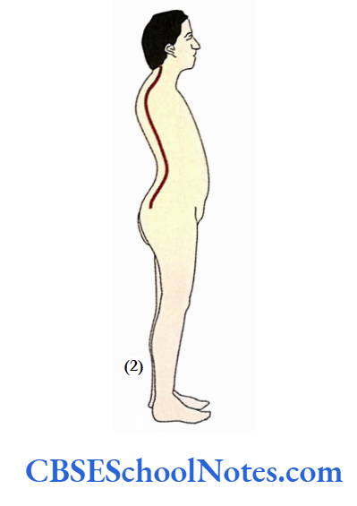
Lordosis
This is due to the abnormal increase in lumbar curvature (increase in the lumbar convexity anteriorly). This results due to the weakness of the abdominal muscles.
Lordosis may also occur in obese people and pregnant ladies. This is due to the shift in the line of gravity because of an increase in the weight of abdominal contents.
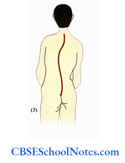
Scoliosis
It is the most common deformity of the vertebral column and is predominantly observed in girls in the teens. In this condition, there is an abnormal lateral curvature associated with the rotation of the vertebrae.
In most cases, the cause of the scoliosis is not known. Known causes are hemivertebra, maldevelopment of one upper limb, and asymmetry in the length of the lower limbs.
Fractures
The fracture of the vertebral column may occur due to forceful flexion or hyperextension of the column.
The forceful sudden flexion of the column may occur due to a fall from the height of the feet or the head. This leads to the impression of fracture and dislocation of one or more successive vertebrae.
This kind of fracture may be associated with the injury to the spinal cord resulting in loss of sensation and paralysis of muscles below the level of injury.
Spondylolysis
In this condition there occurs the breakage (cleft) in the vertebral arch (at lamina between superior and inferior articular processes) of one or both sides. This condition is common in the lower lumbar region. Spondylolysis may result due to excessive mechanical stress.
It is now considered as the fatigue fracture of the lamina. This condition is different from spondylolisthesis (described below). In spondylolysis, there is no displace¬ ment of the vertebral body and it is often asymptomatic (without pain).
Tuberculosis of the Spine
The vertebral bodies are the common sites for the tubercular infection. This is due to the spongy nature of the vertebral body and rich blood supply. Due to the infection, the spongy bone of the body is destroyed and pus is formed. This leads to the collapse of vertebral bodies as the load of the trunk is brought by the upper vertebrae.