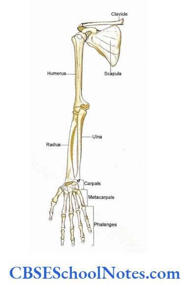
Bones Of The Upper limb
The Upper limb consists of many regions. The following bones are present in various regions of the upper limb
Pectoral Region
It consists of two bones on each side, i.e., clavicle and scapula. These two bones form the pectoral or shoulder girdle. The lateral end of the clavicle articulates with the scapula at the acromioclavicular joint.
The medial end of the clavicle articulates with the sternum (axial skeleton) through the sternoclavicular joint. Thus the pectoral girdle serves to attach the upper limb to the trunk.
Arm
This region consists of a single bone called as humerus. The upper end of this bone articulates with the scapula at the shoulder joint. The lower end of the humerus articulates with two long bones of the forearm, i.e., the radius and ulna at the elbow joint.
Upper Limb Bones
Forearm
The ulna is medial while the radius is laterally placed in the forearm. At their lower ends, they articulate with carpal bones to form wrist joints.
Hand
The skeleton of the hand consists of many bones ie., carpals, metacarpals, and phalanges. These bones form many joints i.e., intercarpal, sternal facet carpometacarpal, metacarpophalangeal, and interphalangeal joints.
Read and Learn More Human Osteology Notes
Bones form many joints i.e., intercarpal, sternal facet carpometacarpal, metacarpophalangeal, and interphalangeal joints.
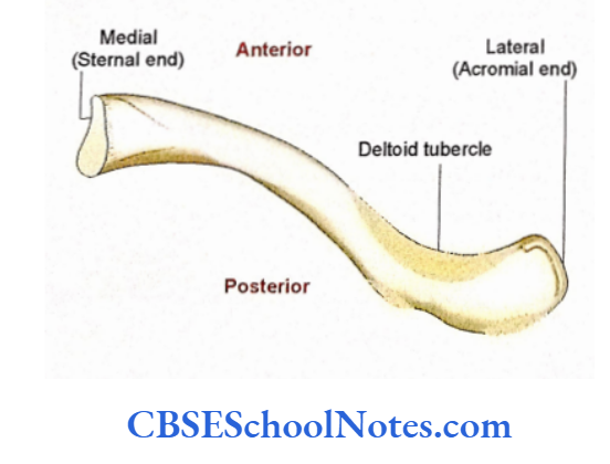
The Clavicle or Clavicle Anatomy
Trapezoid The description of the bone in the following paragraph and the reference will help you to learn the “side determination” and “anatomical position” of bone.
costalFacet for cartilage first Subcostal groove Acromian facet The clavicle (collar bone) is a horizontal or inferior surface of a right-placed long bone, which shows two ends (medial and lateral) and a shaft.
Upper Limb Bones
Clavicle Bone
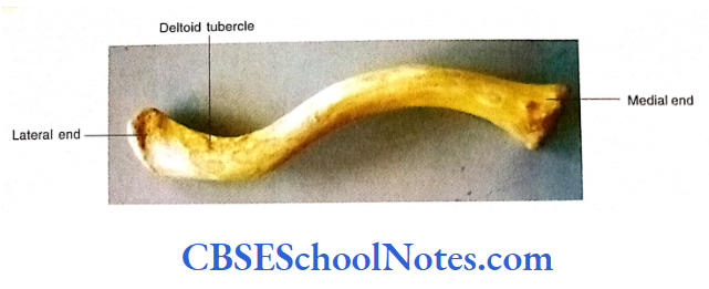
The medial end is thick and rounded Side Determination while the lateral end is broad and flattened. The shaft shows “S” shaped double curvatures, which meet each other at the junction of medial 2/3 and lateral 1/3 of the bone.
The medial 2/3 of the bone is cylindrical and convex forwards, while the lateral 1/3 of the shaft is flattened and concave forwards. The superior surface of the bone is smooth as it is subcutaneous.
On the other hand, the inferior surface is rough near its medial and lateral ends due to the attachment of ligaments. The inferior surface also shows the presence of a shallow groove in its middle third.
Clavicle Anatomy or Clavicle Side Determination
- Hold the bone horizontally in such a way that its rounded thick end is nearer to the median plane (placed medially) and the flattened end is directed laterally.
- Identify the rough inferior surface and smooth superior surface. Keep the inferior surface facing inferiorly (towards the ground).
- The convexity of the medial 2/3 of the shaft should be directed anteriorly.
- In this position, the lateral end will direct the side of the bone.
Clavicle Bone or Clavicle Anatomical Position
Hold the clavicle horizontally in such a way that its medial end is directed slightly forward and inferiorly as compared to its lateral end (which is directed upward and backward).
Clavicle General Features
For ease of understanding the clavicle is divided into medial 2/3 and lateral 1/3.
Medial 2/3 of Clavicle
Hold the bone in an anatomical position and note the following.
- It consists of a cylindrical shaft and a thick rounded medial (sternal) end.
- As the shaft is cylindrical borders are ill-defined (not well marked).
- There are four surfaces, i.e., superior, inferior, anterior, and posterior. In the absence of well-defined borders, these surfaces are continuous with each other.
- The superior surface is mostly smooth. It is covered by skin and platysma muscle.
- The anterior surface of the medial 2/3 of the clavicle is convex anteriorly.
- The posterior surface is smooth and concave posteriorly.
Clavicle Bone
Now turn the bone upside down to the clavicular joint. see the inferior surface.
- The inferior surface has a large rough Particular Features impression near its medial end, which gives attachment to a ligament.
- There is the presence of a shallow groove in the middle third of the inferior surface. This groove is known as the subclavian groove.
The medial end of the clavicle shows a large smooth saddle-shaped articular surface on its medial aspect (sternal surface), which also extends for a short distance on the inferior surface of the bone.
The medial end of the clavicle articulates with the manubrium sterni (at sternoclavicular joint) and also with the first costal cartilage near its inferior surface.
Lateral 1/3 of the Clavicle
As the lateral 1/3 of the clavicle is flat. It shows well-defined anterior and posterior sternohyoid borders and superior and inferior surfaces.
- Hold the bone in the anatomical position and note the following:
- The anterior border is concave forwards and Subclavius shows a small rough area called a deltoid tubercle.
- The posterior border is rough and convex posteriorly.
- The superior surface is mostly smooth except near its anterior and posterior borders, which give attachment to muscles. Now turn the bone upside down to see the inferior surface of lateral1/3.
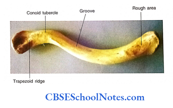
- The inferior surface near its posterior border shows a tubercle known as conoid tubercle.
- A thick ridge extends forward and laterally from the conoid tubercle. This ridge is known as the trapezoid ride.
The lateral end of the bone bears a small oval facet for articulation with the acromian process of the scapula to form an acromioclavicular joint.
Upper Limb Bones
Particular Features Muscles Attached to Bone
Draw the attachments of muscles on the surface of the bone with the help of colored chalk.
Medial 2/3 of Clavicle
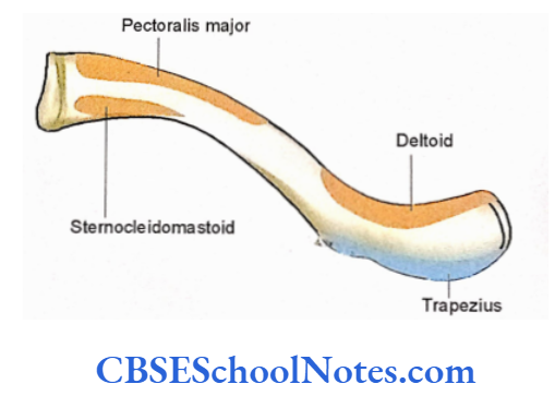
- The superior surface is covered by skin and platysma muscle. The clavicular head of the sternocleidomastoid muscle arises from the medial third of this surface.
- The anterior surface of the medial half of the clavicle gives origin to the clavicular head of pectoralis major.
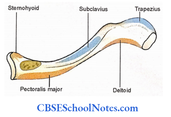
- The muscle sternohyoid is attached near the medial end of the posterior surface of the clavicle.
- On the inferior surface, the subclavius muscle is attached to the subclavian groove.
- The margins of this groove give attachment to clavipectoral fascia.
Lateral 1/3 of the Clavicle
- The anterior border gives origin to the deltoid muscle.
- The posterior border gives attachment to the trapezius muscle.
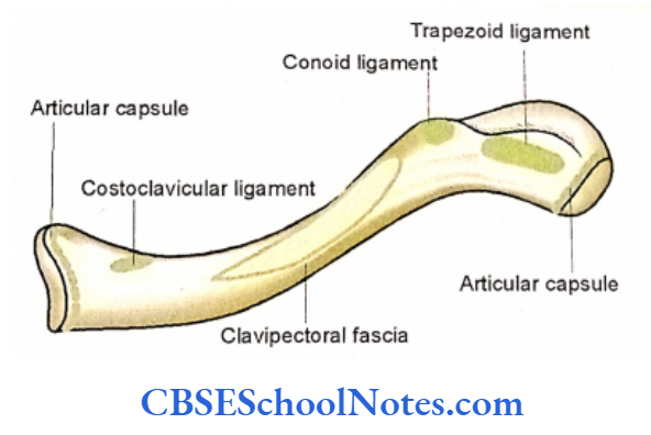
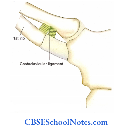
- Costoclavicular ligament
- Ligaments Attached to Bone
- The attachments of the articular capsule at its medial and lateral ends are shown in.
- The costoclavicular ligament is attached to the inferior rough surface near its medial end.
- The interclavicular ligament and the articular disc of the sternoclavicular joint are attached to the medial surface of the medial end.
- Conoid and trapezoid parts of the coracoclavicular ligament are attached on the conoid tubercle and trapezoid ridge respectively on the inferior surface of lateral 1/3.
Upper Limb Bones
Nerves and Blood Vessels Related To Bone
- Three supra-clavicular nerves (medial, intermediate, and lateral) are related to its anterosuperior surface.
- There lasted posterior to the surface trunks of medial brachial 2/3 of the plexus, shaft in the third part of subclavian vessels, and internal jugular vein.
Clavicle Fracture
Peculiarities of the Clavicle
- Though it is a long bone but is placed horizontally.
- Though long bone but ossifies mostly from membrane.
- It is devoid of a medullary cavity.
- The first bone to start ossifying in the body.
Clavicle Clinical Application
The main function of the clavicle is to transmit the forces from the upper limb to the axial skeleton through the sternoclavicular joint.
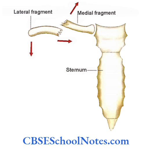
The forces are transmitted from the humerus to the scapula and then from the scapula to the clavicle through the coracoclavicular ligament. If the force, during a fall on the outstretched hand, is greater than the strength of the clavicle, a fracture will result at the junction between two curvatures of bone (medial 2/3 and lateral 1/3).
After the fracture, the shoulder drops, and the lateral fragment of the clavicle is displaced downward due to the weight of the upper limb (gravity).
Clavicle Fracture
The medial fragment of bone is pulled upward due to the action of the clavicular head of the sternocleidomastoid muscle.
The lateral fragment of the bone may also be pulled medially by the action of the pectoralis major.
Thus two fragments may override each other. To approximate the two fractured ends of the bone, a bandage in the shape of a figure of eight is tied.
The clavicle acts as a strut to keep the shoulder laterally. This helps the upper limb to swing away from the trunk.
In a congenital malformation known as cleidocranial dysostosis, the clavicle is either absent or imperfectly developed.
In this condition shoulder joint is not always placed laterally and can be brought in front of the chest.
- The concave posterior surface of medial 2/3 of the bone protects the nerves and vessels (trunks of brachial plexus and subclavian vessels), which go from the root of the neck to the axilla.
Clavicle Fracture
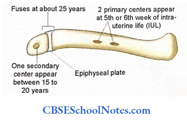
Clavicle Ossification
The clavicle is the first long lone to ossify in the embryo. It is organized by two primary centers and one secondary center. Most of the shaft ossifies by membranous ossification.
The Scapula or Scapula Bone
The description of the scapula is in the following two paragraphs and a reference will help you learn to determine its side and to keep it in the anatomical position. The scapula is also known as the “shoulder blade”.
It is the posterior bone of the shoulder girdle. It lies on the posterolateral aspect of the thorax extending between the 2nd and 7th rib.
The body- of the scapula is thin, flat, and triangular.
Upper Limb Bones
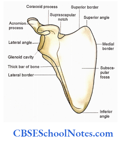
As the body is triangular it has three borders (medial, lateral, and superior) and three angles (superior, lateral, and inferior).
The inferior angle is directed downwards and lies at the level of the 7th rib. Just opposite the inferior angle is a superior border that extends between superior and lateral angles.
Scapula Bone
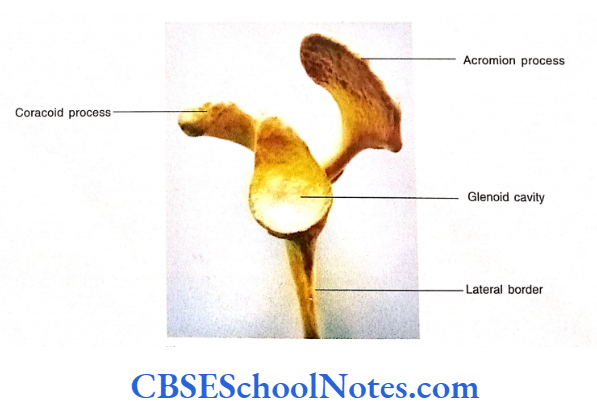
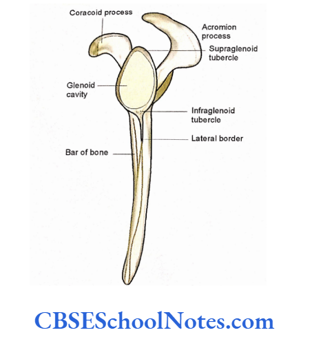
At the lateral angle, there is the presence of a shallow smooth articular fossa called a glenoid cavity, which articulates with the head of the humerus to form a shoulder joint.
The medial border runs between superior and inferior angles and is quite thin. The lateral border is thick and runs between lateral and inferior angles.
The scapula shows two surfaces, i.e., costal and dorsal. The costal (anterior) surface is smooth and forms a shallow fossa called as subscapular fossa.
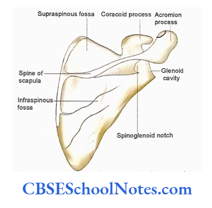
On the other hand dorsal (posterior) surface is convex posteriorly and is divided into supraspinous and infraspinous fossae by the presence of a bony process called as spine of the scapula.
Scapula Bone
It is a thick projecting ridge of bone, which is continuous laterally as the flat expanded acromion process. A beak-like bony process (coracoid process) arises from the upper part of the glenoid cavity.
Scapula Side Determination
Scapula Hold the bone in such a way that:
- The inferior angle is directed downwards and the superior border is directed upwards.
- The costal or anterior surface should face anteriorly and the dorsal (posterior) surface, bearing the spinous process, should face posteriorly.
- The glenoid cavity should face laterally.
- In this position, the direction of a glenoid cavity will determine the side of the bone.
Scapula Anatomical Position
- Hold the bone in such a way that the coastal surface should face antero-medially.
- The glenoid cavity should face anterolaterally and slightly upwards.
Scapula General Features
We shall study the general features of the scapula under four different headings, i.e., surfaces, borders, angles, and processes.
Surfaces
Costal (anterior) surface: The costal surface is slightly concave and forms a large subscapular fossa. There is the presence of a bar of thickened bone near the lateral border extending. between the head of scapula (lateral angle) and the inferior angle.
Dorsal (posterior) surface: The spine of the scapula divides the dorsal surface into a smaller upper supraspinous fossa and a larger infraspinous fossa. Both these fossae are continuous with each other through spinoglenoid notch.
Borders
Medial border: The medial border of the scapula is thin and lies about 5 cm lateral to the spinous processes of the thoracic vertebrae.
Lateral border: This border extends from the lowest part of the glenoid cavity to the inferior angle.
It is a sharp ridge. Close to the lateral border, on the costal surface, there is the presence of a thickened longitudinal bar of the bone. It should not be confused with the lateral border.
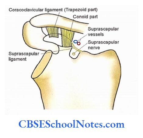
Superior border: It extends between the superior angle and the root of the coracoid process. This border presents a suprascapular notch close to the root of the coracoid process.
Angles
The superior and inferior angles of the scapula are present at the upper and lower ends of the medial border respectively.
The lateral angle of the scapula is the thickest part of the bone where the glenoid cavity is present. This broadened part of the bone is sometimes called as head of the scapula. A constriction between the head and body is called as neck of the scapula.
The glenoid cavity is a shallow, concave, oval fossa. There is a presence of rough areas just above and below the glenoid cavity. These are called supraglenoid and infraglenoid tubercles respectively.
Scapula Processes
The spine of the scapula is a triangular bony process present on the dorsal aspect of the body. Its anterior border is attached to the dorsal surface of the body while its posterior border is free.
This thick posterior border of the spine is also known as the crest of the spine. The lateral border of the spine forms the boundary of the spinoglenoid notch.
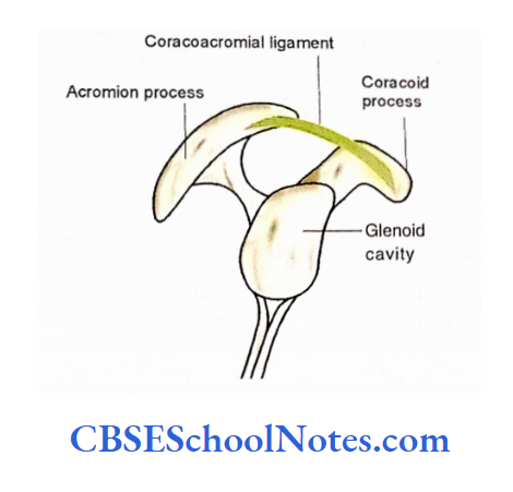
The acromion process is a forward projection from the lateral end of the spine. It is a flat, expanded, subcutaneous process, which overhangs the glenoid cavity.
It has medial and lateral borders and superior and inferior surfaces. It articulates with the lateral end of the clavicle to form an acromioclavicular joint. The coracoid process is so named because it resembles the beak of a crow.
It is present superior to the glenoid cavity and is shaped like a bent finger. In the anatomical position, it projects anterolaterally.
Scapula Particular Features
Muscles Attached to Bone
Draw the attachments of muscles on the surface of the bone with the help of colored chalk as shown in
- Almost the whole of the costal (anterior) surface (except a small part near the neck) gives origin to the subscapularis muscle.
- The medial border on the costal surface gives attachment to the serratus anterior.
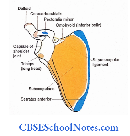
- The inferior belly of the omohyoid is attached to the superior border near the suprascapular notch.
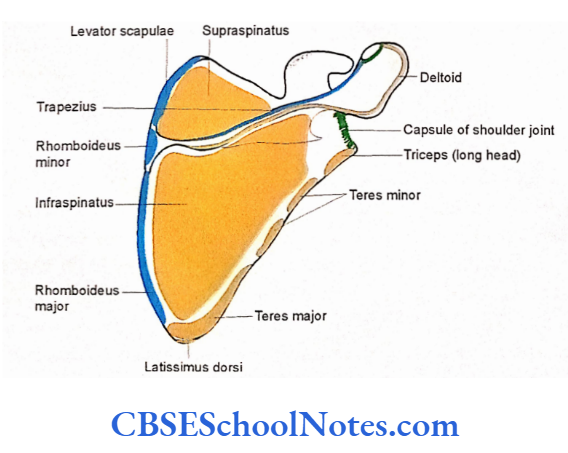
- On the dorsal surface, the supraspinous fossa gives origin to the supraspinatus muscle and infraspinous fossa to the infraspinatus respectively.
- The teres major and teres minor arise from the dorsal surface close to the lateral border.
- Attachments of levator scapulae, rhomboideus minor, and rhomboideus major on the dorsal surface of the medial border.
- The supraglenoid tubercle gives origin to the long head of biceps brachii.
- The long head of the triceps arises from the infraglenoid tubercle.
- The coracobrachialis and short head of biceps arise from the tip of coracoid process.
- Pectoralis minor is inserted on the superior aspect of the coracoid process.
- The deltoid muscle arises from the lower border of the crest of the spine and the lateral border of the acromion process.
- The medial border of the acromion and upper border of the crest of the spine gives insertion to the trapezium.
Ligaments Attached to Bone
- The attachments of the capsule of the shoulder joint and acromioclavicular joints.
- The suprascapular ligament is attached to the superior border above the suprascapular notch
- Three ligaments are attached to the coracoid process, i.e., coracohumeral, coracoacromial, and coracoclavicular.
- One end of the coracoacromial ligament is attached to the lateral border of the coracoid process and the other end is to the tip of the acromian process. The coracohumeral is attached to the root of the coracoid process.
- The conoid part of the coracoclavicular ligament is attached near the root, while the trapezoid part is on the superior aspect of the coracoid process.
Nerves and Blood Vessels Related to the Bone
The suprascapular vessels lie above the suprascapular ligament, while the suprascapular nerve passes deep to it These vessels and nerves then lie in These vessels and nerve then lie in spinoglenoid notch.
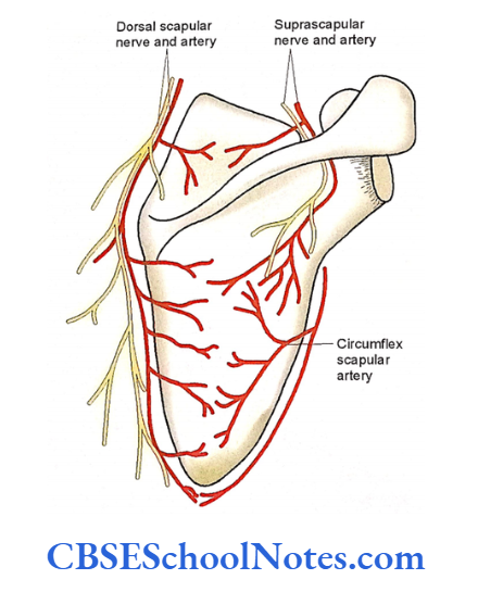
- The circumflex scapular branch of the subscapular artery comes in contact with the lateral border on its dorsal aspect between two heads of teres minor.
- The dorsal scapular nerve and vessels lie close to the medial border.
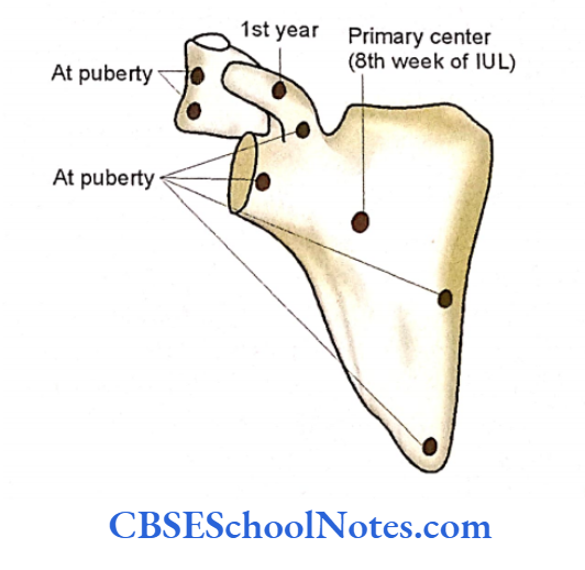
Scapula Ossification
The scapula ossifies by one primary and 7 secondary centers (2 for the coracoid process, 2 for acromion, 1 each for the glenoid cavity, inferior angle, and medial border).
The lower center of the coracoid process fuses with the body by the 16th year while other centers by the end of the 20th year.
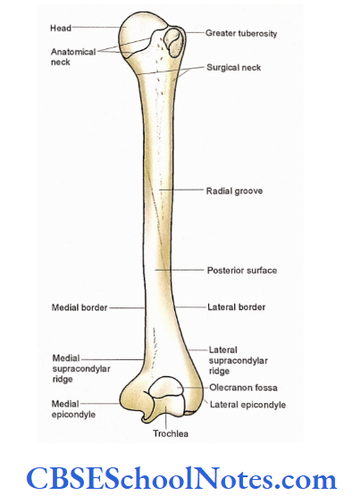
Humerus
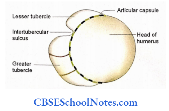
The humerus is the longest bone of the upper limb. It has two ends and a shaft. The proximal (upper) end shows a large, smooth, rounded head that articulates with the glenoid cavity of the scapula to form the shoulder joint. The head is directed medially.
There is the presence of a deep vertical groove {inter-tubercular sulcus or bicipital groove) on the anterior aspect of the upper end.
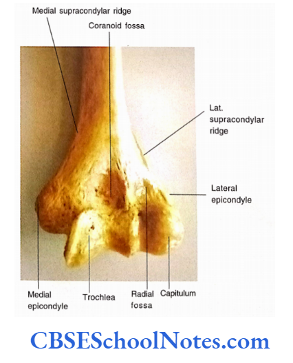
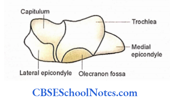
The middle part of the shaft is almost cylindrical. The lower end is expanded from side to side and flattened from before backward. It has two articular structures, i.e., a rounded capitulum and the pulley-shaped trochlea.
On the posterior aspect of the lower end, there is a large olecranon fossa just above the trochlea. The medial and lateral epicondyles are bony non-articular projections at the lower end.
Humerus Side Determination
- You should keep the bone vertical in such a way that the head faces medially.

- The intertubercular sulcus on the anterior aspect of the upper end should face anteriorly.
- The olecranon fossa, at the lower end, should face posteriorly
Humerus Anatomical Position
Hold the bone vertically in such a way that the head faces medially and slightly upwards and backward.
Humerus General Features
We shall study the general features of bone under three headings, i.e., upper end, shaft, and lower end
Humerus Upper end
The upper end consists of the head, neck, greater and lesser tubercles, and an intertubercular sulcus (bicipital groove).
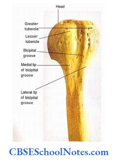
- The head has a convex, rounded, and smooth articular surface.
- The junction of the articular head with the rest of the upper end is called as anatomical neck.
- The junction of the upper expanded end with the shaft is called a surgical neck.
- The lesser tubercle is present on the anterior aspect of the upper end while greater tubercle is present on its anterolateral aspect.
- There is the presence of an intertubercular sulcus between these two tubercles.
- Both the tubercles on their upper surface show the presence of smooth areas for the attachment of muscles.
Humerus Shaft
The shaft is almost cylindrical in the upper half and triangular in the lower half. It presents three borders and three surfaces.
Borders
- Trace the lateral lip of the bicipital groove downward. You will see that it becomes continuous with the anterior border of the shaft.
- The medial border of the bicipital groove is continuous downwards as the medial border.
- If this border is further traced downwards it becomes continuous as medial supracondylar ridge.
- The lateral border of the shaft is well-defined only in the lower part of the shaft where it becomes continuous with the lateral supracondylar ridge.
Humerus Surfaces
- The anteromedial surface is between the anterior and medial border.
- The anterolateral surface is between anterior and lateral borders. Just above the middle of the bone, this surface shows a V-shaped rough area called deltoid tuberosity.
- The posterior surface is situated between lateral and medial borders. In its middle 1/3, it shows the presence of a shallow groove (radial groove) running downwards and laterally.
Humerus Lower End
- The lower end shows the presence of a rounded articular area known as the capitulum. It is placed laterally and articulates with the upper end of the radius.
- The pulley-like trochlea is placed medially and articulates with the trochlear notch of the ulna.
- There is a presence of shallow fossa above the capitulum (known as radial fossa) and coronoid fossa just above the trochlea. When the forearm is flexed, the coronoid fossa and radial fossa receive the coronoid process and head of radius respectively.
- The olecranon fossa is quite large and situated on the posterior surface of the lower end just above the trochlea. It receives the olecranon of the ulna when the forearm is extended.
- If you will trace the medial and lateral supracondylar ridges downwards, you will reach the bony projections on either side of the lower end.
- These rough projections are known as epicondyles. The medial epicondyle is the prominent subcutaneous projection, which you should palpate (feel) in your limb.
Humerus Particular Features
Muscles Attached to Bone
Upper End
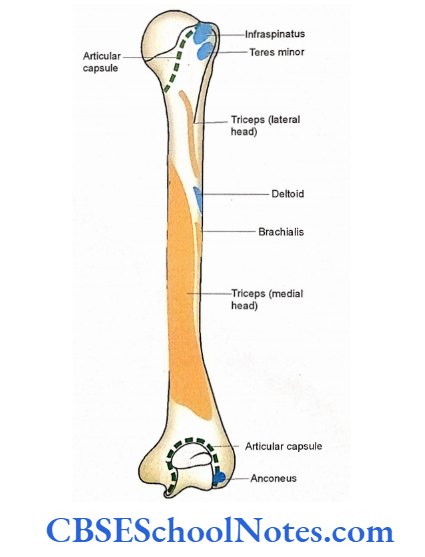
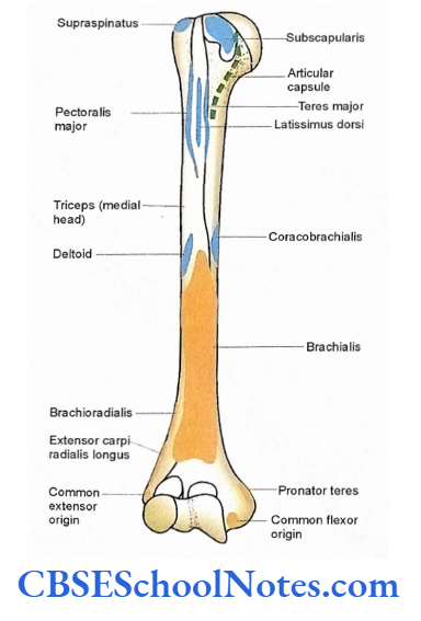
- The three impressions on the surface of the greater tubercle give insertion to supraspinatus, infraspinatus, and teres minor.
- The subscapularis muscle is inserted on the lesser tubercle.
- The tendon of the long head of the biceps passes through the bicipital groove.
Shaft
Shaft Anterior Aspect
- On the anterior aspect of the shaft, the medial lip of the bicipital groove gives attachment to the teres major while the lateral lip to the pectoralis major.
- The floor of the bicipital groove gives insertion to the latissimus dorsi.
- On the V-shaped deltoid tuberosity deltoids are attached.
- The coracobrachialis is attached to the middle of the medial border.
- The brachialis muscle arises from the front aspect of the lower half of the shaft.
- The pronator teres take origin from the medial supracondylar ridge.
- The lateral supracondylar ridge gives origin to the brachioradialis and extensor carpi radialis longus.
Shaft Posterior Aspect
- The lateral head of the triceps arises above the radial groove.
- Below the radial groove, the posterior surface gives origin to the medial head of the triceps.
Shaft Lower End
- The front of the medial epicondyle gives origin to the superficial flexor muscles of the forearm. This origin is known as “common flexor origin”.
- The anterior aspect of the lateral epicondyle gives origin to extensors of the forearm hence known as “common extensor origin”.
- The anconeus originates from the posterior surface of the lateral epicondyle.
Ligaments Attached to Bone
- The attachments of the articular capsule for shoulder joint and elbow joints.
- The ulnar collateral ligament of the elbow joint is attached to the medial epicondyle.
- The radial collateral ligament is attached to the lateral epicondyle.
Nerves and Vessels Related to the Bone
The surgical neck of the humerus is related to the anterior and posterior circumflex humeral vessels and the axillary nerve.
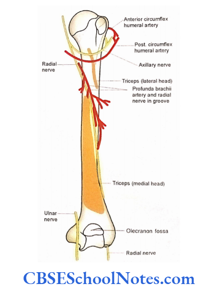
- The radial nerve and profunda/brachial vessels are related to the radial groove.
- The ulnar nerve lies just posterior to the medial epicondyle.
Shaft Clinical Importance
- The common sites of fracture are the surgical neck, middle of the shaft, and supracondylar region.
- The fracture of the surgical neck of the humerus is common in old people. The axillary nerve, which is in direct contact with the neck, may get injured.
- The supracondylar fractures are common in young individuals. The median nerve and brachial artery may get injured as they are related to the lower end.
- The fracture of the middle of the shaft may be due to a direct blow to the arm. Here the radial nerve in the radial groove may get injured.
- The avulsion fractures of the greater tubercle of the humerus are also common (in this fracture the greater tubercle is pulled away from the rest of the upper end). This may result in to fall on the hand while the arm is abducted.
- The fracture of the medial epicondyle is usually associated with the injury of ulnar nerve, as it lies just posterior to the epicondyle.
- As the upper end of the humerus is the growing end, the bone keeps on growing in a young individual even after the amputation of an arm.
- Hence, at the time of amputation of the arm care should be taken that sufficient soft tissue should be left distal to the site of amputation so the bony stump will not penetrate soft tissues.
Shaft Ossification
The upper end ossifies by 3 and the lower end by 4 secondary centers Upper end is the growing end as the secondary center appears first and fuses last (at 20 years).

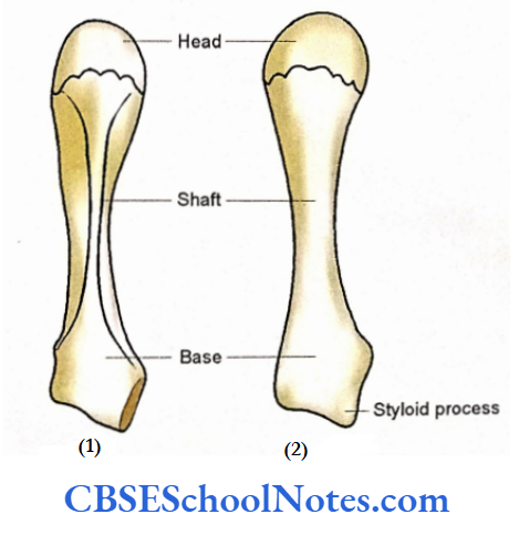
Radius
- The radius is situated on the lateral side (thumb side) of the forearm. Like any other long bone, it also consists of the upper end, shaft, and lower end.
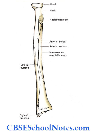
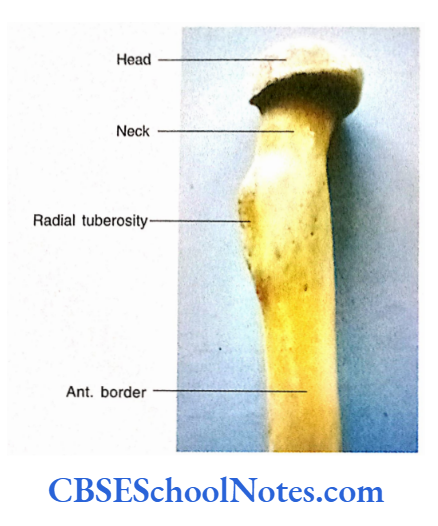
- The upper (proximal) end of the radius is narrow while its lower end is wide. The upper end presents a disc-shaped head, a neck, and medially directed radial tuberosity.
- The shaft of the radius has a laterally directed convexity. The medial border is sharp and called an interosseous border. The lower end is wide and shows anterior, posterior, medial, and lateral surfaces.
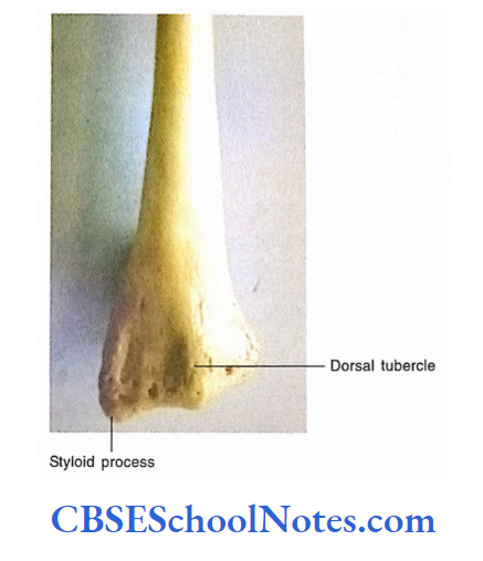
- The posterior surface of the lower end is rough and presents a dorsal tubercle. A projection (styloid process) is present on the lateral aspect of the lower end.
Radius Side Determination
- Keep the bone vertically in such a way that the narrow disc-shaped upper end is directed upward (proximally).
- At the expanded lower end, the dorsal tubercle should face backward and styloid process directed laterally and downwards.
- In this position, a sharp border of the shaft (interosseous border) should be directed medially.
Radius Anatomical Position
Hold the bone vertically in such a way that the head is directed upwards, with radial tuberosity and sharp border medially, and dorsal tubercle posteriorly.
General Features Upper End
The upper end consists of the head, neck, and radial tuberosity.
- The disc-shaped head shows an upper surface, which is slightly concave to articulate with the capitulum of the humerus.
- The circumference of the head articulates medially with the radial notch of the ulna to form the superior radioulnar joint. The rest of the circumference of the head is surrounded by an annular ligament.
- The constriction just below the head is known as the neck.
- There is a prominent elevation just below the medial part of the neck. This is known as radial tuberosity.
Shaft
- The shaft shows 3 borders and three surfaces.
- Trace the anterior border from the anterior aspect of radial tuberosity to the styloid process.
- Trace the posterior border from the posterior Upper-End aspect of the radial tuberosity to the posterior aspect of the lower end Trace the sharp medial (interosseous) border from the lower aspect of radial tuberosity to the posterior margin of the ulnar notch.
- The anterior surface lies between anterior and medial borders while the posterior surface lies between posterior and medial borders.
- The lateral surface is present between anterior and posterior borders. This surface is convex laterally and shows a rough area near the middle part of the shaft.
Lower End
- The lower end is the expanded end and shows anterior, posterior, lateral, and medial surfaces.
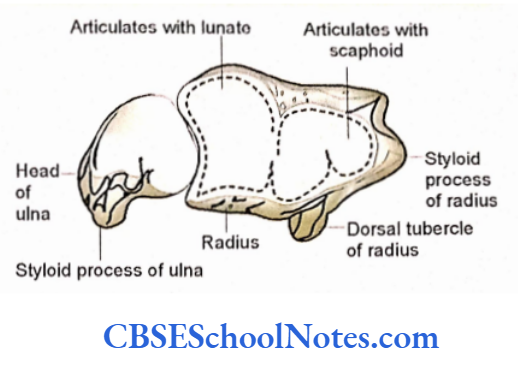
- It also shows an inferior surface, which is smooth (articular) and articulates with the scaphoid laterally and lunate bone medially to form the wrist joint
- The lateral surface when traced downwards, projects as a styloid process.
- The medial surface shows an ulnar notch. It articulates with the head of the ulna to form an inferior radioulnar joint.
Radius Particular Features
Muscles Attached to the Radius
Upper End
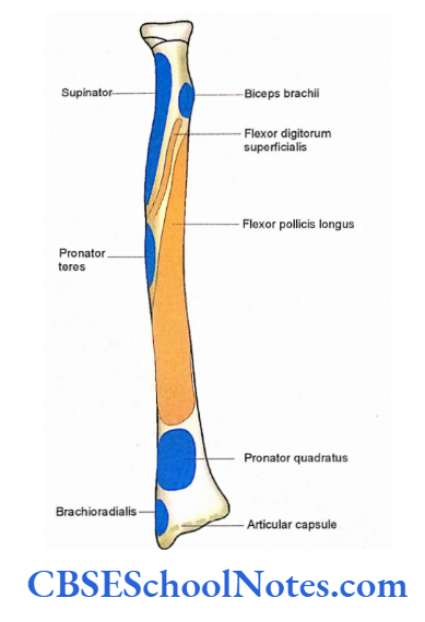
The biceps brachii is inserted on the rough posterior part of the radial tuberosity. The anterior smooth part of the radial tuberosity is in relation with the bursa.
Shaft and Lower End
- The upper part of the anterior border gives origin to the radial head of flexor digitorum superficialis.
- The upper of the anterior surface gives origin to the flexor pollicis longus.
- The pronator quadratus is inserted into the lowermost part of the anterior surface. The attachment also extends on the medial surface of the lower end. posterior surfaces give insertion to the supinator.
- The rough area on the middle of the lateral surface gives insertion to pronator teres.
- The muscle brachioradialis is inserted into the lowest part of the lateral surface just above the styloid process.
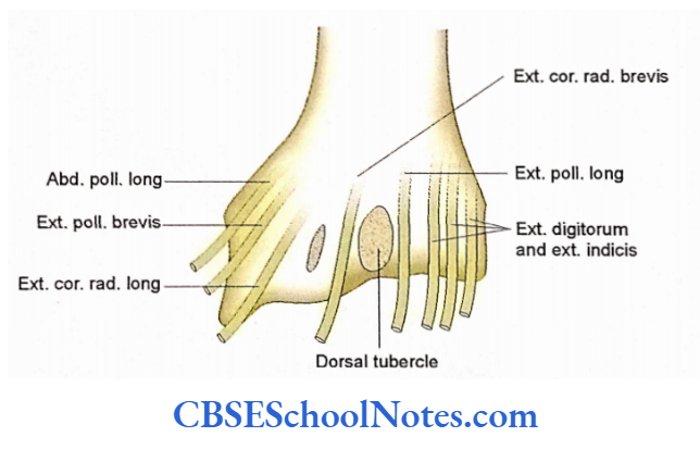
- The abductor pollicis longus and extensor pollicis brevis arise from the posterior surface of the shaft.
Ligaments Attached to the Bone
- Note the attachment of the articular capsule of the wrist joint on the lower end of the radius.
- The interosseous membrane is attached to the lower 3A of the medial border.
- The articular disc of the inferior radioulnar joint is attached to the lower border of the ulnar notch.
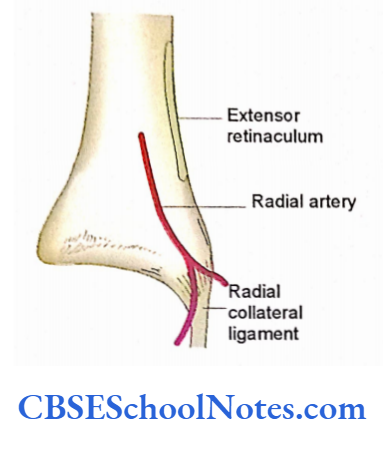
- The anterior border near the lower end gives attachment to the extensor retinaculum.
- The radial collateral ligament of the wrist joint is attached to the tip of the styloid process.
Tendons and Arteries with the Bone
- The relationship of tendons on the dorsal aspect of the lower end
- The radial artery is related to the anterior surface of the lower end. Here the pulsations of the artery can be easily felt by applying pressure against the radial bone.
Radius Clinical Importance
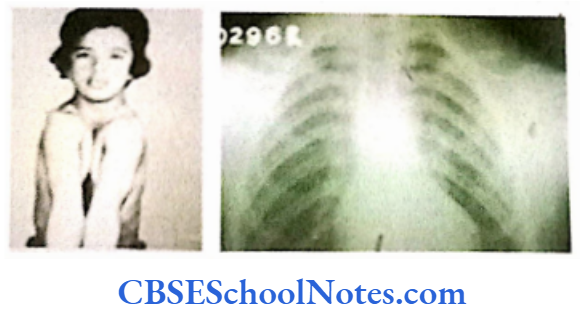
Though the fracture of the radius may occur at its upper end and shaft the fracture of the distal end of the radius is most common, especially in old women.
A fracture of the lower end of the radius is called a Colies’s fracture. The fracture usually results from a fall on an outstretched hand.
The distal fragment overrides and is displaced dorsally. This results in a shortening of radius.
As a result the styloid process of both the bones (radius and ulna) now lies almost at the same level. The clinical deformity is often referred to as “dinner fork deformity.
Radius Ossification
Radius ossifies by one primary and two secondary centers, i.e., one for each end. The lower end is the growing end.
Ulna
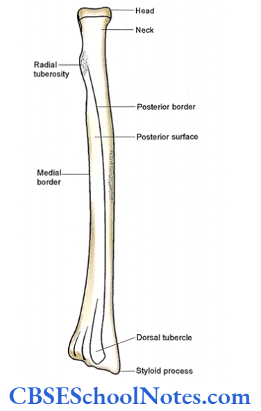
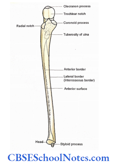
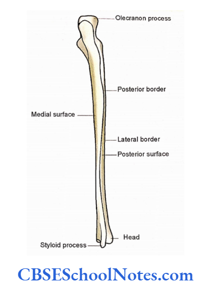
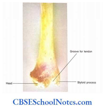
- The ulna is the medial bone of the forearm. It is longer than the radius. The ulna has an upper end, a lower end, and a shaft. The upper end of the ulna is expanded, and irregular and presents a deep notch, which faces anteriorly.
- This notch is called a trochlear notch and it articulates with the trochlea of the humerus at the elbow joint. While its lower end is small and has a tiny styloid process.
- You should remember that the lower end of the ulna is called as head. The shaft has a sharp lateral margin.
- This margin gives attachment to the broad, flat, fibrous connective tissue called interosseous membrane. The ulna is connected with the radius by an interosseous membrane.
Ulna Side Determination
- Hold the bone vertically in such a way that its expanded hook-like end is directed upwards and the small rounded lower end is directed downwards.
- The concavity of the trochlear notch (at the upper end) should face anteriorly.
- The sharp border of the shaft should be directed laterally.
Ulna Anatomical Position
Hold the bone vertically in such a way that the trochlear notch faces anteriorly, the sharp border of the shaft laterally, and the styloid process at the lower end is present posteriorly.
Ulna General Features
Ulna Upper End
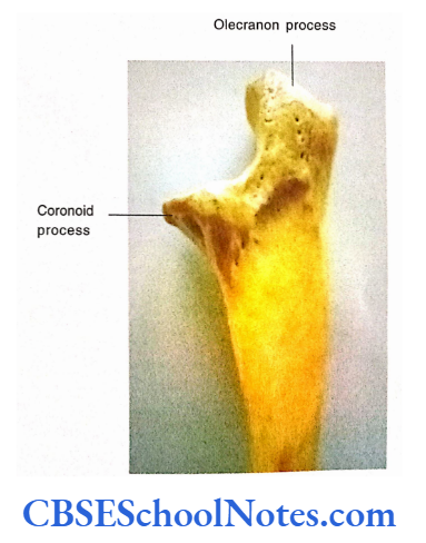
The upper end of the ulna presents the olecranon process, coronoid processes, and two articular surfaces, i.e., trochlear and radial notches.
Olecranon Process
- The olecranon process is the uppermost part of the ulna. It has superior, anterior, posterior, medial, and lateral surfaces.
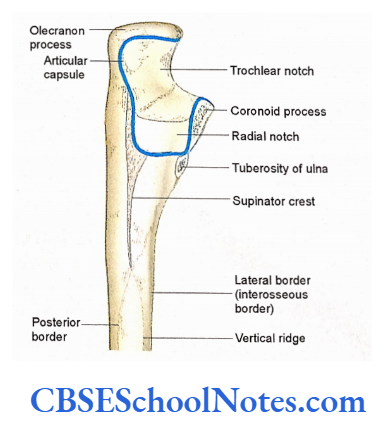
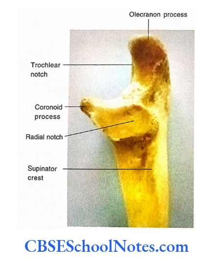
- The anterior surface of the olecranon process forms the upper part of the trochlear notch.
- The posterior surface of this process is triangular and smooth because it lies just beneath the skin.
Coronoid Process
- The coronoid process projects anteriorly from the shaft just below the olecranon.
- It has four surfaces, i.e., superior, anterior, medial, and lateral.
- The superior (upper) surface is almost horizontal and forms the lower part of the trochlear notch.
- The anterior surface is triangular and is rough in its lower part. The rough area is known as the tuberosity of the ulna.
Radial Notch
The lateral surface of the coronoid process has a concave articular area. This is known as a radial notch. It articulates with the head of radius forming the superior radioulnar joint.
Trochlear Notch
- It articulates with the trochlea of the humerus. It is formed by the anterior surface of the olecranon and the upper surface of the coronoid process. There may be a non-articular area(groove) between the two parts.
Lower End
- The lower end (head) is small and rounded.
- Anteriorly and laterally it articulates with the ulnar notch of the radius to form an inferior radioulnar joint.
- The inferior surface is separated from the cavity of the wrist joint by an articular disc.
- The styloid process projects downwards from the posteromedial aspect of the lower end.
Shaft
- The cross-section of the shaft is almost triangular.
- It presents anterior, lateral, and posterior borders and anterior, medial, and posterior surfaces.
- The lateral or interosseous border is sharp. When traced upwards, it becomes continuous with a rough ridge present just posterior to the radial notch. This rough ridge is known as the supinator crest.
- The anterior and posterior borders are rounded. The anterior border begins at the lower end of the tuberosity of the ulna and ends on the anterior aspect of the styloid process.
- The anterior surface lies between the anterior and interosseous border and shows the presence of an oblique ridge in its lower part.
- The posterior surface lies between inter osseous and posterior border. The posterior surface is subdivided into medial and lateral parts by a vertical ridge.
Ulna Particular Features
Muscles Attached to Bone
Upper End
- The triceps is inserted on the posterior part of the upper surface of the olecranon process.
- Brachialis is attached to the anterior surface of the coronoid process and the tuberosity of the ulna.
- The supinator arises from the supinator crest and a triangular area just below the radial notch.
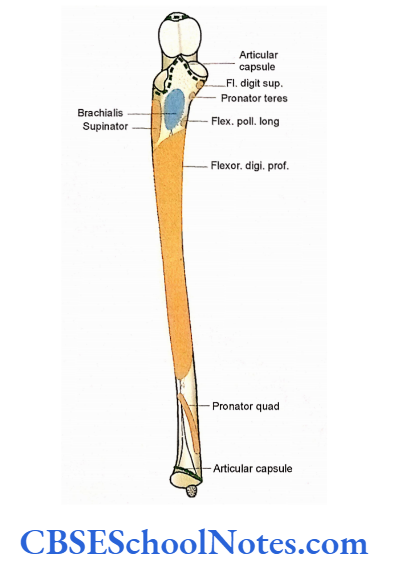
- The medial border of the coronoid process gives origin to the ulnar head of flexor digitorum superficialis above and pronator teres below.
- The anconeus is inserted into the lateral aspect of the olecranon and the upper part of the posterior surface of the shaft.
- The ulnar head of the pronator teres arises from the medial margin of the coronoid process.
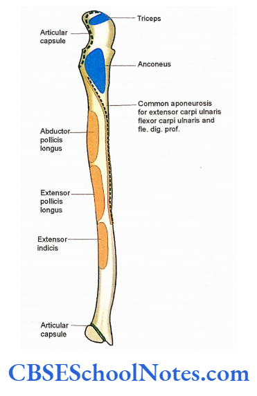
- The ulnar head of the flexor carpi ulnaris arises from the medial side of the olecranon process and from the upper two-thirds of the posterior border through aponeurosis.
- From the same aponeurosis, ulnar head of extensor carpi ulnaris also arise.
Shaft
- Flexor digitorum profundus takes origin from the anterior and medial surface of the shaft On the anterior surface the oblique ridge gives origin to the pronator quadratus
- Three muscles are attached on the posterior surface (between the interosseous border and a vertical ridge) from above downwards i.e., abductor pollicis longus, extensor pollicis long, and extent or indices.
Ligaments Attached to Bone
- The capsular ligament of the elbow joint is attached to the margins of the coronoid and olecranon process.
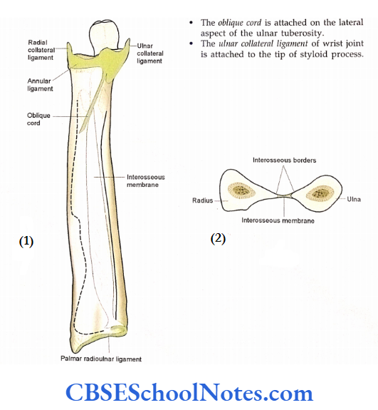
- The capsular ligament of the inferior radioulnar joint is attached close to the articular area.
- The apex of the triangular articular disc is attached to the lateral aspect of the styloid process.
- The interosseous membrane is attached to the sharp interosseous border.
- The annular ligament of a superior radioulnar joint is attached to the anterior and posterior border of the radial notch.
- The oblique cord is attached to the lateral aspect of the ulnar tuberosity.
- The ulnar collateral ligament of the wrist joint is attached to the tip of the styloid process.
Ulna Clinical Importance
- Though the fractures of both the radius and ulna may result due to severe injury isolated fractures of the radius and ulna may also occur.
- The most common fracture of the ulna along with the radius is through the middle of the shaft or at its lower end. Sometimes a fracture of the olecranon can occur due to a fall on the elbow.
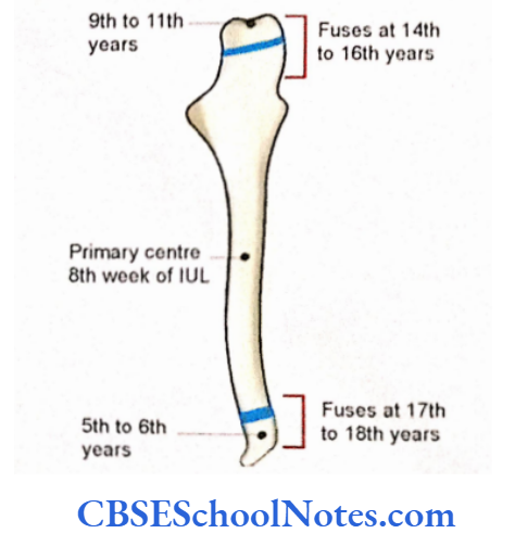
Ulna Ossification
Ulna ossifies by 3 centers, i.e., one primary and two secondary, i.e., one each for upper and lower end. The lower end is the growing end.
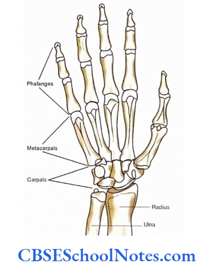
Bones Of The Hand
The bones of the hand consist of carpals, metacarpals, and phalanges.
Carpal Bones
The skeleton of the wrist consists of eight small bones, which are arranged in two transverse rows of four bones each. These bones are called carpal banks. They are named as per their shapes.
The proximal row, from lateral to medial side, consists of:

- Lunate (It is moon-shaped).
- Triquetrum (It has three corners and is pyramidal). Pisiform (It is pea shaped small bone that lies on the anterior surface of the triquetrum).
- The carpal bones in the distal row, from lateral to medial, are:
- Trapezium (table-shaped, four-sided).
- Trapezoid (wedge-shaped).
- Capitate (It is cap-shaped and has a rounded head).
- Hamate (It has a hook-shaped process).
Joints Formed by the Carpal Bones
- The wrist joint is formed between the proximal surfaces of three carpal bones of the proximal row and the lower end of the radius.
- The scaphoid and lunate of the proximal row articulate with the inferior articular surface of the radius, while the triquetrum is related to the articular disc and thus separated from the head of the ulna.
- Carpal bones are joined to one another by interosseous ligaments and intercarpal joints.
- The distal articular surfaces of the carpal bones of the distal row articulate with the metacarpals to form carpometacarpal joints.
Bones of the Hand Clinical importance
- The common injury of the wrist consists of fracture of the scaphoid or lunate.
- The fracture of the scaphoid is the most common. It results in a fall on an outstretched hand. In this condition, the line of fracture passes almost from the middle of the bone dividing the bone into proximal and distal fragments. The patient complains of pain in the scaphoid fossa (lateral side of the wrist.
- As the nutrient arteries scaphoid enter the distal end of the bone, the proximal segment is devoid of the blood supply after fracture. This may result in avascular necrosis (death due to inadequate blood supply) of the proximal fragment of the scaphoid.
- Thus scaphoid fracture heals poorly, i.e., may take several months to heal.
- The fracture of hamate also heals poorly due to the traction produced by the attached muscles.
- As the ulnar nerve lies close to the hook of hamate, it may also get injured due to the fracture of the hamate.
Metacarpals
Metacarpals are miniature long bones, which form the skeleton of the palm. They are five in number and are numbered from the lateral to the medial side. The metacarpal of the thumb is known as the first metacarpal while that of the little finger is known as the fifth (V) metacarpal.
Each metacarpal bone consists of a proximal base, an intermediate shaft, and a distal head.

- The proximal ends or bases of the metacarpal are irregularly expanded and articulate with the carpal bones. The distal ends (heads) are rounded and articulate with the proximal phalanges.
- The shaft of each metacarpal is curved with the concavity looking forward. It has three surfaces, i.e., medial, lateral, and posterior.
- The first metacarpal is easily distinguished from the rest because it is the thickest and shortest of all the metacarpals. The articular surface at its base is concavoconvex which articulates with trapezium.
- The third metacarpal can also be differentiated from the rest because it has a styloid process on the lateral side of its base.
Joints Formed by the Metacarpals
- The distal articular surfaces of the carpal bones of the distal row articulate with the metacarpals to form carpometacarpal joints.
- The trapezium articulates with the first metacarpal, the trapezoid with the second, the capitate with the third, and the hamate with the fourth and fifth metacarpals.
- The distal articular surfaces of metacarpals articulate with the proximal articular surfaces of proximal phalanges to form carpometacarpal joints.
Phalanges
- Phalanges are the bones of digits. Each digit has three phalanges (i.e., proximal, middle Flexor carpi, and distal) except the first (for thumb), which has only two phalanges.
- Each phalanx has a proximal base, a body, and a distal head. The proximal phalanges are the longest and most articulate with the head of metacarpals at their bases to form metacarpophalangeal joints.
- The middle phalanges are intermediate in size and articulate at their bases with the heads of proximal phalanges to form proximal interphalangeal joints.
- The distal phalanges are smallest, flattened, and expanded at their distal ends (non-articular) to form the nail beds.
Particular Features of the Bones of Hand
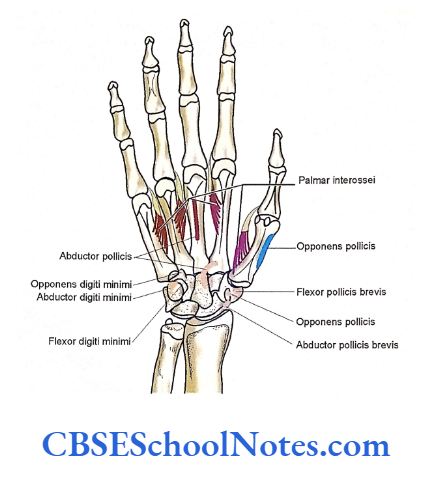
- Insertion of forearm muscles on the palmar surface of the hand.
- The flexor carpi ulnaris is attached on the pisiform bone. From this bone abductor digiti minimi also takes origin.
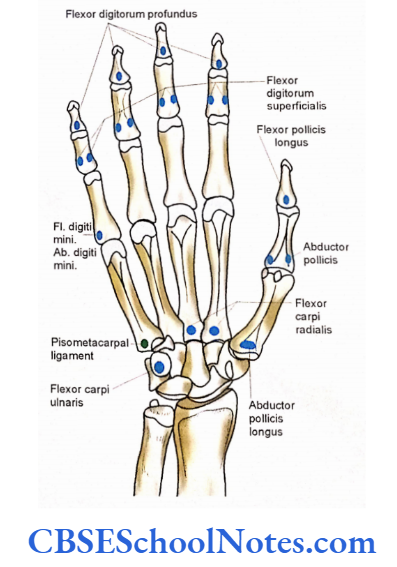
- Flexor carpi radialis is inserted on the base of the 2nd and 3rd metacarpals on their palmar surface.
- The flexor digitorum profundus is inserted on the bases of distal phalanges, while flexor digitorum superficialis is inserted on both the sides of middle phalanges of all the fingers except that of the thumb.
- The flexor pollicis longus is inserted on the base of the distal phalanx of the thumb, on its palmar surface.
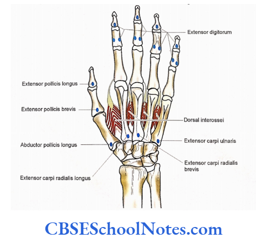
Insertion of forearm muscles on the dorsal aspect of the hand and dorsal interossei.
- The extensor carpi ulnaris is attached to the base of the fifth metacarpal; the abductor pollicis longus to the base of the first; the extensor carpi radialis longus to the base of the second; extensor carpi radialis brevis to the bases of 2nd and 3rd metacarpal.
Attachments of Ligaments
- The attachment of carpal bones with each other (due to their shape) presents a concavity forward.
- The attachment of the flexor retinaculum medially to the hook of the hamate and pisiform bone and laterally to the tubercles of the scaphoid and trapezium
form an osteofascial carpal tunnel. - Through this tunnel passes the tendons, vessels, and nerves from the forearm to the hand.
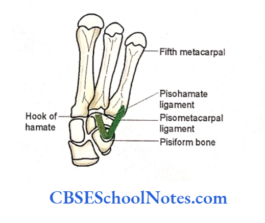
- Pisiform bone gives attachment to two ligaments, i.e., first extending from pisiform to the base of the fifth metacarpal (pisometacarpal) and second from pisiform to the hook of the hamate (pisohamate).
- The medial end of the extensor retinaculum is attached to the triquetral and pisiform bones.
- The lateral margins of each phalanx give attachment to the fibrous flexor sheath.