
Bones Of The Skull Mandible
The mandible is the bone of the lower jaw.
General Features of Mandible Body
Mandible Body
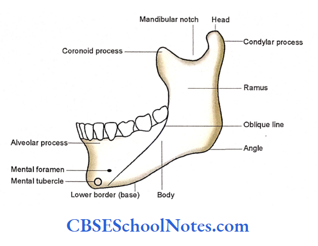
The body is U-shaped and has two surfaces and two borders. It consists of right and left halves united in the median plane at the symphysis menti. The symphysis menti is a faint ridge on the upper part of the midline.
Mandible Body Upper Border
The upper border of the mandible is also known as alveolar border. It bears sockets for the teeth.
Mandible Body Lower Border
- This border is also known as the base of the mandible. Posteriorly it is continuous with the lower border of the ramus of the mandible.
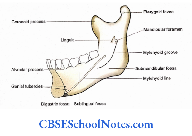
- On either side of the midline, there is the presence of a digastric fossa for the attachment of the anterior belly of the digastric.
Mandible Body External Surface
- At the lower part of the midline (at symphysis menti) a triangular mental protuberance is present.
- The upper angle of the triangle lies at the lower end of the symphysis menti.
- The mental tubercles are present at the lower angles of this triangle.
- A faint ridge is present on the external surface of the body extending upwards and backward from the mental tubercle up to the anterior margin of the ramus. This is called an oblique line.
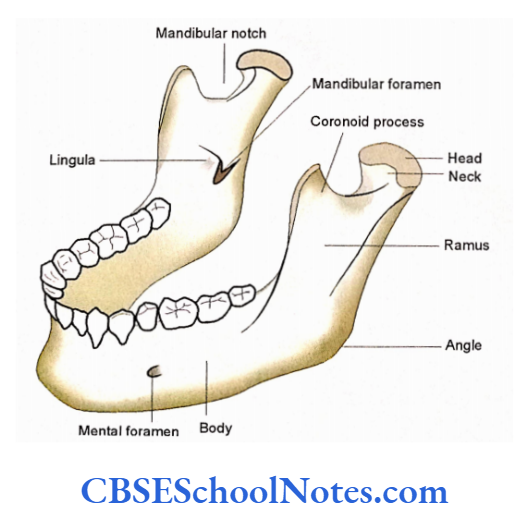
- The mental foramen, in an adult, is present below the second premolar tooth midway between the upper and lower borders.
Mandible Body Internal Surface
- The mylohyoid line is oblique and extends diagonally downwards and forwards from just below the alveolar border (behind the 3rd molar tooth) to the lower part of symphysis menti.
- The sublingual fossa is a smooth area above the anterior part of the mylohyoid line for the lodgement of the sublingual salivary gland.
- The submandibular fossa is a concave area present below the posterior part of the mylohyoid line. It comes in contact with the superficial part of the submandibular gland.
- The posterior part of the symphysis menti on either side of the midline presents a pair of elevations called superior and inferior genial tubercles. These tubercles are present above the anterior end of the mylohyoid line.
Read and Learn More Human Osteology Notes
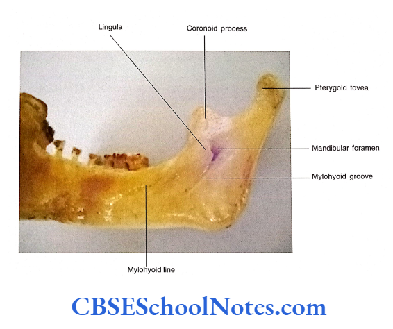
Ramus of Mandible
The ramus of the mandible projects upwards from the posterior part of the body. It has four borders (anterior, posterior, upper, and lower), two surfaces (lateral and medial), and two processes (coronoid and condylar).
Ramus of Mandible Borders
- The upper border forms a notch, i.e., the mandibular notch. This border displays a triangular coronoid process anteriorly and a condylar process posteriorly.
- The lower border is the backward continuation of the base of the body of the mandible.
- The anterior border of the ramus is sharp and continuous below with the oblique line on the lateral surface of the body of the mandible.
- The posterior border meets with the lower border of the ramus to form the angle of the mandible. This border is in continuation above with the condylar process
Ramus of Mandible Processes
- The coronoid process is flat and triangular, which projects upwards from the anterosuperior part of the ramus. It lies at the junction of the upper (mandibular notch) and anterior border of the ramus.
- The condylar process projects upwards from the posterosuperior part of the ramus.
- The upper end of the condylar process is expanded and forms the head of the mandible. While the lower part of the condylar process is constricted and called the neck.
- The head of the mandible bears an articular surface that articulates with the mandibular fossa of the temporal bone to form the temporomandibular joint.
- The anterior surface of the neck shows a depression called pterygoid fovea for attachment of lateral pterygoid muscle.
Ramus of Mandible Surfaces
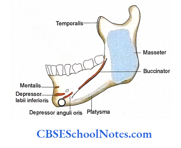
- The lateral surface is rough and flat in its anteroinferior part for the attachment of the masseter.
- The medial surface presents a mandibular foramen close to its center. It leads into a mandibular canal which opens at the mental foramen.
- The lingula is a tongue-shaped projection near the anterior margin of the mandibular foramen. There is the presence of a mylohoid groove just behind the lingula.
- This groove runs forward and downwards from the mandibular foramen to the inner surface of the body of the mandible.
- The area above the angle of the mandible is rough for the attachment of the medial pterygoid muscle.
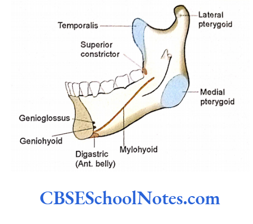
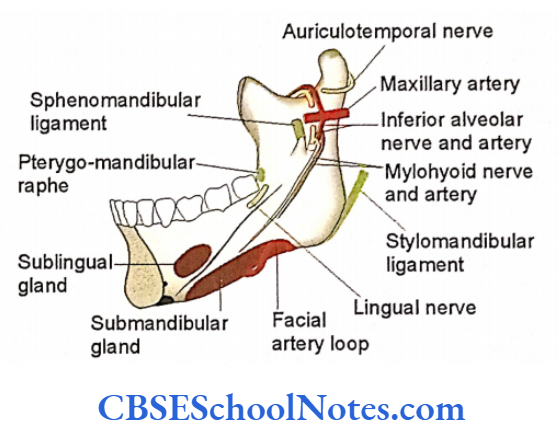
Particular Features of Mandible Body
Attachments of muscles are shown for the external surface and internal surface. Relations of nerves, blood vessels, glands, and ligaments.
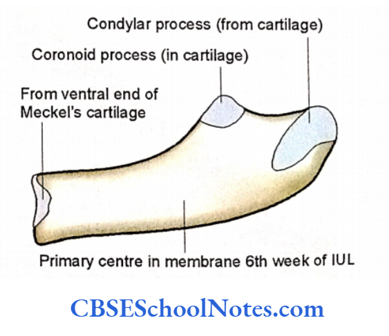
Mandible Body Ossification
- The part of the bone between the mental foramen and mandibular foramen ossifies in the membrane (fibrous envelop of the Meckel’s cartilage of the first branchial arch).
- The part of the body in front of the mental foramen and the part of the ramus above the mandibular foramen ossify in cartilage.
- Each half was replaced by a bony union at the end of the first year of postnatal mandible ossifying in 6th week of IUL.
- At birth, the mandible consists of two halves of the mandibular body united by the fibrous joint at symphysis menti. This fibrous joint is aged.
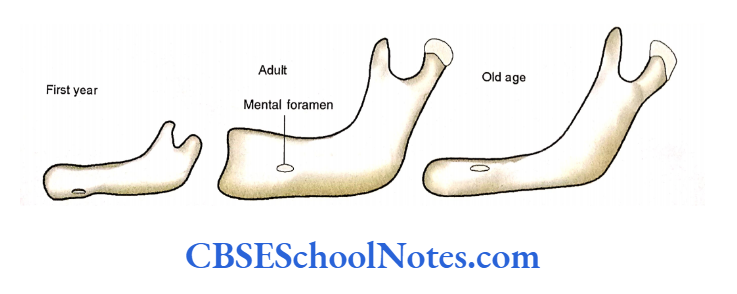
Age Changes In Mandible
The bony features of the mandible change with age (children, adults, and old age).
Frontal Bone
- The frontal bone forms most of the part of the anterior cranial fossa. The bone consists of squamous (main) parts and orbital parts.
- The squamous part forms the forehead and orbital parts form the major part of the roof of each orbit.
Frontal Bone Squamous (Main) Part
This part of the bone has an external surface and internal surface, right and left temporal surface, nasal part, and zygomatic processes.
Frontal Bone External Surface
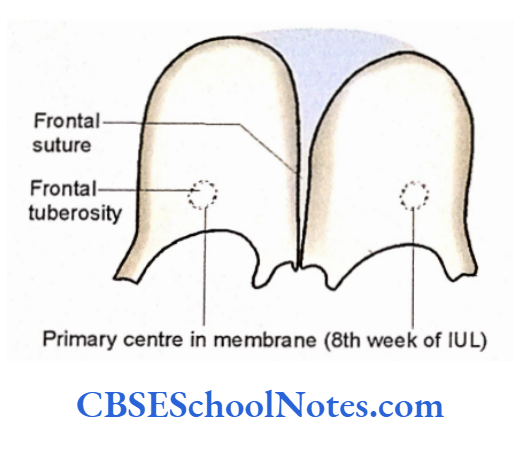
- The external surface of the squamous part forms the forehead. This surface presents frontal eminence about 3 cm above the supraorbital margin.
- The other features seen on this surface are the glabella, superciliary arches, and supraorbital margins.
- The supraorbital notch or foramen is seen on the supraorbital margin. Occasionally the metopic suture may be seen in the region of the glabella.
Frontal Bone Temporal Surface
This forms a small surface on the right and left sides below the temporal lines
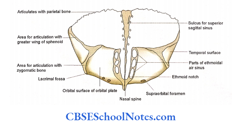
Frontal Bone Internal Surface
- This surface is concave and shows the impressions of cerebral sulci and gyri.
- It shows a median groove (sagittal sulcus) for the superior sagittal sinus.
- At the anterior end of the groove, there is the presence of a median ridge called the frontal crest. Just below the frontal crest is the presence of foramen caecum.
Frontal Bone Nasal Part
- The nasal part is the downward projection of the squamous part of bone between two superior orbital margins.
- The lower serrated margin of this part is concave downwards and known as the nasal notch.
- The nasal notch bears a median projection, the nasal spine which articulates with the nasal bones in front and behind with the perpendicular plate of the ethmoid.
- From medial to lateral, the nasal bone, frontal process of maxilla, and lacrimal bone articulate with each half of the nasal notch.
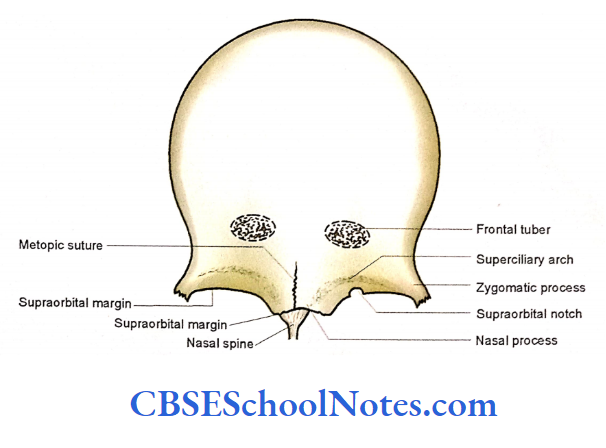
Zygomatic Process
The zygomatic process passes downwards and laterally from the lateral end of the superior orbital margin.
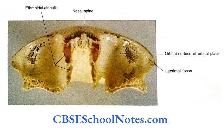
The Orbital Part
The orbital part consists of right and left orbital plates separated from each other by a wide notch called an ethmoid notch.
- The notch is occupied by the ethmoid bone.
- The orbital surface faces downwards. It is smooth and forms the roof of the orbit.
- The inferior aspect of the orbital plate, just lateral to a notch, shows the depressions of ethmoidal air cells. More anteriorly it shows the opening of the frontal air sinus.
- There is the presence of lacrimal fossa at its anterolateral angle.
- The trochlear fovea is present, at its anteromedial part.
- The upper surface of the orbital plate forms the floor of the anterior cranial fossa and shows the impressions for sulci and gyri.
Articulations of the Frontal Bone
The following bones articulate with the squamous part of the frontal bone:
- Posteriorly: Parietal bones (at the coronal suture), greater wings of the sphenoid.
- Anteriorly: Zygomatic bone, nasal bone, frontal process of maxilla, lacrimal bone, and perpendicular plate of the ethmoid.
- The following bones articulate with the orbital part:
- Posteriorly: Lesser wings of sphenoid.
- Medially: Orbital plate of ethmoid.
Frontal Bone Ossification
- It ossifies in the membrane.
- Two halves of the frontal bone begin to fuse at the age of the first post-natal year. The fusion is completed by the end of eight years.
- If the suture persists in adults then it is known as a metopic suture.
Parietal Bone
The parietal bones are quadrilateral in shape and form the roof and side walls of the cranial cavity.
Parietal Bones General Features
Each one has:
- Four borders: Superior, inferior, anterior and posterior.
- Two surfaces: External and internal.
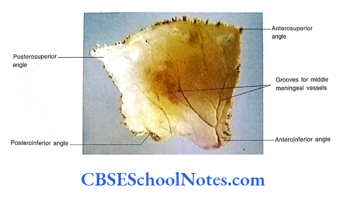
- Four angles: Anterosuperior, anteroinferior, posterosuperior and posteroinferior.
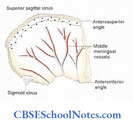
The external surface is convex. The external surface shows the parietal eminence, parietal foramen, and superior and inferior temporal lines.
On the concave internal surface, the vascular grooves (for middle meningeal vessels) run upwards and backward from the anteroinferior angle. A shallow groove for the sigmoid sinus is seen at its posteroinferior angle.
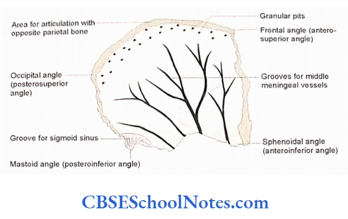
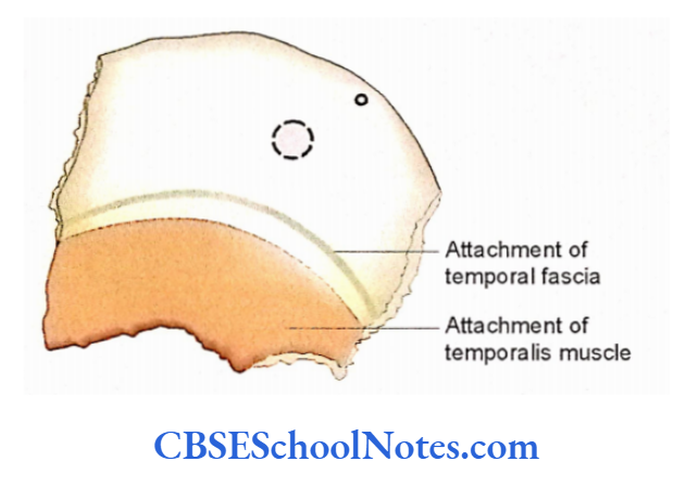
Parietal Bones Particular Features
The attachments and relations of the bone with the soft tissues
Parietal Bones Articulations
Articulations of the parietal bone are shown in
Parietal Bones Side Determination
Hold the bone in such a way that the convex external surface faces laterally. The angle showing the vascular groove at the internal surface should be kept antero-inferiorly.
Parietal Bones Anatomical Position
The superior border should be kept towards the median plane, the antero-inferior angle downwards and forwards.
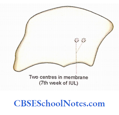
Parietal Bones Ossification
It ossifies in the membrane
Parietal Bones Occipital Bone
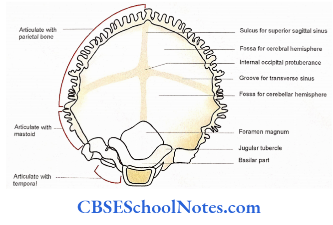
- This bone is present in the posterior part of the skull. The presence of the large foramen magnum helps in dividing the bone into four parts.
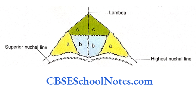
- A part above and behind the foramen magnum is an expanded curved plate of bone, i.e., a squamous part. It shows a superior angle and two lateral angles.
- The superior angle lies at lambda and each lateral angle at asterion.
- A part in front of frame n magnum is called the basilar part.
- A pair of condylar parts are present on either side of the foramen magnum.
Parietal Bones Anatomical Position
Hold the bone in such a way that the foramen magnum should lie in the horizontal plane and the basilar part should be directed forwards and upwards.
Parietal Bones General Features
Squamous Part
- The squamous part shows a convex external and a concave internal surface: one superior angle two lateral angles and two borders i.e., lambdoid and mastoid borders.
- The external surface shows the presence of the external occipital protuberance and external
occipital crest. - The highest nuchal line and the superior nuchal line run laterally from the external occipital protuberance, on the right and left sides. The inferior nuchal line runs laterally from the middle of the external occipital crest.
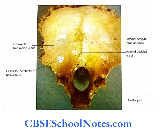
- The internal surface of the squamous part shows the presence of internal occipital protuberance in the middle.
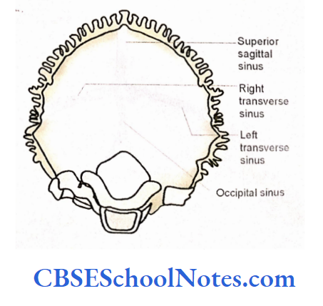
- A wide median groove for the superior sagittal sinus is present above the protuberance. Grooves for transverse sinuses are present on either side of the protuberance. An internal occipital crest runs downwards in the midline from the internal occipital protuberance.
- Borders—The lambdoid border articulates with the parietal bone at the lambdoid suture and the mastoid border articulates with the mastoid bone at the occipito¬ mastoid suture.
Basilar Part of the Occipital Bone
- It extends forward and upwards from the anterior margin of the foramen magnum. This part has three surfaces (anterior, superior, and inferior), and two lateral borders.
- The anterior surface extends upto the body of the sphenoid bone and forms a primary cartilaginous joint (basi-sphenoid joint), which is replaced by the bone at about 25 years of age.
- The superior surface slopes downwards and backward and forms a part of the clivus.
- The inferior surface bears a pharyngeal tubercle The lateral border of the basilar part articulates with the posterior border of the petrous part of the temporal bone.

The Condylar Part of the Occipital Bone
- It presents superior and inferior surfaces. The inferior surface of the condylar part presents an occipital condyle. The hypo-glossal or anterior condylar canal opens just above the anterolateral side of the occipital condyle.
- Just behind the condyle is the condylar fossa, which may show the opening of the posterior condylar canal.
- The jugular process is a quadrilateral plate present lateral to the posterior half of the occipital condyle.
- The upper surface of the jugular process is deeply grooved for the sigmoid sinus. The superior surface of the condylar part shows a jugular tubercle.
The Foramen Magnum
For structures passing through the foramen magnum
Foramen Magnum Particular Features
The soft tissue relation
Foramen Magnum Articulations
The following bones articulate with the occipital bone:
- Parietal (at lambdoid suture).
- Mastoid (at occipitomastoid suture).
- Sphenoid (at the basisphenoid suture, which gets ossified at about the age of 25 years).
Foramen Magnum Ossification
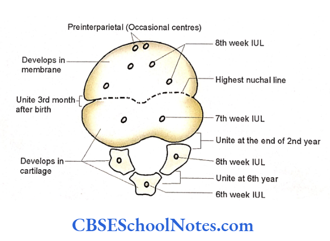
The squamous part, above the highest nuchal line, ossifies in the membrane. The rest of the bone ossifies in cartilage.
The part above the highest nuchal line may not unite with the rest of the bone and is then called interparietal bone.
Zygomatic Bone
It is situated on the upper lateral part of the face and forms the prominence of the cheek. Each zygomatic bone has a body and two processes (a frontal and a temporal process)
Zygomatic Bone General Features
The frontal process is directed upwards to articulate with the zygomatic process of the frontal bone.
The temporal process extends backward to articulate with zygomatic process of the temporal bone and forms the zygomatic arch. Anteromedially the body articulates with the maxilla.
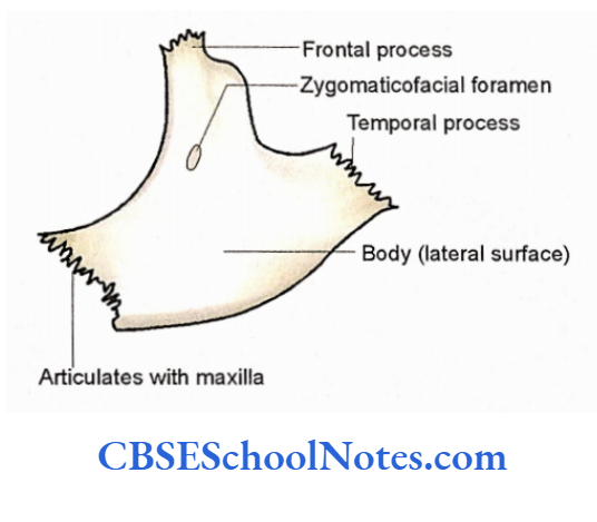
General Features Body
- The body has three surfaces and five borders. The surfaces are lateral, temporal arid orbital, while bordes are anteroinferior, anterosuperior (orbital), posteromedial, posterosuperior and postero-inferior.
- The lateral surface is convex and presents zygomaticofacial foramen. This surface forms the prominence of the cheek.
- The orbital surface forms the part of the lateral wall and floor of the orbit. It has the zygomatico-orbital foramen.
- The temporal surface is directed posteromedially. It forms the anterior wall of the temporal fossa. The zygomaticotemporal foramen is present on this surface.
Zygomatic Bone Particular Features
The attachments of muscles on the zygomatic bone.
The following bones articulate with the zygomatic bone:
- Frontal, maxilla, greater wing of the sphenoid, and the zygomatic process of temporal.
- Side Determination and Anatomical Position
Hold the bone in such a way that:
- The frontal process should look upwards.
- The smooth concave orbital margin should face upwards and medially.
- The smooth lateral surface should face laterally.
- The direction of the lateral surface will determine the side of the bone
Zygomatic Bone Ossification
The bone ossifies in the membrane
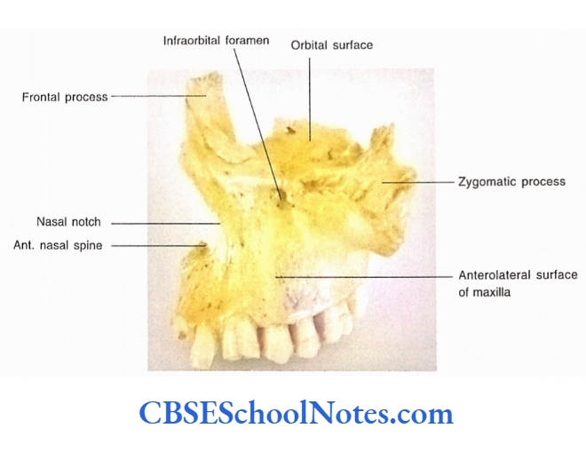
Maxilla
The right and left maxillae take part in the formation of the whole of the upper jaw and part of the hard palate.
Maxilla General Features
Each maxilla consists of a body and four processes, i.e., an alveolar process, a zygomatic process, a frontal process, and a palatine process.
General Features Body
The body is pyramidal and encloses a large maxillary air sinus. Inferiorly, the body bears an alveolar process for the attachment of teeth.
The body possesses four surfaces: anterolateral, posterior, orbital, and nasal.
Anterolateral Surface
It is directed forward and laterally. Above, the anterior surface is limited by the infra¬ orbital margin.
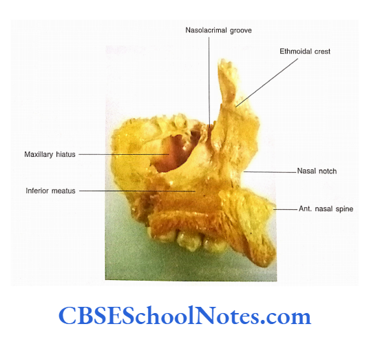
Medially, this surface presents a nasal notch. Below the infraorbital margin, the infraorbital foramen is present. This surface also presents canine eminence, incisive fossa, and canine fossa.
Posterior Surface
It faces backward and laterally. This surface forms the anterior wall of the infratemporal fossa hence, also known as the infratemporal surface.
Inferiorly, this surface shows the maxillary tuberosity, which articulates with the pyramidal process of the palatine bone. Posterior superior alveolar foramina are seen on the surface.
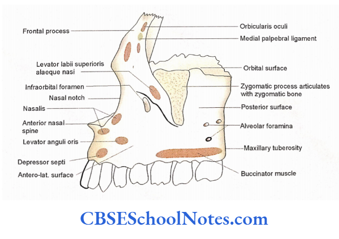
Orbital Surface
It forms the major part of the floor of the orbit. Posteriorly, it forms the anteromedial border of the infraorbital fissure.
There is the presence of an infraorbital groove near the center of the orbital surface. This groove leads to the infraorbital canal anteriorly, which opens on the anterior surface as infraorbital foramen.
Nasal (Medial) Surface
- This surface takes part in the formation of the lateral wall of the nasal cavity.
- The maxillary air sinus opens on this surface as a large maxillary hiatus
- Posterior to the hiatus there is a groove, which is converted into a greater palatine canal by the perpendicular plate of the palatine bone.
- Anteroinferior to the hiatus is the area of the inferior meatus.
- Superior to the inferior meatus is the nasolacrimal groove.
- There is the presence of a ridge anterior to the nasolacrimal groove. It is called the conchal crest, which articulates with the inferior nasal concha.
Nasal (Medial) Surface Processes
Zygomatic Process This strong process extends laterally from the body to articulate with the body of the zygomatic bone.
Zygomatic Process – Frontal Process
It extends upwards and medially where its tip articulates with the nasal part of the frontal bone. Anteriorly this process articulates with the nasal bone and posteriorly with the lacrimal bone.
The label surface of this process has a ridge called as a lacrimal crest, The medial surface is marked by an ethmoidal cress.
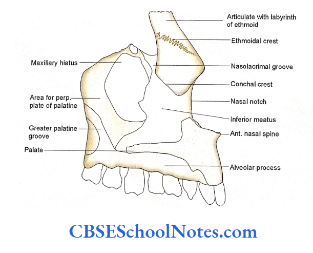
Alveolar Process
It extends downwards and carries the sockets for the teeth of the upper jaw.
Palatine Process
It is a horizontal plate, that projects medially from the nasal surface of the body of the maxilla. Medially it articulates with the palatine process of the opposite maxilla to form the anterior % of the hard palate.
It has superior and inferior surfaces and two free borders, i.e., medial and posterior. Posteriorly, it articulates with the anterior border of the horizontal plate of the palatine bone.
Anatomical Position and Side Determination Mold the bone in such a way that:
- The medial border of its palatine process should be in the median plane.
- The frontal process should point upwards.
- Zygomatic process laterally.
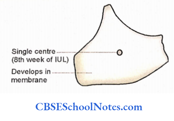
Ossification
It ossifies in the membrane from a single centre which appears in the 6th week of JUL.
Ethmoid
It is a single, irregular midline bone situated between two orbits, in the ethmoidal notch of the frontal bone.
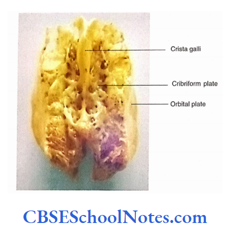
The parts, of the ethmoid can be appreciated by seeing a coronal section of the bone. It consists of a midline perpendicular plate, and two cuboidal bony masses on either side (labyrinth), which are connected to the perpendicular plate by a horizontal cribriform plate.
Perpendicular Plate
At the upper end of the perpendicular plate crista galli is present. The crista galli is seen on the floor of the anterior cranial cavity.
The perpendicular plate takes part in the formation of the median nasal septum.
Labyrinths
The right and left labyrinths are fragile, pneumatic bone.
- It consists of many small air spaces (ethmoidal air sinuses). The ethmoidal air sinuses are arranged in the anterior, middle, and posterior groups.
- The walls of these air spaces are very thin and incomplete. The labyrinth is present between nasal and orbital cavities.
- The medial wall of the labyrinth is a part of the lateral wall of the nasal cavity and is bounded by the medial plate.
From this medial plate, there arises superior and middle conchae. Deep to middle concha there is the presence of a hook-like uncinate process and a rounded elevation, the bulla ethmoidale
The lateral wall of the labyrinth is formed by the orbital plate. The orbital plate forms the part of the medial wall of the orbit.
Cribriform Plate
The cribriform plate is the horizontal plate with numerous perforations.
It connects the labyrinths with the median perpendicular plate. The cribriform plate is seen on the floor of the anterior cranial fossa on either side of the crista galli. It also forms the roof of the nasal cavity.
Articulations
- The perpendicular plate articulates with vomer, septal cartilage, frontal, nasal, and sphenoid bone.
- The cribriform plate articulates with the orbital plate and sphenoid bone.
- Labyrinth articulates with frontal, sphenoid, maxilla, lacrimal, perpendicular plate of palatine, and inferior nasal concha.
Ossification
The ethmoid bone ossifies in a cartilaginous capsule. There appear three centers, i.e., one for the perpendicular plate and one each for the labyrinth. The centers for the labyrinth appear labyrinth. The centers for the labyrinth appear All centers unite to form a single bone by the end of the third year of age.
Palatine Bone
It is situated between the maxilla and pterygoid process. It is L-shaped in appearance and consists of perpendicular and horizontal plates. It also has three processes, pyramidal, orbital, and sphenoidal.
Horizontal Plate
The horizontal plate forms the posterior part plate of palatine and inferior nasal concha anterior, posterior, lateral, and medial. The posterior border is free and projects backward in the midline to form the posterior nasal spine.
Perpendicular Plate
- It has two surfaces, maxillary and nasal, and four borders, i.e., anterior, posterior,
- All centers unite to form a single bone by greater palatine which is converted into the greater palatine canal with the help of the maxilla. This surface also forms the medial wall of the pterygopalatine fossa.
- The nasal is L-shaped in appearance (i.e., ethmoidal crest, which articulates with the middle concha of the ethmoid bone, and conchal crest articulates with the inferior concha).
- The right and left pterygoid processes. The posterior border of the perpendicular plate articulates with the medial pterygoid Body plate.
- The superior border of the perpendicular plate bears an orbital process and a sphenoidal process.
- Between these two processes, there is the presence of a sphenopalatine notch, which is converted into sphenopalatine foramen with the body of the sphenoid.
Pyramidal Process
- The pyramidal process is directed backward and laterally from the junction of horizontal and perpendicular plates.
- This process fits into a pterygoid fissure of pterygoid processes. Its inferior surface presents lesser palatine foramina.
Orbital Process
It is present at the anterior end of the upper border of the perpendicular plate. It forms the posterior part of the floor of the orbit.
Sphenoidal Process
It is situated at the posterior end of the upper border of the perpendicular plate. This process is grooved to complete the palatinovaginal canal.
Sphenoid Bone
It is a single, irregular, pneumatic bone situated posterior to the ethmoid and frontal bone. It forms the middle part of the base of the skull.
Sphenoid Bone General Features
The sphenoid consists of:
- A centrally placed body
- A pair of greater and lesser wings.
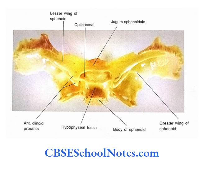
Sphenoid Bone Body
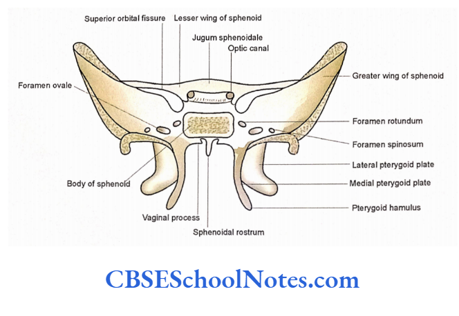
- The body of the sphenoid is cuboidal in shape and contains a pair of sphenoidal air sinuses. It has six surfaces (i.e., superior, inferior, anterior, posterior, and right and left lateral surfaces).
- The superior surface of the body bears the hypophyseal fossa. It forms the posterior part of the anterior cranial fossa (jugum sphenoidale, sulcus chiasmaticus, and tuberculum sellae) and the central part of the middle cranial fossa (hypophyseal fossa, dorsum sellae, and posterior clinoid process).
- The inferior swap of the body forms the posterior part of the roof of the nasal cavity and the roof of the nasopharynx. It shows a median ridge called a rostrum, which articulates with the grooved margin of the vomer.
- The anterior surface of the sphenoid articulates with the ethmoid. It presents a median ridge called a sphenoidal crest for articulation with the perpendicular plate of the ethmoid bone. On either side of the crest lies a thin plate of bone called sphenoidal concha. The sphenoidal foramen lies above and medial to the concha.
- The posterior surface is rough and articulates with the basilar part of the occipital bone.
- The upper part of the lateral surface is seen in the floor of the middle cranial fossa and shows a shallow groove the carotid sulcus. The lower part of the lateral surface unites with the greater wing and medial pterygoid plate.
Sphenoid Bone Wings
Sphenoid Bone Greater Wings
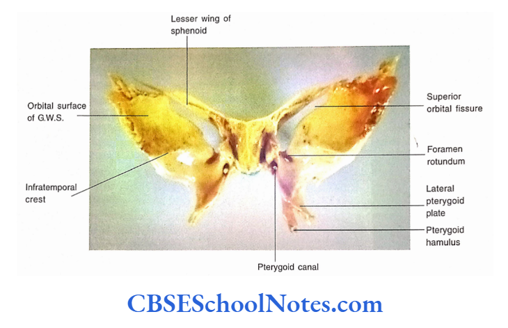
- Each greater wing has three surfaces, i.e., cerebral (upper), orbital, and lateral.
- The cerebral surface forms a part of the middle cranial fossa and is related to the temporal lobe of the cerebrum.
- Anteromedially, it has a sharp free margin, which forms the inferolateral boundary of the superior orbital fissure. This surface shows the presence of foramen rotundum, ovale, and spinosum.
- The lateral surface of the greater wing of the sphenoid is divided into the upper temporal surface and lower infratemporal surface.
- The infratemporal surface forms the roof of infratemporal fossa. This surface shows the opening of foramen ovale and spinosum.
- This surface also shows the presence of the spine of the sphenoid, which is present posterolateral to the foramen spinosum. At the junction of the temporal and infratemporal surfaces, there is the presence of an infratemporal crest.
- The orbital surface is quadrilateral in shape and forms part of the lateral wall of the orbit. This surface concerns the superior orbital fissure (above) and inferior orbital fissure.
Sphenoid Bone Lesser Wings
It extends laterally from the anterosuperior part of the body of the sphenoid.
- It is connected to the body by anterior and posterior roots. In between the two roots and the body of the sphenoid, there is the presence of the optic canal.
- The lesser wing consists of two surfaces (superior and inferior) and two borders (anterior and posterior).
- The superior surface forms the posterior-most part of the floor of the anterior cranial fossa. The inferior surface forms the superior border, of the superior orbital fissure.
- The anterior border articulates with the posterior border of the orbital plate, while the posterior border is free.
- The medial end of the posterior border ends in the anterior clinoid process.
Pterygoid Processes
- The pterygoid processes, on either side, extend downwards from the junction of the root of the greater wing and body of the sphenoid.
- Each process consists of lateral and medial pterygoid plates, which are separated posteriorly from each other by pterygoid fossa.
- Anteriorly both the plates are continuous with each other and form the posterior wall of the pterygopalatine fossa.
- Following three foramina open anteriorly in the pterygoid process—foramen rotundum, pterygoid canal, and palatinovaginal canal
Sphenoid Bone Articulations
- Body of sphenoid Ethmoid bone (anteriorly), basilar part of the occipital (posteriorly)
- The lesser living of the Orbital plate of the frontal bone sphenoid anteriorly
- Greater wing of Frontal bone (anterosphenoid medially)
- Zygomatic bone (anterolaterally)
- Parietal bone (superiorly)
- Squamous temporal bone (posterolateral) Petrous temporal (posteriorly) Pterygoid process Maxilla (anteriorly) Perpendicular plate of palatine (Medial pterygoid plate)
- Pyramidal process of palatine (Lower ends of pterygoid plates).
Sphenoid Bone Ossification
The sphenoid gets ossified partly in the membrane and partly in cartilage.
Sphenoid Bone Temporal Bone
The temporal bone is situated on each side of the base and side of the skull.
This bone consists of the following parts:
- Squamous.
- Petrosal.
- Mastoid.
- Tympanic.
- Styloid process.
Sphenoid Bone Squamous Part
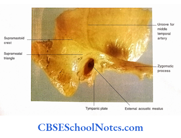
- It is a thin plate-like bone. It forms the anterosuperior part of the bone. It has external (temporal) and internal (cerebral) surfaces and a superior and an anteroinferior border.
- The external surface (on the lateral aspect) shows the zygomatic process, roots of the zygomatic process, supramastoid crest, temporal lines, and supra-meatal triangle.
- The external surface (on the inferior aspect) is made up of a small infratemporal suiface, articular tubercle, mandibularfossa and scfuamotympanic fissure.
- The internal or cerebral surface is concave and shows an impression of the gyri of the cerebrum and blood vessels.
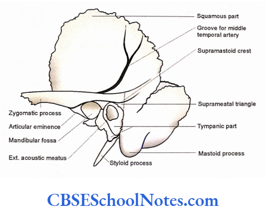
Mastoid Part of Sphenoid Bone
- It lies posterior to the squamous part. It shows external and internal surfaces and a posterior border.
- It bears a downward extending conical projection, the mastoid process.
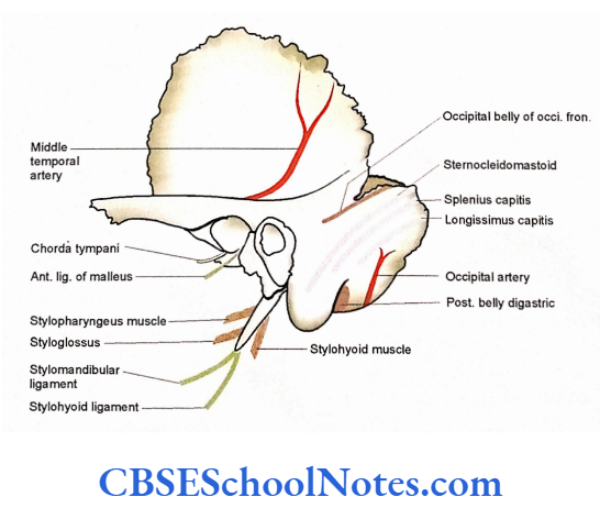
- The outer surface is smooth and may show the opening of the mastoid foramen near its posterior border.
- The inner surface is marked by a vertical groove for the sigmoid sinus.
- The medial surface of the mastoid process shows a deep groove called the mastoid notch and a shallow groove for the occipital artery.
Petrous Part of Sphenoid Bone
- This part of the bone is triangular and present at the base of the skull.
- It has a base, an apex, three borders (superior, anterior, and posterior), and three surfaces (anterior, posterior, and inferior). The bone contains the internal and middle ear cavities
Base of Sphenoid Bone
- The base of the petrous bone is directed backward and laterally and fuses with the squamous and mastoid parts.
Apex of Sphenoid Bone
It is directed forward and medially. It forms the posterior margin of the foramen lacerum.
Borders of Sphenoid Bone
- The superior border demarcates the middle cranial fossa from the posterior cranial fossa. It is grooved by the superior petrosal sinus.
- The medial part of the anterior border articulates with the greater wing of the sphenoid and the lateral part fuses with the squamous part.
- The medial part of the posterior border forms a sulcus for the inferior petrosal sinus.
- The lateral part forms the boundary of the jugular foramen.
Surfaces of Sphenoid Bone
The anterior surface of the petrous bone forms the posterior wall of the middle cranial fossa and shows the following features from medial to the lateral side trigeminal impression, hiatus for greater petrosal nerve, hiatus for lesser petrosal nerve, arcuate imminence, and tegmen tympani.
The posterior surface forms the anterior wall of the posterior cranial fossa and shows the opening of the internal acoustic meatus.
The inferior surface of the petrous bone is rough and presents the lower opening of the carotid canal and depression of the jugular fossa in front of the jugular foramen.
The Tympanic Part of Sphenoid Bone
- It is a curved plate of bone, situated anterior and inferior to the external acoustic meatus. It forms the non-articular posterior wall of the mandibular fossa.
- This plate forms the anterior, inferior, and lower part of the posterior wall of the external acoustic meatus.
- The superior border of the tympanic plate meets the squamous temporal bone at the squamotympanic fissure. The inferior border forms a sheath for the base of the styloid process.
- The rough lateral margin of the tympanic plate gives attachment to the cartilaginous part of the external acoustic meatus.
The Styloid Process of Sphenoid Bone
The styloid process is a conical projection of about 2.5 cm long. It is present on the inferior aspect of the temporal bone.
It is directed downwards forward and medially. The root of the styloid process is ensheathed by the tympanic plate. The stylomastoid foramen is situated between the styloid and mastoid process.
Side Determination and Anatomical Position Hold the bone in such a way that:
- The zygomatic process faces anteriorly.
- External acoustic meatus laterally.
- The mastoid process faces backward and downwards
Articulations of Sphenoid Bone
Squamous part – With greater wing of sphenoid anteriorly With the parietal bone superiorly With the head of the mandible at the temporomandibular joint.
Petrous part – With the greater wing of the sphenoid anteriorly With the occipital bone posteriorly
Mastoid part- With the parietal bone superiorly With the occipital bone posteriorly and medially.
Zygomatic Process- With the temporal process of zygomatic bone.