
Disorders Of Bones and Joints
Rheumatoid Arthritis
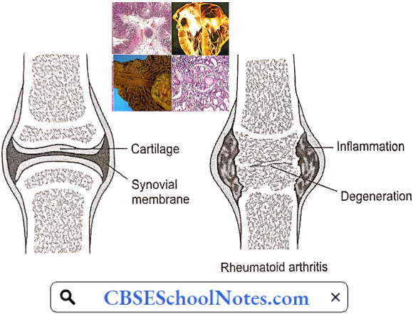
Rheumatoid arthritis (RA) is characterized by inflammation and swelling of the synovial membrane of the joint, with subsequent destruction of articular structures. RA affects about 1% of the Indian population.
Rheumatoid Arthritis Aetiology: Rheumatoid arthritis is an autoimmune disorder of the synovial joints and other organs of the body.
Rheumatoid Arthritis Symptoms: Signs and symptoms of rheumatoid arthritis may include
- Tender, warm, swollen joints
- Joint stiffness that is usually worse in the mornings
- Fatigue
- Fever
- Loss of appetite
Read and Learn More Pathophysiology
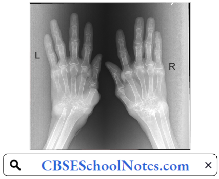
Early rheumatoid arthritis tends to affect smaller joints first—particularly the joints that attach fingers to the hands and toes to the feet. As the disease progresses, symptoms often spread to the wrists, knees, ankles, elbows, hips, and shoulders.
In most cases, symptoms occur in the same joints on both sides of the body. Loss of movement and erosion of the joint surface lead to deformity and loss of function of the joint. About 40 percent of the people who have rheumatoid arthritis also experience signs and symptoms in other body systems, cardiovascular system in particular.
Rheumatoid Arthritis Pathophysiology: RA typically manifests with signs of inflammation of the synovial membrane. The affected joint is swollen, warm, painful, and stiff, particularly early in the morning on waking. The resulting inflammation thickens the synovial membrane, which can eventually destroy the cartilage and bone within the joint.
- As the pathology progresses, the inflammatory activity leads to tendon damage as well as erosion and destruction of the joint surface, which increases the range of movement and leads to deformity. Gradually the joint loses its shape and alignment.
- Destruction and fusion of bones of the wrist (carpal bones) is commonly observed. The skin, bones, and muscles adjacent to the joints atrophy from disuse and destruction.

Rheumatoid Arthritis Risk Factors
- Sex: Women are more likely than men to develop rheumatoid arthritis.
- Age: Rheumatoid arthritis can occur at any age, but it most commonly begins in middle age.
- Family History: The risk of developing RA is greater, if there is a family history of the disease.
- Smoking: Cigarette smoking increases the risk of developing rheumatoid arthritis. Moreover, smoking also appears to be associated with greater disease severity.
Rheumatoid Arthritis Complications
- Osteoporosis, partly because of drugs used in the treatment of the disorder.
- Coronary artery disease
- Eye inflammation
- Stroke
- Anemia
- Lung fibrosis
- Depression
Osteoarthritis
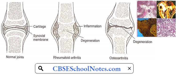
Osteoarthritis is the most common chronic musculoskeletal disorder. Epidemiological studies estimate that it affects about 15% of the world’s population. In contrast to the inflammatory cause of rheumatoid arthritis, osteoarthritis is a degenerative disease.
It occurs when the protective cartilage that cushions the ends of your bones wears down over time. The most common joints affected by osteoarthritis are the small joints of the hands and feet, the hip joint, and the knee joint.
Osteoarthritis Symptoms
- Pain or aching in the joint during activity, after a long activity, or at the end of the day.
- Joint stiffness usually occurs first thing in the morning or after resting.
- Limited range of motion that may go away after movement.
- Clicking or cracking sound when a joint bends.
- Swelling around a joint.
- Muscle weakness around the joint.
- Joint instability or buckling (knee gives out).
Osteoarthritis Aetiology: Osteoarthritis occurs when the cartilage that cushions the ends of bones in the joints gradually deteriorates. Cartilage is a firm, slippery tissue that enables nearly frictionless joint motion. Eventually, if the cartilage wears down completely, the bone will mb on bone.
Osteoarthritis Pathogenesis: Earlier, osteoarthritis has been considered to be a disease involving wear and tear of articular cartilage. Remnants of degenerated cartilage are found in the joint. Recent research has indicated that tire condition involves the entire joint.
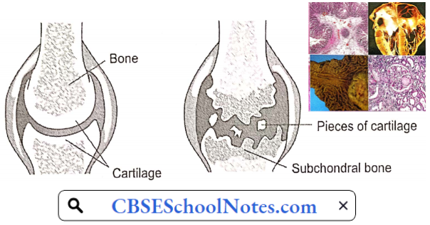
The loss of articular cartilage is the primary change, but a combination of cellular changes and biomechanical stresses causes several secondary changes, including subchondral bone remodeling, the formation of new bone at the joint margins called osteophytes and degenerative changes in the joint capsule, ligaments, and periarticular muscles.
Osteoarthritis Risk Factors: Factors that can increase the risk of osteoarthritis include
- Older Age: The risk of osteoarthritis increases with age.
- Sex: Women are more likely to develop osteoarthritis, though it is not clear why.
- Obesity: Carrying extra body weight contributes to osteoarthritis. The greater the body weight, the greater is the risk of osteoarthritis. Increased weight adds stress to weight¬bearing joints, such as hips and knees.
- Joint Injuries: Injuries, such as those that occur when playing sports or from an accident, can increase the risk of osteoarthritis.
- Repeated Stress On The Joint: If the job or a sport places repetitive stress on a joint, that joint might eventually develop osteoarthritis.
- Genetics: Some people inherit a tendency to develop osteoarthritis.
Osteoarthritis Complications
- Osteoarthritis is a degenerative disease that worsens over time, often resulting in chronic pain. Joint pain and stiffness can become severe enough to make daily tasks difficult.
- Depression and sleep disturbances can result from the pain and disability of osteoarthritis.
Osteoporosis
Osteoporosis is characterized by reduced bone mass and the disruption of normal bone architecture. The result is the fragility of bone that is easily fractured. Osteoporosis occurs in both men and women, but it sets in early and is more severe in women.
Around the world, 1 in 3 women and 1 in 5 men aged 50 years and over are at risk of an osteoporotic fracture. As explained below, Indians have lower bone mass as adults and hence are more prone to develop osteoporosis.

Osteoporosis seen in cases with immobilization or hormonal disorders such as Cushing syndrome or thyrotoxicosis is known as secondary osteoporosis. Senile osteoporosis in men and postmenopausal osteoporosis is far more common.
Osteoarthritis Symptoms: There typically are no symptoms in the early stages of bone loss. But once bones have been weakened by osteoporosis, signs and symptoms include:
- Back pain, caused by a fractured or collapsed vertebra
- Loss of height over time
- A stooped posture
- A bone that breaks much more easily than expected
Hip fractures often are caused by a fall and can result in disability and even an increased risk of death within the first year after the injury.
Osteoarthritis Aetiology And Risk Factors: The exact cause of senile/postmenopausal osteoporosis is not known. However, certain risk factors have been identified
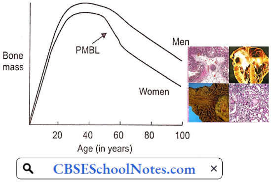
- Peak Bone Mass: Peak bone mass has been identified as one of the most important factors in the pathogenesis of osteoporosis. Maximum bone mass is attained in both men and women at around the age of 30 years. A failure to attain optimal bone strength by this point is one factor that contributes to osteoporosis later in life.
- Bone is a living tissue. All the time some part of the bone is being resorbed and new bone is laid out. This process is known as bone remodeling. After the age of 30 years, the remodeling process involves slightly greater bone resorption than bone deposition.
- Thus, age-related bone loss gradually sets in. By the age of 60 years in men, and approximately a decade earlier in women, the loss of bone mass becomes significant. Osteoporosis sets in earlier in those with low peak bone mass.
- The racial difference in peak bone mass is reflected in the incidence of severe osteoporosis. In the American population, peak bone mass is significantly greater in blacks than whites. The incidence of fractured femoral neck (severe osteoporosis) is significantly greater in whites than blacks.
- Indians in general have low peak bone mass. Twin and family studies have shown that genetic factors play an important role in regulating bone mineral density and bone turnover.
- Gonadal Hormonal Deficiency: Ageing and loss of gonadal function are the 2 most important factors contributing to the development of osteoporosis. Estrogen or testosterone deficiency, regardless of age of occurrence, results in accelerated bone loss.
- The exact mechanisms of this bone loss is not clear. Whatever the mechanism, ultimately there is an increased bone resorption, which outpaces bone formation. Studies have shown that bone loss in women accelerates rapidly in the first years after menopause.
- Blood gonadal hormone levels decline with age both in males and females. However, the postmenopausal precipitous fall in blood estrogen levels observed in females does not occur in blood testosterone levels in males; blood testosterone levels in males decline more gradually.
- Dietary Calcium And Vitamin D: Dietary calcium and vitamin D intake during the first three decades of life is an important factor since it determines the value of peak bone mass.
- Exercise: At any age, immobilization with decreased weight bearing leads to rapid bone resorption. Physical activity, muscle strength, and body weight seem to be important determinants of bone strength. Osteoporotic subjects in general are less muscular and have lower body weight.
- Excessive Intake Of Acids: High acid intake, particularly in the form of a high protein diet may contribute to the development of osteoporosis; bone is involved in buffering the acid load. Acidosis may also increase the osteoclastic activity by a direct action.
Osteoarthritis Complications: Bone fractures, particularly in the spine or hip, are the most serious complications of osteoporosis.
Gout
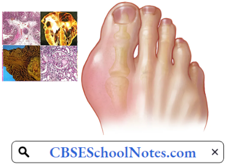
Gout is characterized by sudden, severe attacks of pain, swelling, redness, and tenderness in the joints, often the joint at the base of the big toe. In Western countries, gout occurs in 3-6% of men and 1-2% of women. In India, the prevalence of gout is much less, around 0.1%.
Gout Symptoms And Signs: The signs and symptoms of gout almost always occur suddenly, and often at night. They include
- Intense Joint Pain: Gout usually affects the large joint of the big toe, but it can occur in any joint. Other commonly affected joints include the ankles, knees, elbows, wrists, and fingers. The pain is likely to be most severe within the first four to 12 hours after it begins.
- Inflammation And Redness: The affected joint or joints become swollen, tender, warm, and red.
- Lingering Discomfort: After the most severe pain subsides, sore joint discomfort may last from a few days to a few weeks. Paler attacks are likely to last longer and affect more joints.
- Limited Range Of Motion: As gout progresses, movement in the joint becomes limit
Gout Causes: Clout occurs when urate crystals accumulate in a joint, causing inflammation and intense pain Urate crystals can form when a patient has a high serum uric acid level.
Gout Pathogenesis: Uric acid is normally produced in our body in the catabolism of purines. Purines are also found in certain foods, such as organ meats and seafood. Other foods also promote higher levels of uric acid, such as alcoholic beverages, especially beer, and drinks sweetened with fructose.
- Normally, uric acid remains dissolved in the blood and excreted by the kidneys into the urine. But sometimes serum uric acid rises due to either greater uric acid production in the body, or more often, decreased excretion by the kidneys.
- When this happens, uric acid can be deposited as sharp, needle-like urate crystals in a joint or surrounding tissue that cause pain, inflammation, and swelling.
- These crystals initiate the inflammatory process by being engulfed by synovial phagocytic cells leading to the release of lysosomal enzymes and the production of inflammatory chemokines. The chemotactic factors produced by monocytes and mast cells and the local vasodilatation stimulate neutrophilic chemotaxis.
- Also, endothelial cell activation further aggravates the inflammatory response and migration of neutrophils. The chemokines attract neutrophils into the synovial tissue and the synovial fluid.
The way to ultimately correct the underlying metabolic problem of hyperuricemia and the crystal deposition is to lower the serum urate level and dissolve the crystal deposits by medicines. This will stop both the acute attacks and the progressive joint damage.

Gout Risk Factors: Factors that increase the uric acid level in the body include
- Diet: Eating a diet rich in meat and seafood and drinking beverages sweetened with fruit sugar (fructose) increases levels of uric acid. Alcohol consumption, especially beer, also increases the risk of gout.
- Obesity: If one is overweight, the body produces more uric acid.
- Medical Conditions: Certain diseases increase the risk of gout. These include hypertension, diabetes, metabolic syndrome, and heart and kidney diseases.
- Certain Medications: The use of thiazide diuretics—commonly used to treat hypertension—and low-dose aspirin also can increase uric acid levels.
- Family History Of Gout
- Age And Sex: Gout occurs more often in men, primarily because women tend to have lower uric acid levels. After menopause, however, women’s uric acid levels approach those of men. Men are also more likely to develop gout earlier—usually between the ages of 30 and 50, whereas women generally develop signs and symptoms after menopause.
Gout Complications: People with gout can develop more severe conditions, such as
- Joint Destruction: If left untreated, gout can cause erosion and destruction of a joint.
- Urate Tophi: Untreated gout may cause deposits of urate crystals to form under the skin in nodules called tophi. Tophi can develop in several areas such as your fingers, hands, feet, elbows, or Achilles tendons.
- Kidney Stones: Urate crystals may collect in the urinary tract of people with gout, causing kidney stones.