
Blood Supply Of The Brain And Spinal Cord
The brain is supplied by a pair of vertebral and a pair of internal carotid arteries. In the cranial cavity, both pairs of arteries divide extensively into many branches, some of which are anastomose with each other.
The spinal cord is about a 45-cm long structure; hence, many arteries supply it. The spinal cord is supplied by branches of vertebral arteries and also by segmental arteries, i.e. intercostal and lumbar segmental arteries.
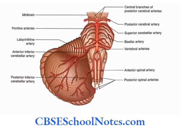
Vertebral Arteries. Each vertebral artery enters the cranial cavity through the foramen magnum after piercing the dura and arachnoid mater.
It curves around the ventrolateral aspect of the medulla and at the lower border of pons it joins with its fellow, from the other side, to form a median basilar artery. The branches of the vertebral artery are given in the following text.
Read and Learn More Neuroanatomy
- Posterior spinal artery: The posterior spinal artery on either side passes inferiorly along the spinal cord, among the dorsal rootlets of spinal nerves.
- Posterior inferior cerebellar artery: The posterior inferior cerebellar artery is the largest branch of the vertebral artery and arises near the lower end of the olive. Had) artery runs posteriorly on either side of the medulla. 1 lore, it supplies the choroid plexus of the fourth ventricle and reaches the cerebellum to supply the posterior part of its inferior surface
- Anterior spinal artery: The anterior spinal artery arises near the inferior border of the pons. It descends anteromedially in front of the medulla and joins its fellow, from the opposite side, to form a single trunk. It is the single trunk of the anterior spinal artery that runs in the anterior median fissure throughout the length of the spinal cord and supplies it
Basilar Artery and Its Branches: The basilar artery runs cranially in the median groove on the pons.
At the superior border of the pons, it divides into two terminal branches:
- Right and
- left posterior cerebral arteries.
The branches of the basilar artery are as follows:
Arteries of the labyrinth: On each side, these arteries arise close to the inferior border of pons and run along with the vestibulocochlear nerve towards the internal ear.
Anterior inferior cerebellar arteries: These arteries arise from the lower part of the basilar artery, on each side, and supply the anterior part of the inferior surface of the cerebellum.
Pontine arteries: These are many small branches that supply pons and the adjacent parts of the brain.
Superior cerebellar artery: On each side, the superior cerebellar artery arises near the upper border of the pons. It supplies the upper pons, lower midbrain and superior surface of the cerebellum.
Posterior cerebral arteries: These arteries are two large terminal branches of the basilar artery, which arise at the superior border of the pons.
Each artery breaks up into branches to supply the temporal and occipital lobes.
The branches of the posterior cerebral arteries are as follows:
Central branches: These are numerous small branches which enter the cerebrum through the posterior perforated substance between two crue
Internal Carotid Artery (ICA). After passing through the cavernous sinus, the ICA pierces the dura mater medial to the anterior clinoid process.
Here, the artery lies immediately inferior to the anterior perforated substance and lateral to optic chiasma.
The ICA has two major branches—anterior and middle cerebral arteries which are its terminal branches and middle cerebral arteries which are its terminal branches ICA also gives smaller branches—ophthalmic, posterior communicating and anterior choroidal arteries.
Anterior cerebral artery: The anterior cerebral artery is a smaller terminal branch of the ICA. After its origin, the right and left anterior cerebral arteries come close to each other and are joined by the anterior communicating artery.
The anterior cerebral artery now ascends in the longitudinal fissure and runs posteriorly along the corpus callosum. The artery gives central and cortical branches.
Central branches: They form the anteromedial group which enters deep inside the cerebral hemisphere by passing through the anterior perforated substance and lamina terminalis. These branches supply the rostrum, septum pellucidum and corpus.
Middle cerebral artery: The middle cerebral artery is a larger terminal branch and a more direct continuation of the ICA. It runs in the stem of the lateral sulcus, between the frontal and temporal lobes, and then turns laterally in the posterior ramus of the lateral sulcus. It gives central and cortical branches.
Central branches: They form the anterolateral group and enter the deeper part of the cerebral hemisphere through the anterior perforated substance. These central branches are grouped into medial and lateral striate arteries.
The medial striate arteries ascend through the lentiform nucleus and supply the lentiform nucleus, internal capsule and caudate nucleus The lateral striate arteries ascend through the external capsule.
Then, they turn medially to supply the lentiform and caudate nuclei along with the internal capsule.
One of the larger lateral striate arteries is more susceptible to rupture; hence, it is called the artery of cerebral haemorrhage (Charcot’s artery of cerebral haemorrhage).
Cortical branches: They emerge in the superolateral surface of the cerebral hemisphere between the lips of the lateral sulcus.
These cortical branches supply most of the superolateral surface and the adjacent part of the orbital surface, tentorial surface and temporal pole.
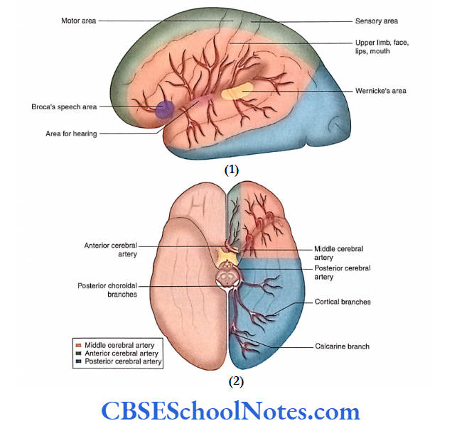
This artery is responsible for supplying the motor and premotor areas, primary sensory and auditory areas in both hemispheres. The left cerebral hemisphere supplies Wernicke’s language area and Broca’s area of speech.
Occlusion of Cortical Branches of the Middle Cerebral Artery
- The thrombosis or occlusion of the cortical branches of the middle cerebral artery will lead to motor paralysis and sensory loss of the opposite half of the body above the leg.
- Involvement of motor and sensory speech areas of the left cerebral hemisphere (in right-handed persons) will result in aphasia (loss of speech).
- The thrombosis of the middle cerebral artery may also involve the auditory area with little hearing loss, as auditory impulses are projected to both the cerebral hemispheres.
- Occlusion of central branches may lead to hemiplegia
because of infarction of motor fibres in the internal capsule.
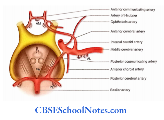
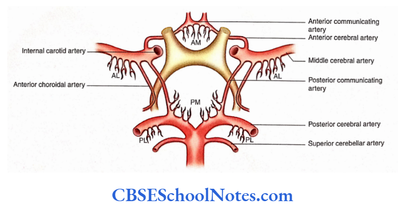
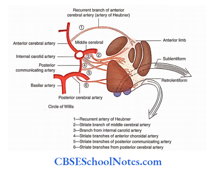
Circulus Arteriosus Circle Of Willis
Circulus arteriosus is a polygonal circle of arteries, present in the interpeduncular fossa. It is formed by posterior cerebral, posterior communicating, internal carotid, anterior cerebral and anterior communicating arteries.
Formation: The circulus arteriosus is bounded posteriorly by two posterior cerebral arteries that communicate with the middle cerebral artery on each side by the posterior communicating branch.
Anteriorly, two anterior cerebral arteries communicate with each other through the anterior communicating artery.
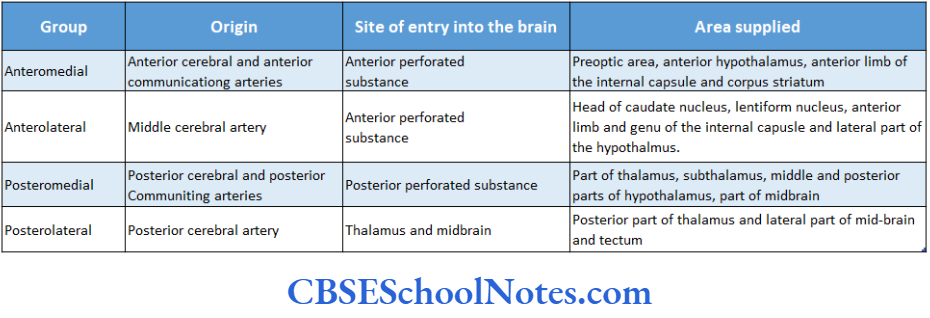
Branches: The circle of Willis gives origin to many slender central branches.
These arteries arise in four groups:
Anteromedial, Anterolateral, Posteromedial and Posterolateral.
These branches immediately pierce the surface of the brain (mostly through anterior and posterior perforated substances) to supply its internal parts such as the corpus striatum, thalamus, internal capsule and midbrain.
These arteries do not anastomose significantly within the brain substance. As these arteries act as end arteries, the damage to the branches entering the substance of the brain leads to the destruction of the brain tissue.
Significance of Circle of Willis
This arterial anastomosis acts as a route through which blood entering the ICA or the basilar artery may be distributed to any part of the cerebral hemisphere.
If one of the major arteries forming the circle of Willis is blocked, then this arterial anastomosis provides an alternative route through which blood can be supplied to the area of the blocked artery.
Arterial Supply Of The Individual Parts Of The Brain
After studying the major arteries and their branches supplying the brain, we shall now study the arterial supply Of the individual parts of the brain as follows
- Arterial supply of the cerebral cortex
- Arterial supply of the internal structures of the cerebral hemisphere
- Arterial supply of brainstem
- Arterial supply of the cerebellum
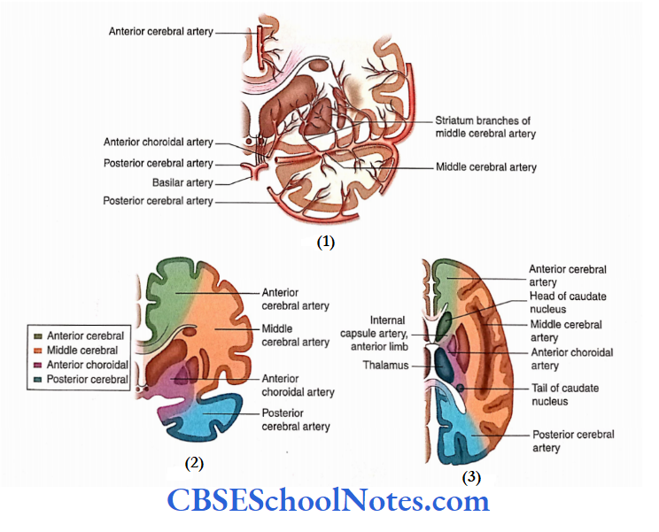
Arterial Supply Of The Cerebral Cortex
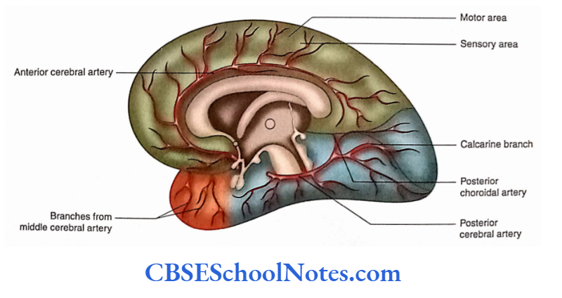
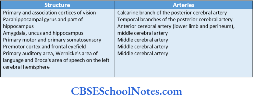
- The cortical branches of three cerebral arteries (anterior, middle and posterior) ramify on the medial, superolateral and inferior surfaces of the cerebral cortex and supply it
- Arterial Supply of Motor and Sensory Areas supply most of the superolateral surface and supply most of the superolateral surface and the temporal pole.
- The middle cerebral artery is responsible for supplying the motor and premotor areas, primary sensory and auditory areas, in both hemispheres. In the left cerebral hemisphere, it supplies Wernickes’ language area and Broca’s area of speech.
- The cortical branches of the anterior cerebral artery supply the motor and sensory areas for the leg and perineum of the opposite side. These areas are present on the medial surface of the cerebral hemisphere.
- The cortical branches of the posterior cerebral arteries supply the visual cortex (area 17) in the occipital lobe.
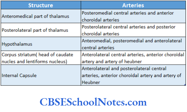
Arterial Supply Of The Internal Structures Of The Cerebral Hemisphere
The central branches enter the cerebral hemisphere and supply the thalamus, hypothalamus, corpus striatum and internal capsule.
Arterial Supply of Brainstem
The medulla is supplied by the anterior and posterior spinal arteries and the posterior inferior cerebellar arteries.
The pons are supplied by the basilar artery, anterior inferior cerebellar arteries and superior cerebellar arteries. The midbrain is supplied by the basilar, superior cerebellar and posterior cerebral arteries.
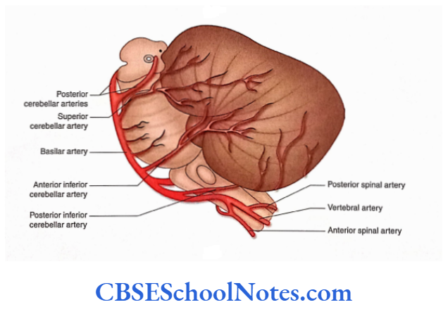
Arterial Supply Of Cerebellum
The superior and inferior surfaces of the cerebellum are supplied by the superior cerebellar and inferior cerebellar arteries.
Both of these arteries are branches of the basilar artery. The inferior surface is also supplied by the posterior inferior cerebellar artery which is a branch of the vertebral artery.
Venous Drainage of the Brain
The veins draining the brain are thin-walled because their walls are devoid of muscles. They are also without valves as functionally they have to drain towards the direction of gravity.
These veins drain their blood into dural venous sinuses after piercing through the arachnoid mater and inner layer of the dura mater.
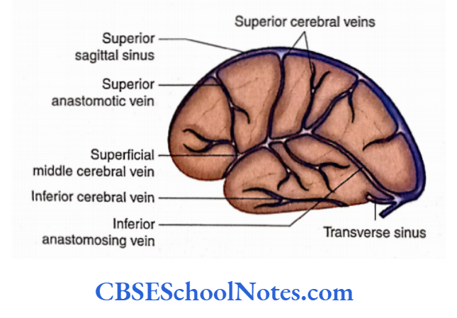
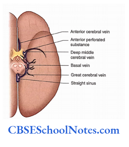
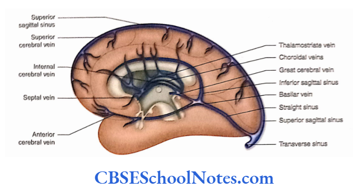
The cerebral veins are classified into external and internal cerebral veins.
The external cerebral veins lie in the subarachnoid space on the surface of the cerebral hemisphere. These are superior, middle and inferior cerebral veins.
The internal cerebral veins, on the other hand, drain the internal parts of the cerebrum and ultimately pour their blood into the great cerebral vein.
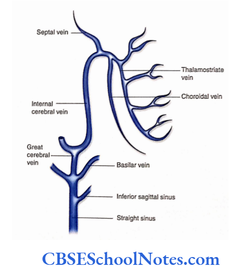
The internal cerebral vein is formed near the interventricular foramen by the union of the thalamostriate vein and choroid veins.
Internal Cerebral Veins
The internal cerebral vein is formed near the interventricular foramen by the union of the thalamostriate vein and choroid veins.
The right and left internal cerebral veins run posteriorly in the transverse fissure where they unite beneath the splenium to form the great cerebral vein.
This vein receives the right and left basal veins and then opens into the straight sinus.
Blood-Brain Barrier
The endothelial lining of brain capillaries does not permit some substances to pass from blood to brain tissue.
The barrier protects the delicate brain tissues from harmful (toxic) substances, lie blood-brain barrier is highly permeable to water, glucose, lipid-soluble substances, O2, C02 and drugs such as alcohol, coffee, nicotine and anaesthetics.
This barrier is, however, an obstacle to delivering drugs such as antibiotics and cancer drugs.
The following structures form the blood-brain barrier:
- The endothelial lining of capillary: There is the presence of tight junctions between endothelial cells.
- The basal lamina of endothelium
- The perivascular end feet of astrocytes on the basal lamina

The barrier is mainly formed by the endothelial cells resting on the basal lamina and not by the astrocytes as they do not fully surround the capillary. However, the foot processes stimulate the formation of tight junctions between endothelial cells.
Blood Supply Of The Spinal Cord
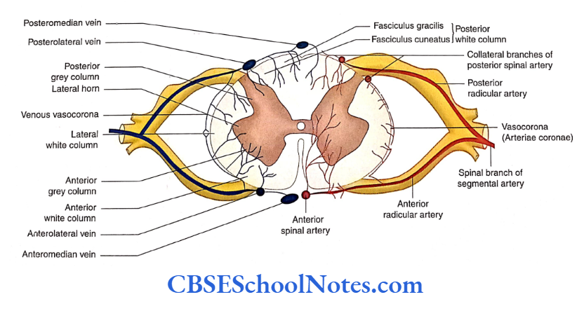
The spinal cord is a long structure present in the vertebral canal. Because of its length, the spinal cord is supplied by many arteries. It is drained by longitudinally arranged veins which further drain into radicular veins.
Arterial Supply
The arteries supplying the spinal cord can be grouped into the following:
- Branches of the vertebral arteries, i.e. one anterior and two posterior spinal arteries.
- Multiple spinal branches of segmental arteries
- Radicular arteries
Branches of the vertebral arteries: As the right and left vertebral arteries lie on the anterolateral surface of the medulla, each artery gives two branches that supply the spinal cord. These are the anterior and posterior spinal arteries.
The anterior two-thirds of the spinal cord is supplied by the anterior spinal artery while the posterior one-third is by the posterior spinal arteries.
Segmental arteries: As the spinal cord is a long structure, many segmental arteries supply it.
The spinal branches of the segmental arteries (vertebral, deep cervical, posterior intercostals and lumbar arteries) enter the vertebral canal through the corresponding intervertebral foramen.
Radicular arteries: Each spinal artery divides into anterior and posterior radicular arteries, which run towards the spinal cord along the ventral and dorsal nerve roots, respectively.
The total number of anterior radicular arteries varies from 12 to 17 (mostly six in cervical, two to four in thoracic and two to three in lumbar segments).
Out of these radicular arteries, a single anterior radicular artery is very large and is called the arteria radicularis magna (artery of Adamkiewicz) which usually arises from the lower thoracic or upper lumbar level.
Venous Drainage
Six longitudinally running veins that drain the spinal cord are as follows:
- One along the anterior median fissure
- One along the posterior median sulcus
- Two along the line of right and left ventral nerve rootlets
Two along the line of right and left dorsal nerve rootlets All these veins freely communicate with each other and are drained by anterior and posterior radicular veins.
Blood Supply Of The Brain And Spinal Cord Summary
- The brain has a rich blood supply. It is supplied by a pair of vertebral arteries and a pair of internal carotid arteries.
- The branches of vertebral arteries in the cranial cavity are posterior spinal, posterior inferior cerebellar and anterior spinal arteries.
- Two vertebral arteries join each other to form a median basilar artery at the lower border of the pons.
- The branches of the basilar artery are the artery of the labyrinth, anterior inferior cerebellar artery, pontine arteries and superior cerebellar arteries. It terminates in two posterior cerebral arteries.
- The circle of Willis is formed by the posterior cerebral, posterior communicating, anterior cerebral and anterior communicating arteries.
- The circle of Willis gives origin to many central branches. These arteries arise in four groups:
- Anteromedial,
- Anterolateral,
- Posteromedial and
- Posterolateral.
- Various surfaces of the cerebral cortex are supplied by the cortical branches of the anterior, middle and posterior cerebral arteries.
- The superolateral, medial and inferior surfaces are supplied by all three cerebral arteries:
- Anterior, Middle and Posterior cerebral arteries.
- The corpus striatum and internal capsule are supplied by central branches of the anterior and middle cerebral arteries.
- The thalamus is supplied by the central branches of the posterior cerebral artery and basilar arteries.
- The veins of the brain are grouped as external cerebral veins and internal cerebral veins.
- The veins of the brain drain into intracranial dural venous sinuses.
- The blood-brain barrier is formed by capillary endothelial cells and their basement membrane.
- The spinal cord is supplied by branches of vertebral arteries, namely the anterior spinal artery and two posterior spinal arteries.
- The spinal cord is also supplied by multiple spinal branches of segmental arteries.
- Six longitudinally running veins drain the venous blood from the spinal cord.
Blood Supply Of The Brain And Spinal Cord Multiple Choice Questions
Question 1. Which of the following is not a branch of the vertebra artery?
- Posterior spinal arteries
- Anterior spinal artery
- Superior cerebellar artery
- Posterior inferior cerebellar artery
- Anterior inferior cerebellar artery
Answer: 3. Posterior inferior cerebellar artery
Question 2. Which of the following are the branches of the basilar artery?
- Anterior inferior cerebellar artery
- Pontine artery
- Superior cerebellar artery
- Posterior cerebral artery
- All of the above
Answer: 5. All of the above
Question 3. The Circle of Willis is formed by the following arteries except
- Posterior cerebellar artery
- Post communicating artery
- Anterior cerebral artery
- Middle cerebral artery
- Anterior communicating artery
Answer: 4. Anterior communicating artery
Question 4. Which of the following arteries supply the internal capsule?
- Anterolateral central arteries
- Posterolateral central arteries
- Anterior choroidal artery
- Artery of Heubner
- All of the above
Answer: 5. All of the above
Question 5. Which of the following are the group of central arteries?
- Anteromedial
- Anterolateral
- Posteromedial
- Posterolateral
- All of the above
Answer: 5. All of the above
Question 6. Which of the following is not a tributary of the internal cerebral vein?
- Septal vein
- Thalamostriate vein
- Choroidal vain
- Great cerebral vein
Answer: 4. Great cerebral vein