
Basal Nuclei
The basal nuclei, or the basal ganglia, are large masses of grey matter situated in the subcortical regions.
They are considered a part of the extrapyramidal system. This is because the motor functions of basal nuclei are independent of the activity of the pyramidal system. They play an important role in voluntary movements.
Parts Of Basal Nuclei
Basal nuclei consist of the following parts or nuclei:
- Caudate nucleus
- Lentiform nucleus, further divided into putamen and globus pallidus
- Subthalamic nucleus
- Substantia nigra
All the above nuclei are grouped under the heading ‘basal nuclei’.
Read and Learn More Neuroanatomy
This is because all of them are interconnected to form a single functional unit. A lesion in any of these nuclei results in motor disorders.
1. Caudate nucleus: The caudate nucleus is an arch-shaped mass of grey matter that surrounds the thalamus.
It is present in the lateral ventricle divided into three parts:
- Head,
- Body and
- Tail
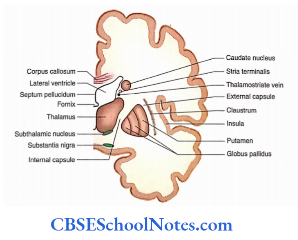
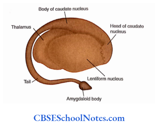
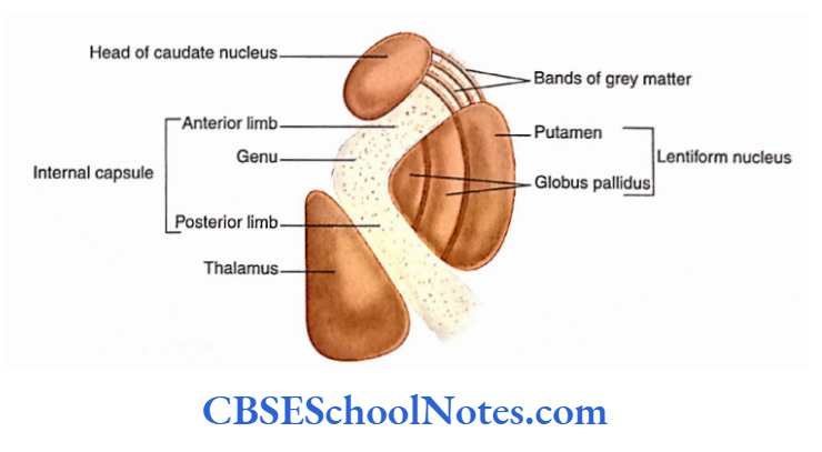
Head: The head of the caudate nucleus is a large, rounded mass present on the floor of the anterior horn of the lateral ventricle.
Laterally, the head is related to the anterior limb of the internal capsule and putamen.
Bands of grey matter, running across the anterior limb of the internal capsule, connect the head of the caudate nucleus to the putamen.
Body: The body of the caudate nucleus is narrow and long and forms the central part of the caudate nucleus.
It lies on the floor of the central part of the lateral ventricle.
Tail: The tail of the caudate nucleus is formed by the body of the caudate nucleus as it curves downwards and forwards, ‘I lie lall Is long and slender and lies In the roof of die Inferior horn of die lateral ventricle. Anteriorly, the tail hi continuous with the amygdaloid body.
2. lentiform nucleus: The lentiform nucleus Is a wedge-shaped (convex) mav of grey matter that lies deeply hurled in the cerebral hemisphere,
Unlike the caudate nucleus, the lentiform nucleus is not related to the lateral ventricle.
Medially, the die lentiform nucleus is related to the internal capsule and laterally to the external capsule.
Antcroinfcriorly, the heads of caudate and lentiform nuclei are continuous with each other.
The lentiform nucleus is further divided into two parts:
- Medial ylobus pallidum and
- Lateral putamen.
3. Subthalamic nucleus: The subthalamic nucleus lies just below the thalamus and above the substantia nigra. It is a small, biconvex mass of grey matter that lies intermedia! to Globus pallidum.
4. Substantia nigra: The substantia nigra is a large motor nucleus present in the midbrain. The dopamine synthesized by the substantia nigra is used as a neurotransmitter in the corpus striatum.
The deficiency of dopamine leads to a condition called Parkinson’s disease characterized by diminished movements.
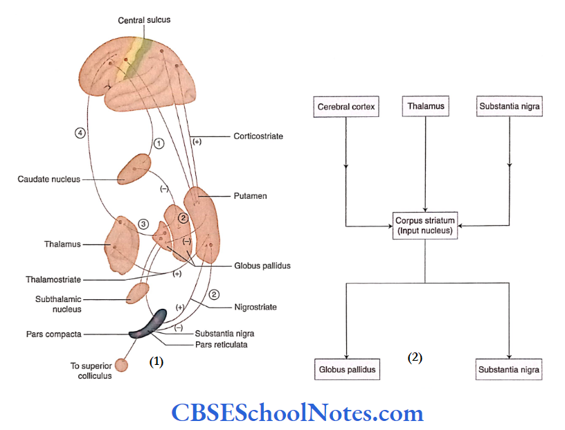
The caudate nucleus and putamen are histologically identical and are collectively known as corpus striatum.
It is recent in phylogeny; hence, it is also called neostriatum. The Globus pallidum, phylogenetically, is an old structure; hence, it is called palaeostriatum.
Connections Of Basal Nuclei
- The corpus striatum (caudate nucleus and putamen receives almost all the inputs (afferents) of the basal nuclei,
- The axons of the striatum project to globus pallidus and substantia nigra.
- The axons from globus pallidus and substantia nigra constitute the major output (efferent) from the basal nuclei.
Connections of Corpus Striatum (Input Nucleus)
Afferents
The afferents of basal nuclei terminate in the corpus striatum (i.e. caudate nucleus and putamen.
From the cerebral cortex: Corticostriate fibers especially from the frontal and parietal lobes, terminate in various parts of the striatum. Thus, information regarding motor movements reaches the basal nuclei from the premotor and motor areas of the cerebral cortex.
From the thalamus: Thalamostriate fibers originate from the intralaminar nuclei of the thalamus and project to the striatum
From the substantia nigra: Nigrostriate fibers originate in the pars compacta of the substantia nigra and terminate in the putamen. These fibers use dopamine as a neurotransmitter.
Efferents
The efferents from the corpus striatum go to the output nuclei—globus pallidus and substantia nigra. These afferents are called striatopallidal and striatonigral fibers, respectively.
Both striatopallidal and striatonigral fibers are inhibitory and use GABA as their neurotransmitter.
To globus pallidus: Striopallidal fibers begin in the putamen and caudate nucleus and terminate in both the outer and the inner parts of globus pallidus.
To substantia nigra: Strionigral fibers originate in the putamen and terminate in both parts of the substantia nigra (pars reticulata and pars compacta).
Connections of Output Nuclei
The output nuclei (globus pallidus and substantia nigra) receive their afferents from the corpus striatum.
Efferent Connections
The efferent connections of globus pallidus and substanti nigra go to the thalamus, subthalamic nucleus, superior colliculus, reticular nuclei and habenular nucleus.
To the thalamus: terminate in the thalamus. The axons from the nuclei of the thalamus then terminate in the motor areas of the cerebrum. Thus, basal nuclei exert their influence on the motor areas of the cerebral cortex through the thalamus.
To the subthalamic nucleus: The fibers of globus pallidus terminate in the subthalamic nucleus. The fibers from the subthalamic nucleus, in turn, terminate on the internal segment of the globus pallidus.
To the superior colliculus: The fibres ofsubstantia nigra terminate on the superior colliculus. This connection controls eye movements
To the habenular nucleus: The fibers from the pallidum terminate in the habenular nucleus. The basal nuclei can modify the limbic system through these connections.
Functions Of Basal Nuclei
The basal nuclei have motor as well as cognitive and behavioral functions.
Motor Functions
The basal nuclei are a part of the extrapyramidal system and are concerned with regulation of the voluntary muscular activities. The important motor functions of
basal nuclei are as follows
Programming of voluntary movements: Instructions for learned muscular movements are stored in basal nuclei.
Regulation of automatic movements: The caudate nucleus and putamen control the automatic movements of skeletal muscles, such as swinging the arm while walking and laughing in response to a joke.
Regulation of muscle tone: The globus pallidus helps regulate the muscle tone required for a specific body movement.
Effect on the functions of reticular formation: This nucleus is involved in stereotyped motor functions such as locomotion.
Cognitive and Behavioural Functions
The basal nuclei also have a role in cognition (reasoning judgment and memory), mood, and non-motor behavior.
Basal Nuclei: Part of the Extrapyramidal System
- The basal nuclei receive their inputs from the cerebral cortex; these inputs carry information regarding voluntary movements.
- After programming the voluntary movements, the basal nuclei send their output back to the motor areas of the frontal cortex through the thalamus (via the thalamocortical circuit).
- Thus, the motor functions of the basal nuclei are mediated through their actions in the motor areas of the frontal cortex.
- This activity of basal nuclei is independent of the pyramidal motor system. Therefore, basal nuclei are included in the category of the extrapyramidal system.
Lesions Of Basal Nuclei
The lesions of basal nuclei lead to movement disorders characterized by involuntary movements, muscular rigidity, and immobility without paralysis.
Movement Disorders of Basal Nuclei
The movement disorders of basal nuclei are classified into two major classes:
1. Hypokinetic disorders: for example Parkinsons disease
2. Hyperkinetic disorders: Excessive and abnormal motor activity leading to involuntary movements
The involuntary movements arc of several types:
- Athetosis: Slow, sinuous, writhing movements of limbs
- Chorea: Quick, jerky, involuntary random movements of limbs and orofacial structure
- Ballism: Violent, large amplitude, proximal limb movements
Parkinson’s Disease
Parkinson’s disease is a chronic, progressive, nervous disorder. It is more common in people aged over 60 years, especially in males.
The exact cause of the disease is unknown. The disease is characterized by tremors, muscular weakness, rigidity, and a peculiar gait. This disease is also known as paralysis agitans/parkinsonism.
Symptoms
Fine tremor (pill-rolling tremor): It appears as if the patient is rolling a pill between the tips of the thumb and the index finger. This is a kind of resting tremor.
Muscle stiffness, making it difficult to start moving. This is due to increased muscle tone
Slowness of movement and absence of arm swinging during walking
Stooped posture (the body bent forward): In this posture, the elbows, knees, and back are flexed.
The gait of the patient is stiff due to the rigidity of muscles and joints.
Pathology
Parkinson’s disease results due to a deficiency of the neurotransmitter ‘dopamine’ in the striatum.
The deficiency occurs due to degeneration of dopaminergic neurons in the pars compacta of substantia nigra.
Hemiballismus
- Hemiballismus results from a small stroke lesion in the subthalamic nucleus or due to a lesion in its connections with globus pallidus.
- In a normal condition, the subthalamic nucleus is responsible for smooth voluntary movement while its lesion leads to involuntary, often violent, movement of the opposite limb.
- These movements resemble the movements of throwing; hence, it is ballism.
Chorea
Chorea is of two different types:
- Sydenham’s chorea and Huntington’s chorea. Sydenham’s chorea occurs in children. The disease is transient and recovery is full.
- The disease is associated with rheumatic fever. The proteins on the surface of the streptococcal bacteria are similar to the proteins on the membrane of neurons of the caudate and lentiform nuclei.
- Therefore, most antibodies also combine with the membranes of the neurons of basal nuclei. This results in Sydenham’s chorea.
- Huntington’s chorea is a rare genetic disorder. The onset of the disease occurs in the third to fifth decades of life.
- This disease is characterized by heritability, chorea, psychiatric disturbances, dementia, and death within 15-20 years of onset.
- The choreiform movements are involuntary movements of limbs and twitching of the face. Later, the patient becomes immobile and unable to speak or swallow.
Basal Nuclei Summary
- Basal nuclei are large masses of grey matter situated in subcortical regions. They play a role in normal voluntary movement.
- Basal nuclei consist of a caudate nucleus, lentiform nucleus, subthalamic nucleus, and substantia nigra.
The lentiform nucleus consists of two parts:
- Putamen and Globus pallidus. The putamen and caudate nucleus together form the striatum.
Substantia nigra is present in the midbrain and consists of two parts:
- Pars reticulata and
- Pars compacta.
The striatum (caudate nucleus and putamen) receives almost all the inputs (afferents) of basal nuclei; hence, it is called the input nucleus. It receives fibers from the cerebral cortex, thalamus, and substantia nigra.
The axons of the striatum project to globus pallidus and substantia nigra; hence, these are considered output nuclei of basal nuclei.
The efferents from globus pallidus and substantia nigra go to the thalamus, subthalamic nucleus, superior colliculus, and habenular nucleus.
The functions of basal nuclei include programming of normal voluntary movements, control of abnormal involuntary movement, regulation of nuclei zone, and control of reticular formation. Basal nuclei are also involved in memory and thought.
Parkinson’s disease results due to a deficiency of the neurotransmitter ‘dopamine’ in the striatum.
Dopamine is synthesized in pars compacta of substantia nigra and transmitted to striatum through nigrostriatal fibers.
Basal Nuclei Multiple Choice Questions
Question 1. Which of the following facts are true about basal nuclei?
- They are large masses of grey matter situated in subcortical regions
- They are a major component of the motor system
- They belong to the extrapyramidal system
- The lesion of basal nuclei leads to involuntary movements, muscular rigidity, and immobility without paralysis
- All of the above
Answer: 3. The lesion of basal nuclei leads to involuntary movements, muscular rigidity, and immobility without paralysis
Question 2. Basal nuclei consist of the following except
- Caudate nucleus
- Lentiform nucleus
- Subthalamic nucleus
- Red nucleus
- Substantia nigra
Answer: 4. Substantia nigra
Question 3. The corpus striatum consists of
- Putamen and caudate nucleus
- Lentiform nucleus
- Globus pallidus and caudate nucleus
- Caudate nucleus and substantia nigra
Answer: 1. Putamen and caudate nucleus
Question 4. Which of the following fact(s) about the lentiform nucleus is/are false?
- It is not related to the lateral ventricle
- Anteroposteriorly, the caudate and lentiform nuclei are continuous with each other
- It is deeply buried in the cerebral hemisphere
- Medially, it is related to the external capsule and laterally to the internal capsule
Answer: 4. Medially, it is related to the external capsule and laterally to the internal capsule
Question 5. Which ofthe following statement(s) about the subthalamic nucleus is/are correct?
- It lies just below the thalamus and above the substantia nigra
- It is closely connected with the globus pallidus
- It contains glutaminergic neurons
- It is involved in the smooth movements of different parts of the body
- All of the above
Answer: 5. All of the above
Question 6. The following statements regarding substantia nigra are correct except
- It is a large motor nucleus present in the midbrain
- It contains glutaminergic cells
- The neuromelanin
- The dopamine is synthesized by the substantia nigra
- Deficiency of dopamine leads to Parkinsons disease
Answer: 2. It contains glutaminergic cells
Question 7. Parkinson’s disease has the following symptoms:
- Fine tremor
- Muscle stiffness
- Slowness of movements
- Stooped posture
- All of the above neurons of the substantia nigra contain
Answer: 5. All of the above neurons of substantia nigra contain
Question 8. Following are the motor functions of basal nuclei except
- They are part of the extrapyramidal system and concerned with somatic muscular movements
- They are concerned with programming of voluntary movements
- They control the reticulospinal tract
- The caudate nucleus and putamen control the automatic movements of skeletal muscles
- They are concerned with the maintenance of equilibrium and posture
Answer: 5. They are concerned with the maintenance of equilibrium and posture.