Ascending Descending Tracts Of The Spinal Cord
- The ascending and descending fibers in the spinal cord are organized in the form of ‘tracts’ or ‘fasciculus’.
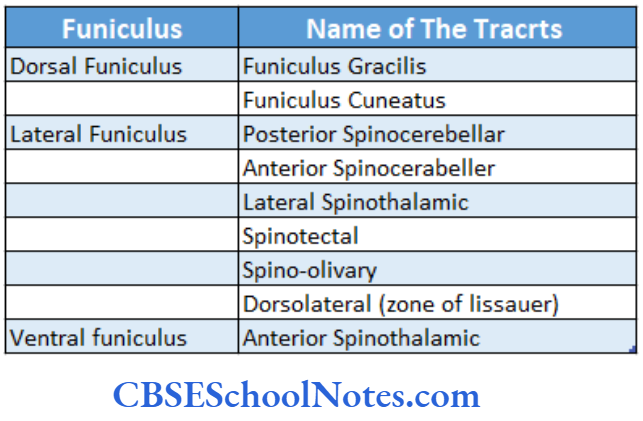
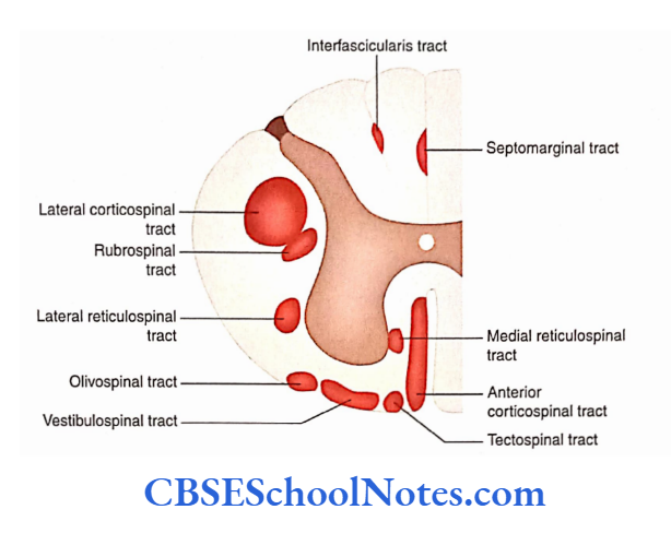
- These tracts are present in the anterior, lateral, and posterior funiculi (columns of white matter) of the spinal cord.
Ascending Tracts
Ascending Tracts
- The ascending tracts or the sensory tracts in the spinal cord carry somatic and visceral sensations.
- The somatic sensations include pain, touch, temperature, vibration, and pressure.
- The visceral sensory impulses are mainly in the form of pain and stretch sensations. Most visceral sensory and some somatic sensory impulses do not reach the level of consciousness.
Read and Learn More Neuroanatomy
Sensory Pathway
The sensations are first received by receptors, which are sensory end organs. Each receptor receives a stimulus and transforms it into a nerve impulse.
The nerve impulses are then carried through a pathway, which usually consists of three sensory neurons arranged in sequence:
- First-order,
- Second-order and
- Third-order neurons.
First-Order Neuron
The first-order sensory neurons are located in the dorsal root ganglia (DRG) of the spinal nerves and the corresponding root ganglion of the cranial nerves.
Second-Order Neuron
The cell bodies of second-order sensory neurons lie in the dorsal grey column (horn) of the spinal cord.
The axonal processes of the second-order sensor)7 neurons, as a general rule, cross to the opposite side before reaching the thalamus.
Hence, sensations from the left side of the body go to the right thalamus and vice versa.
As a result, the left side of the brain receives sensory information from the right side of the body while the right side of the brain senses the left side of the body.
Ascending Tracts
Third-Order Neuron
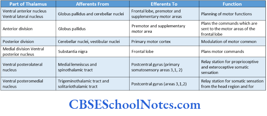
The cell bodies of third-order sensory neurons are located in the thalamus.
The axons of third-order sensory neurons from the thalamus project to the sensor)7 cortex of the cerebrum (postcentral gyrus; areas 3, 1, 2). Thus, general sensations are perceived in the postcentral gyrus in Various ascending tracts.
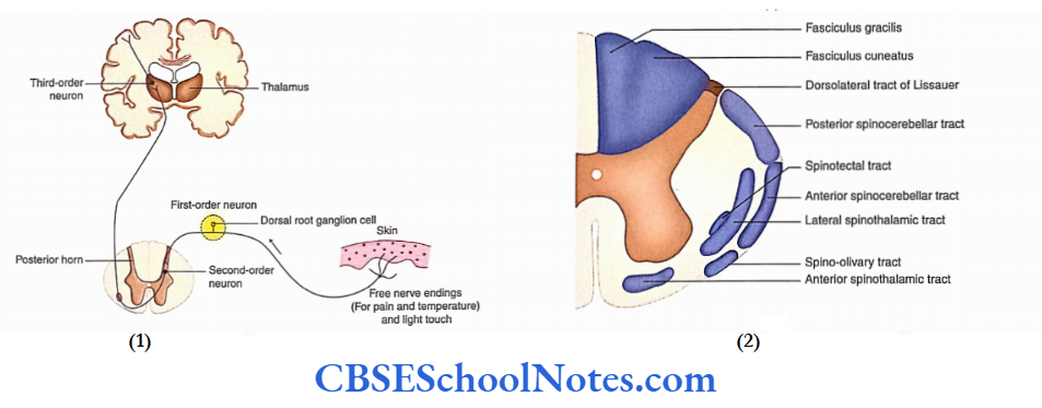
Ascending Tracts Of The Dorsal White Column
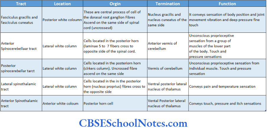
The ascending tracts located in the dorsal white column or funiculus of the spinal cord are fasciculus gracilis and fasciculus cuneatus.
Fasciculus Gracilis and Fasciculus Cuneatus
Fasciculus gracilis and fasciculus cuneatus carry sensations of conscious proprioception, fine touch, vibration and wo-point discrimination.
The lower spinal cord shows only fasciculus gracilis while the upper spinal cord shows both gracilis and cuneatus fasciculi.
Ascending Tracts
Sensory Pathway
The sensory pathway involves the participation of three sets of neurons: First-order, Second-order, and third-order sensory neurons.
First-Order Sensory Neurons
They lie in DRG. The central processes of the dorsal nerve root ascend in the dorsal funiculus without relaying in the posterior horn. The fibers of gracile and cuneate fasciculi terminate on the cell bodies of gracile and cuneate nuclei, respectively.
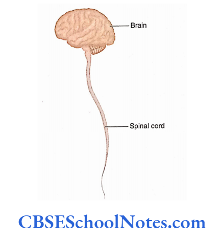
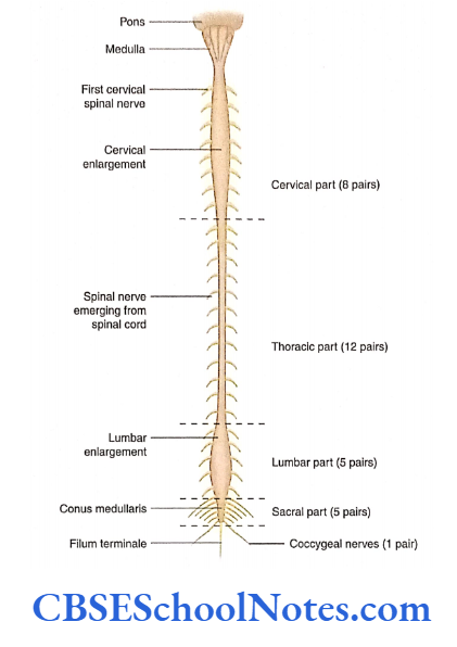
Spinal Cord
Second-Order Sensory Neurons
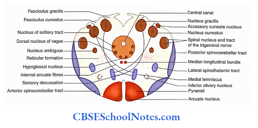
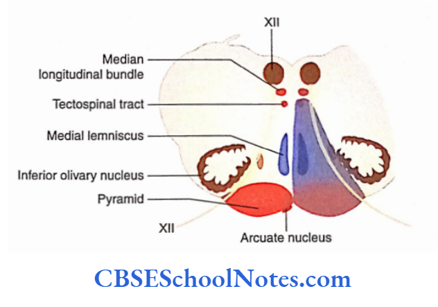
Nuclei gracilis and cuneatus contain second-order sensory neurons. These nuclei are located in the medulla. Their axons curve ventromedially as internal arcuate fibers.
These fibers decussate in the midline with the fibers arising from the nuclei located on the opposite side.
After decussation, these fibers then turn upwards to form a medial lemniscus.
This decussation is known as great sensory decussation. The fibers of the medial lemniscus ascend through the medulla, pons, and midbrain to terminate in the ventral-posterolateral nucleus of the thalamus.
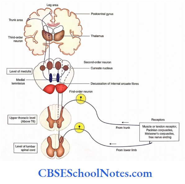
Third-Order Sensory Neurons
They are located in the ventral-posterolateral nucleus of the thalamus. The axons of third-order neurons end in the sensory area of the cerebral cortex (postcentral gyrus; areas 2, 1, 3).
Lesions of the Posterior Column (Tabes Dorsalis)
- The lesion of the posterior column will diminish or abolish the discriminating tactile and kinaesthetic sense.
- The loss of sense of position in the lower extremities greatly affects the equilibrium, stance, and gait.
- Usually, posterior column lesions are associated with syphilis infection.
- The degenerative disease of the posterior column due to syphilis is called tabes dorsalis.
Ascending Tracts
Ascending Tracts Of The Lateral White Column
The major ascending tracts of the lateral white column or funiculus are described as follows:
- Posterior spinocerebellar tract
- Anterior spinocerebellar tract
- Lateral spinothalamic tract
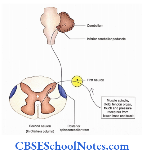
Posterior Spinocerebellar Tract
The posterior or dorsal spinocerebellar tract is uncrossed and carries unconscious proprioceptive impulses from joints, muscles, and tendons.
This tract forms a part of the neural pathway that carries these impulses from the lower limb and caudal part of the body to the cerebellum.
Posterior Spinocerebellar Tract Origin, Course, and Termination
The pathway involves two sets of neurons. This is in contrast to the sensory pathways going to the cerebral cortex, which usually consists of three neurons.
Posterior Spinocerebellar Tract First-Order Neurons
These are located in the DRG of the spinal nerves
Posterior Spinocerebellar Tract Second-Order Neurons
- The second-order neurons of this tract are large neurons of Clarke’s column. This nucleus extends from C8 to L3 spinal segments.
- Though these fibers are second-order neurons, they do not cross to the opposite side.
- These fibers ascend as a posterior spinocerebellar tract in the lateral white column of the spinal cord and end in the cerebellum through the inferior cerebellar peduncle.
Second-Order Neurons Functions
- The function of the dorsal spinocerebellar tract is to convey unconscious proprioceptive impulses from a single (individual) muscle.
- This information is useful in posture maintenance and body movements.
- These tracts also carry unconscious exteroceptive impulses (touch and pressure) to the cerebellum, which are utilized for the coordination of posture.
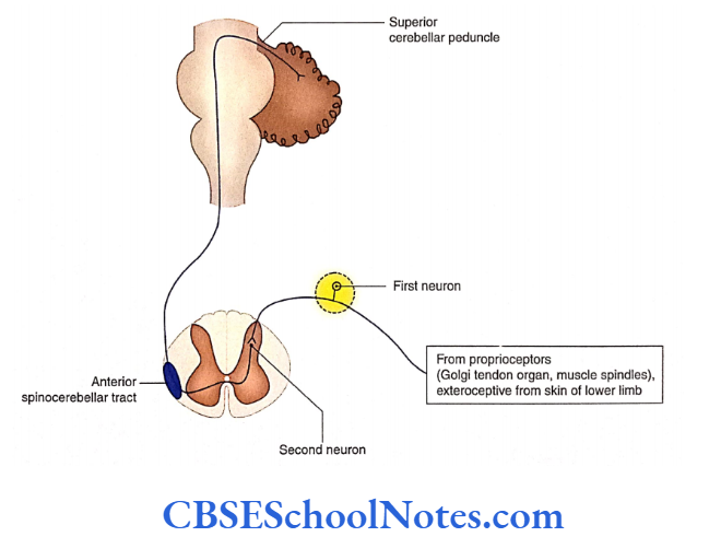
Anterior Spinocerebellar Tract
The anterior or ventral spinocerebellar tract is a crossed tract carrying proprioceptive and exteroceptive impulses from the lower limbs.
- Anterior Spinocerebellar Tract Origin, Course, and Termination
There are two sets of neurons which are involved in this pathway.
Ascending Tracts
- Anterior Spinocerebellar Tract First-Order Neuron
This lies in the DRG of the coccygeal, sacral, and lumbar spinal nerves.
- Anterior Spinocerebellar Tract Second-Order Neuron
The cell body of the second-order neuron is located in the dorsal horn.
The axons of the second-order neuron immediately cross to the opposite side and give origin to the ventral spinocerebellar tract.
Anterior Spinocerebellar Tract Function
Similar to a dorsal spinocerebellar tract, this tract also carries unconscious proprioceptive and exteroceptive impulses from the lower limbs.
Spinal Cord
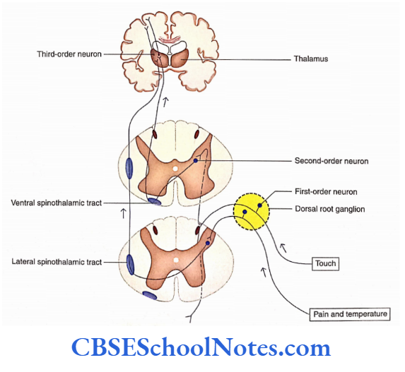
Lateral Spinothalamic Tract
The lateral spinothalamic tract is a crossed tract, which carries the sensations of pain and temperature. It arises at all levels of the spinal cord namly cervical thoracic lumbar and sacral.
Lateral Spinothalamic Tract Origin, Course, and Termination
Lateral Spinothalamic Tract First-Order Neurons
These lie in the DRG of spinal nerves. The central process of the DRG cells then makes synaptic contacts in the neurons of substantia gelatinosa, whose main function is to modify the pain and temperature impulses.
Thus, the neurons of substantia gelatinosa only act as interneurons These interneurons then project on the neurons of second order.
Lateral Spinothalamic Tract Second-Order Neurons
These are situated in laminae 1, 4, and 5 (of the nucleus proprius). Their dendrites synapse with substantia gelatinosa cells and their axons form the lateral spinothalamic tract.
These axons soon cross over to the opposite side in ventral white commissure and ascend upwards in the lateral funiculus as the lateral spinothalamic tract. This tract, after ascending through the brainstem, terminates in the VPL nucleus of the thalamus.
There may be some awareness of pain and temperature at the level of the thalamus.
Lateral Spinothalamic Tract Third-Order Neurons
These are situated in the VPL nucleus of the thalamus. Their axons project to the primary sensory cortex ofthe cerebral hemisphere.
Lateral Spinothalamic Tract Functions
- This tract conveys pain and temperature (hot and cold) sensations from the limbs and trunk.
- Unilateral damage to the spinothalamic tract leads to loss of pain and temperature (hot and cold) sensations from the skin of the opposite side of the body, below the lesion.
- As the fibers of pain lie anterior and superficial to the fibers of temperature in this tract, the cutting of the anterolateral white column cuts the pain fibers.
- This surgical procedure is called cordotomy, which is usually performed at the cervical level to relieve the severe pain associated with malignancy (cancer) of the neck or thorax.
Ascending Tracts of the Ventral White Column
The important ascending tract present in the ventral white column is the anterior or ventral spinothalamic tract.
Spinal Cord
Anterior or Ventral Spinothalamic Tract
This tract conveys touch, pressure, and itch sensations. The first-order neurons, after entering the spinal cord, may ascend or descend for new segments in the dorsolateral tract of Lissauer before terminating into posterior horn cells. The details of this tract.
Descending Tracts
The descending tracts of the spinal cord begin from the brain and terminate at different levels in the spinal cord. These tracts are motor in nature. These tracts are grouped
as pyramidal and extrapyramidal tracts.
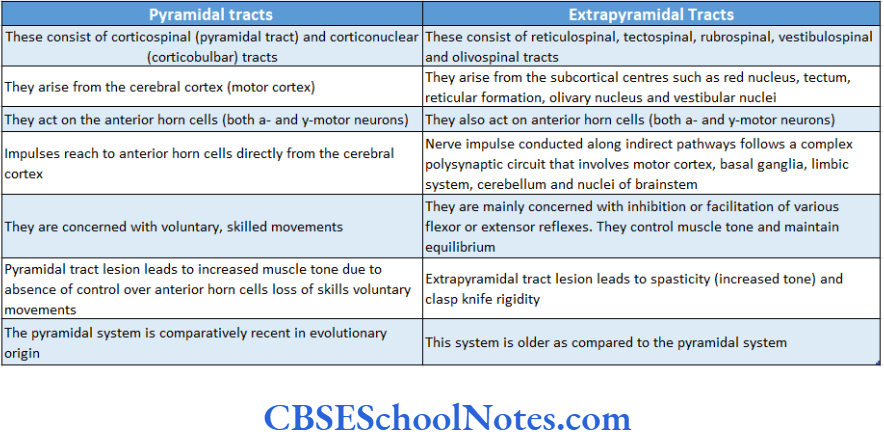
Pyramidal tracts: These tracts originate in the cerebral cortex and play a major role in controlling precise voluntary muscular movements.
Extrapyramidal tracts: These tracts originate mainly from the subcortical regions of the brain (corpus striatum, reticular nuclei, vestibular nuclei, olivary nuclei, etc.) and play an important role in integration for regulation of muscular movements and muscular tone.
The descending tracts are organized in the form of columns in the white matter of the spinal cord.
Pyramidal Tracts
The pyramidal tracts carry impulses that regulate precise and voluntary muscular movements. These tracts consist of fibers that originate from the motor cortex of the cerebrum.
The pyramidal tracts include the following:
- Corticonuclear, also called corticobulbar
- Corticospinal
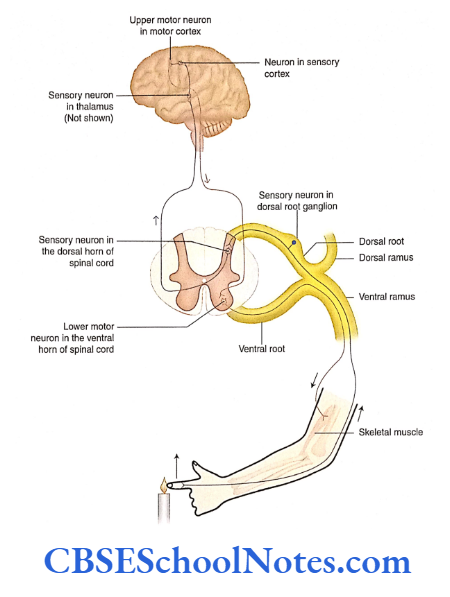
These tracts form a part of the motor pathway that extends from the brain to the skeletal muscles.
The pathway consists of two sets of motor neurons that are referred to as upper motor neurons and lower motor neurons, respectively (see in the following text).
Upper and lower motor neurons
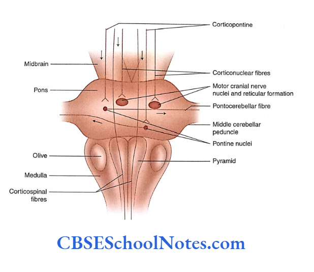
The upper motor neurons are situated in the cerebral cortex. These neurons send their axons to the different cranial nerve nuclei (motor neurons) in the brainstem, through corticobulbar/corticonuclear tracts.
The axons of the upper motor neurons also project to the anterior horn cells (motor neurons) at different levels of the spinal cord through corticospinal tracts.
The lower motor neurons are situated in the brainstem (cranial nerve nuclei) and the spinal cord (neurons located in the anterior horn).
The lower motor neurons receive impulses from the pyramidal tracts (corticonuclear and corticospinal), and these impulses are then transmitted along the axons of lower motor neurons to the skeletal muscles. Thus, the pyramidal system controls the voluntary movements of the body.
This system of descending fibers is known as the pyramidal system because the corticospinal fibers occupy the pyramid of the medulla.
It may be noted that though corticonuclear fibers are confined to the brainstem (i.e. they do not occupy the pyramid), however, they are also included in the category of the pyramidal system.
Corticonuclear or Corticobulbar Tracts
The fibers of these tracts arise from the neurons of the motor cortex, in the cerebrum, and end in the motor neurons of cranial nerve nuclei (located in the brainstem). However, before terminating in the cranial nerve nuclei these fibers cross to the opposite side.
Corticospinal Tract
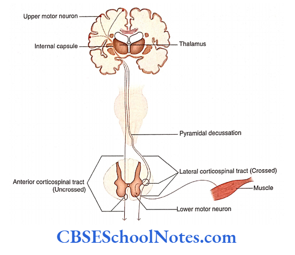
The fibers of this tract arise from the cells of the cerebral cortex and end in the anterior horn cells of the spinal cord.
- The corticospinal tract consists of axons arising mainly from the cells of the precentral gyrus (motor area; area 4), from giant-sized Betz cells.
- These fibers subsequently descend through the posterior limb of the internal capsule, cerebral peduncle of the midbrain, pons, and medulla. In the upper part
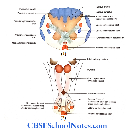
of the medulla, the corticospinal fibers form an elevated bundle known as a pyramid. - Below the level of the pyramid, about 75-90% of corticospinal fibers cross towards the opposite side. This crossing forms the pyramidal decussation.
- The fibers that have crossed to the opposite side descend downwards in the lateral funiculus of the spinal cord as the lateral corticospinal tract.
- The rest of the fibers of the pyramid remain uncrossed and descend downwards in the anterior funiculus as the anterior or ventral corticospinal tract.
Lateral Corticospinal Tract
This tract is present at almost all the levels of the spinal cord (up to the fourth sacral segment).
As it gives fibers to the anterior horn cells ofthe same side at all levels, it progressively diminishes in size from above downwards.
Ventral Corticospinal Tract
This tract consists of only 10-25% of uncrossed fibers of the pyramidal tract. This tract is present in the cervical and upper thoracic regions of the spinal cord and is not found below the mid-thoracic spinal segments.
These fibers terminate on the anterior horn cells of the opposite side by crossing at various levels.
Ventral Corticospinal Tract Functions
The corticospinal tract is concerned with the contraction of skeletal muscles; hence, it controls the voluntary and skilled movement of the body.
Lesion of Corticospinal Tract
- The damage to the corticospinal tract is the damage to the upper motor neurons of the pyramidal system. This leads to paralysis (loss of motor power) of the skeletal muscles.
- The part of the body paralyzed will depend upon the site of the lesion.
- The lesion at the level of the internal capsule results in the paralysis of the opposite side of the body (hemiplegia). A lesion of the tract in the spinal cord will lead to paralysis of the skeletal muscle below the level of the lesion on the same side.
Extrapyramidal Descending Spinal Tracts
The extrapyramidal system is responsible for regulating muscle tone as well as posture and equilibrium of the body.
The major descending tracts of the spinal cord included in the extrapyramidal system are as follows:
- Tectospinal
- Rubrospinal
- Vestibulospinal
- Olivospinal
- Reticulospinal
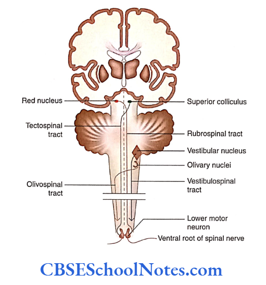
Tectospinal Tract
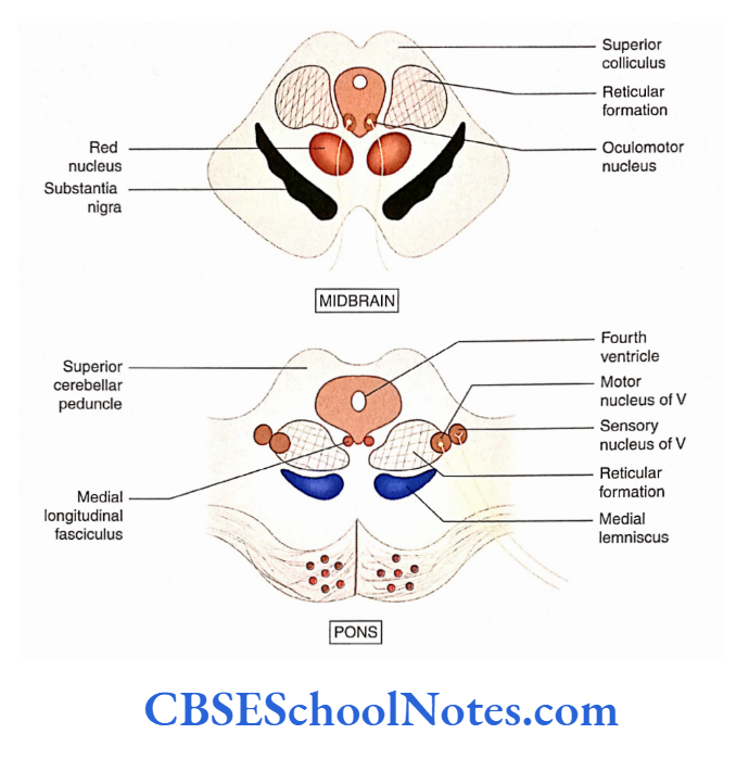
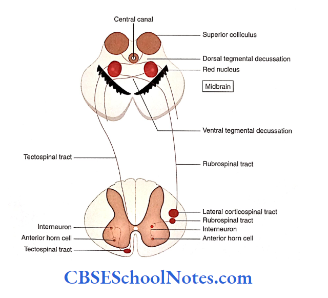
- The first-order neurons are situated in the superior colliculus. Axons from these neurons cross to the opposite side in front of periaqueductal grey and form dorsal tegmental decussation.
- After decussation, the fibers descend downwards in the spinal cord in the anterior funiculus.
- They ultimately synapse with the motor neurons of the ventral horn (second-order neurons), which innervate muscles of the neck.
Tectospinal Tract Function
The tectospinal tract conveys impulses for reflex postural movements in response to visual and auditory stimuli.
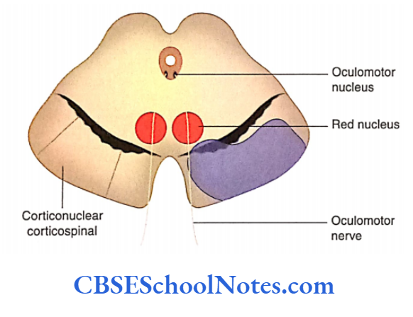
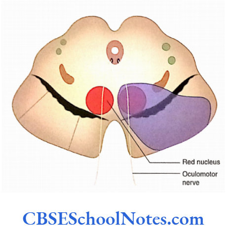
Rubrospinal Tract
- The first-order neurons of this tract lie in the red nucleus of the midbrain. The axons soon decussate in the tegmentum of the midbrain (ventral tegmental decussation) and cross to the opposite side to descend downwards in the lateral funiculus.
- The fibers of this tract synapse with the second-order neurons (alpha motor neurons of the ventral horn).
Rubrospinal Tract Functions
The rubrospinal tract exerts facilitator}’ influence on flexor muscle tone and inhibitory influence on extensor muscle tone.
Vestibulospinal Tract
- The vestibulospinal tract is concerned with the maintenance of the body’s equilibrium.
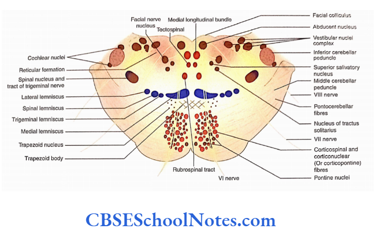
- This tract conveys the vestibular and cerebellar influences to the spinal cord. The vestibular nuclei are situated at the junction of the pons and medulla.
- The vestibulospinal tracts originate from the lateral and medial vestibular nuclei.
- The fibers of the lateral vestibulospinal tract extend throughout the spinal cord. The tract is facilitatory to motor neurons supplying extensor muscles.
- The medial vestibulospinal tract only reaches the upper thoracic region. This tract is inhibitory to the muscles of the neck and back.
Olivospinal Tract
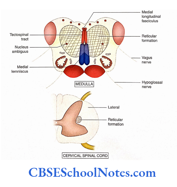
The olivary nucleus is situated deep to the olive in the medulla. This tract descends from the inferior olivary nucleus to neurons of the anterior horn in the spinal cord. The details of this tract are not known.
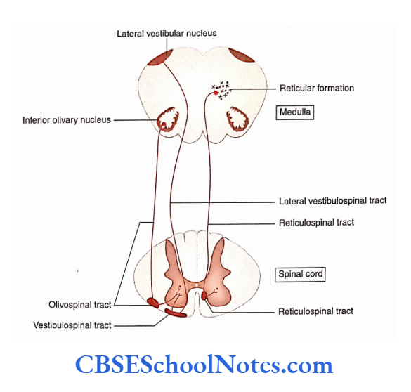
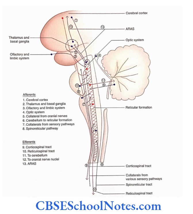
Reticulospinal Tract
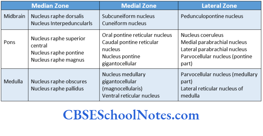
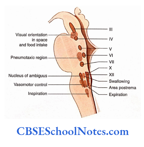
- A reticular formation is a group of scattered nerve cells and nerve fibers that are present in the midbrain, pons, and medulla. This tract arises from the neurons situated in the reticular formation of the brainstem.
- The axons of this tract descend in the anterior funiculus of the spinal cord.
- This tract reaches all the levels of the spinal cord. The fibers of this tract terminate on the alpha and gamma motor neurons, either directly or indirectly through interneurons.
Reticulospinal Tract Functions
The reticulospinal tract is facilitatory to the muscles of the trunk and limbs and helps in the maintenance of body posture. It also modifies the transmission of pain impluses.
Pyramidal and Extrapyramidal Tracts: A Comparison
Spinal Cord Trauma
- Trauma to the spinal cord occurs as a result of vertebral fractures. Most of these injuries are due to automobile or motorcycle accidents, sports-related injuries, falls from a height gunshot, and stab wounds. The trauma may result in complete transection, incomplete transection, or hemisection of the spinal cord.
- Trauma is the most common cause affecting the spinal cord. It occurs as a result of a fracture of the vertebral column (spine).
Complete transection of the spinal cord: It results in complete loss of motor control (paralysis) and loss of sensations at and below the level of injury to the spinal cord.
Hemisection of the spinal cord (Brown-Sequard syndrome): In this lesion, the lateral half of the cord is damaged. Extensive paralysis of muscles on the same side below the level of injury occurs while extensive sensory loss occurs on the opposite side (normal side). This phenomenon is called the BrownSequard syndrome.
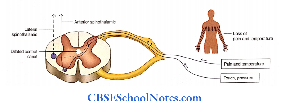
Syringomyelia: In this disease, dilatation of the central canal occurs, which leads to damage to decussating fibers of the spinothalamic tract in the cervical to upper thoracic segments of the cord.
Loss of pain and temperature sensations occurs in hands and arms bilaterally while the sense of touch is retained.
Complete Transection of the Spinal Cord
The complete transection of the spinal cord results in immediate loss of motor control (paralysis) and loss of sensation (anesthesia) at and below the level of injury to the spinal cord
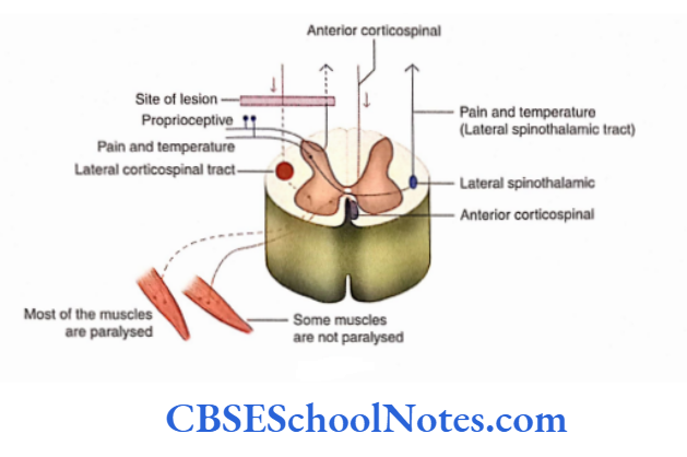
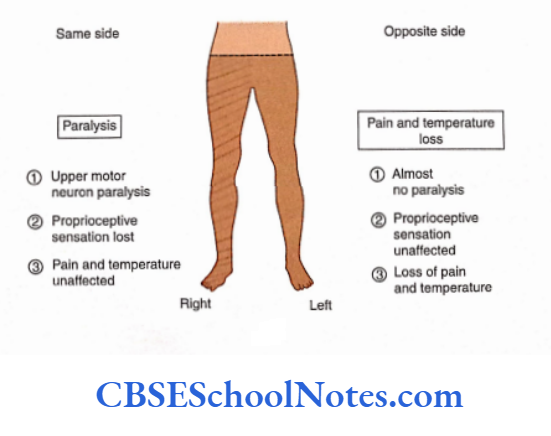
Hemisection of the Spinal Cord (Brown-Sequard Syndrome)
In this lesson on the spinal cord, the lateral half of the cord is damaged (lateral hemisection;. This usually happens due to gunshot injury. The clinical features of this injury include minimal changes in autonomic functions but extensive changes in the somatic nervous system.
Changes below the level of hemisection of the spinal cord
1. On the same side of injury
- Proprioceptive sensations such as vibration, kinaesthetic, fine touch, and tactile localization are lost on the same side of the lesion due to damage to the fasciculi gracilis and cuneatus (dorsal white column).
- As the spinothalamic tract crosses to the opposite side, the sensations of pain, temperature, and crude touch remain unaffected.
- As the lateral corticospinal tract (crossed pyramidal tract) gets damaged, all the skeletal muscles below the level of injury are paralyzed. The paralysis is of the UMN type.
- However, the anterior corticospinal tract (uncrossed pyramidal tract) which runs on the opposite side escapes injury. Therefore, some muscles on the side of the injury are not paralyzed.
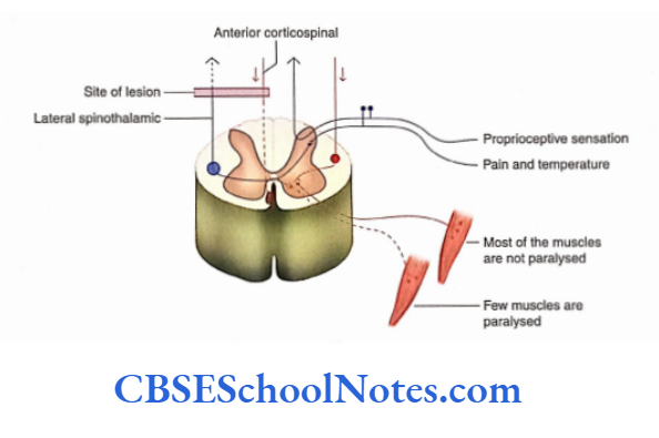
2. On the opposite side of injury
- There is no loss of proprioceptive sensation.
- Loss of pain, temperature, and crude touch below the level of injury because of the spinothalamic tract.
- Carrying these sensations crosses to the opposite side and gets damaged.
- Usually, there is no noticeable paralysis of muscles on this side.
- Damage to the direct pyramidal tract (anterior corticospinal tract) may lead to paralysis of a few muscles on this side.
- To summarise the changes in the preceding text, it may be said that, below the level of lesion, extensive paralysis of muscles occurs on the same side of injury, while extensive sensory loss occurs on the opposite side (normal side). This phenomenon is called the Brown-Sequard syndrome.
Degenerative And Demyelinating Disorders Of The Spinal Cord
Syringomyelia
- Syringomyelia is a chronic disease characterized by the formation of cavities in the central canal of the spinal cord.
- This results in the dilatation of the central canal and damage to the nervous tissue (spinothalamic tract) surrounding the central canal.
- At the beginning of the disease, loss of pain and thermal sensations in the hands and arms bilaterally with the preservation of touch sensation occurs.
- Touch is retained as it has a double pathway. Fine touch ascends in the dorsal white column.
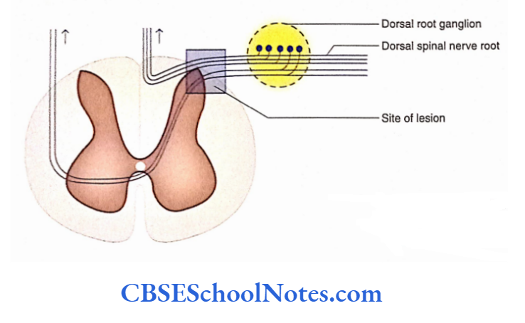
Tabes Dorsalis
- Tabes dorsalis is a disease that leads to sensory disturbances, usually in the lower limbs. This disease is caused due to immunological reactions in patients suffering from syphilis.
- Because of the immunological reaction, degeneration in the central processes of spined ganglion cells occurs.
- The site of the lesion is located on the dorsal spinal nerve root, just lateral to its entrance into the spinal cord.
Ascending Descending Tracts Of The Spinal Cord Summary
- The ascending and descending fibers in the spinal cord are organized in the form of ‘tracts’.
- A tract is defined as ‘the bundle of fibers having the same origin, course, termination and function’.
- The ascending tracts of the spinal cord carry sensory information to the brain while the descending fibres carry motor information from the brain towards the peripheral tissues.
- These tracts are present in the anterior, lateral, and posterior funiculi of the spinal cord.
Ascending Tracts
- The sensory nerve impulses, in ascending tracts, are usually carried through three sensory neurons arranged in sequence: first-order, second-order, and third-order neurons.
- The first-order sensory neuron is located in the dorsal root ganglion of the spinal nerve and third-order neurons are located in the thalamus.
- As a general rule, the axons of the second-order sensory neurons cross to the opposite side before reaching the thalamus.
1. Ascending tracts in the dorsal white column (funiculus)
Fasciculi gracilis and cuneatus: These tracts carry the sensations of conscious proprioception—fine touch, vibration, and two-point discrimination.
The first-order sensory neurons lie in the dorsal root ganglion Their central processes ascend in the dorsal funiculus without relay in the posterior horn.
These fibers terminate on second-order sensory neurons present in gracile and cuneate nuclei Fibres of the second-order neurons (internal arcuate fibers) decussate in the midline to form medial lemniscus.
These fibers ascend through the medulla, pons, and midbrain to terminate on third-order neurons in the thalamus. The third-order neurons project on the cerebral cortex.
2. Ascending tracts in the lateral white column
Posterior spinocerebellar tract: It carries unconscious proprioceptive impulses, touch, and pressure sensation from the lower limb and caudal part ofthe body to the cerebellum. This pathway consists of only two neurons.
The first-order sensory neurons lie in the dorsal root ganglion Their central processes end in the nucleus dorsalis (Clarke’s column) of the dorsal grey horn of the spinal cord The axons of the second-order neurons form the dorsal spinocerebellar tract. This tract ascends on the same side tract ends in the vermis of the cerebellum.
Anterior spinocerebellar tract: It carries unconscious proprioceptive and exteroceptive impulses. There are two sets of neurons involved in this pathway.
The first-order neurons lie in the dorsal root ganglion Their central processes end in dorsal grey column cells The axons of the second-order neuron immediately cross to the opposite side and give origin to the ventral spinocerebellar tract.
This tract runs through the medulla, pons, midbrain, and superior cerebellar peduncle to terminate in the opposite vermis of the cerebellum Thus, fibers of the second-order neurons cross twice and reach the ipsilateral cerebellar hemisphere.
Lateral spinothalamic tract: It is a crossed tract, which carries sensations of pain and temperature. The first-order sensory neurons are located in the dorsal root ganglion The central process of these neurons makes synaptic contact with substantia gelatinosa The second-order neurons are situated in the nucleus proprius.
The axons of second-order neurons soon cross to the opposite side to form the lateral spinothalamic tract The tract after ascending through the brainstem terminates in the thalamus The third-order neurons of the thalamus project on the sensory cortex of the cerebrum.
3. Ascending tract in the ventral white column
Ventralspinothalamic tract: It conveys the sensations of touch, pressure, and itch.
The first-order neurons are in the dorsal root ganglion → The second-order neurons are located in the dorsal horn of the spinal cord →The axons of these cells cross to the opposite side to form the ventral spinothalamic tract The tract terminates in the thalamus from where third-order neurons send their axons to the cerebral cortex.
Descending Tracts
1. Pyramidal tracts: These tracts play an important role in the control of precise voluntary muscular movements. These tracts begin from the brain (cerebrum) and terminate at different levels of the spinal cord. The pyramidal pathway consists of two sets of neurons:
- Upper motor neurons (humans) and
- Lower motor neurons (lmns).
Corticonuclear tracts: The first-order neurons are located in the motor cortex which sends their axons to cranial nerve nuclei. These axons constitute the corticonuclear tract. The axons of second-order neurons innervate the skeletal nucleus.
Corticospinal tracts: They are concerned with the control of voluntary and skilled movements ofthe body.
The first-order motor neurons (UMN) are located in the cerebral cortex The axons of these neurons pass through the midbrain, pons, and medulla as the pyramidal tract and form a pyramid in the medulla Below the level of the pyramid, 75-90% fibers cross towards the opposite side and form the lateral corticospinal tract.
The rest of the fibers of the pyramid remain uncrossed and descend as the ventral corticospinal tract The second-order motor neurons (LMN) are located in the anterior horn of the spinal cord.
Fibers of both corticospinal tracts (ventral and lateral corticospinal) make synaptic contact with the anterior horn cells of the opposite side.
2. Extrapyramidal tracts: These descending tracts originate mainly from the subcortical regions of the brain and terminate at different levels of the spinal cord.
The extrapyramidal system is responsible for regulating muscle tone as well as posture and equilibrium of the body. These tracts are not involved in voluntary control over skeletal muscles.
Tectospinal tract: It is concerned with visuospinal reflexes. The first-order neurons are situated in the superior colliculus.
Axons of this tract cross to the opposite side in tegmental decussation and terminate on the motor neurons of the ventral horn (second-order neurons) of the cervical spinal cord Axons of motor neurons innervate muscles of the neck.
Rubrospinal tract: It is concerned with the regulation of tone in the flexor muscles. The first-order neurons are situated in the red nucleus Axons of these neurons cross to the opposite side in ventral tegmental decussation and form the rubrospinal tract The second-order neurons are located in the ventral horn cells of the spinal cord.
Vestibulospinal tract: It is concerned with the maintenance of body equilibrium. The first-order neurons are located in medial and lateral vestibular nuclei Axons of these neurons descend on the same side Fibres of the tract terminate on second-order neurons (ventral horn cells).
Olivospinal tract: The first-order neurons are situated in the inferior olivary nucleus The second-order neurons of this tract are situated in the anterior horn of the spinal cord.
Reticulospinal tract: It is concerned with regulating the muscle tone in antigravity muscles. The first-order neurons are situated in the reticular formation of the brainstem.
Axons of these neurons form two tracts:
Medial reticulospinal and lateral reticulospinal The second-order neurons are alpha and gamma motor neurons of the spinal cord.